. 2019 Jun 20;10(4):229-242.
doi: 10.1080/21501203.2019.1631896. eCollection 2019.

Synthesis and role of melanin for tolerating in vitro rumen digestion in Duddingtonia flagrans, a nematode-trapping fungus

Affiliations

Synthesis and role of melanin for tolerating in vitro rumen digestion in Duddingtonia flagrans, a nematode-trapping fungus

Deivid França Freitas et al. Mycology. .

Abstract

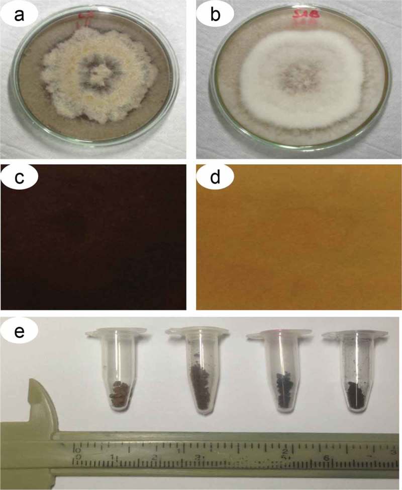
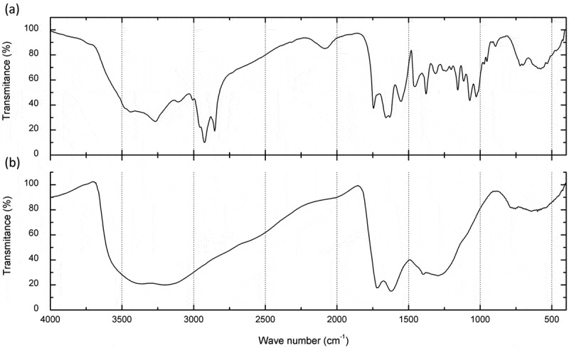
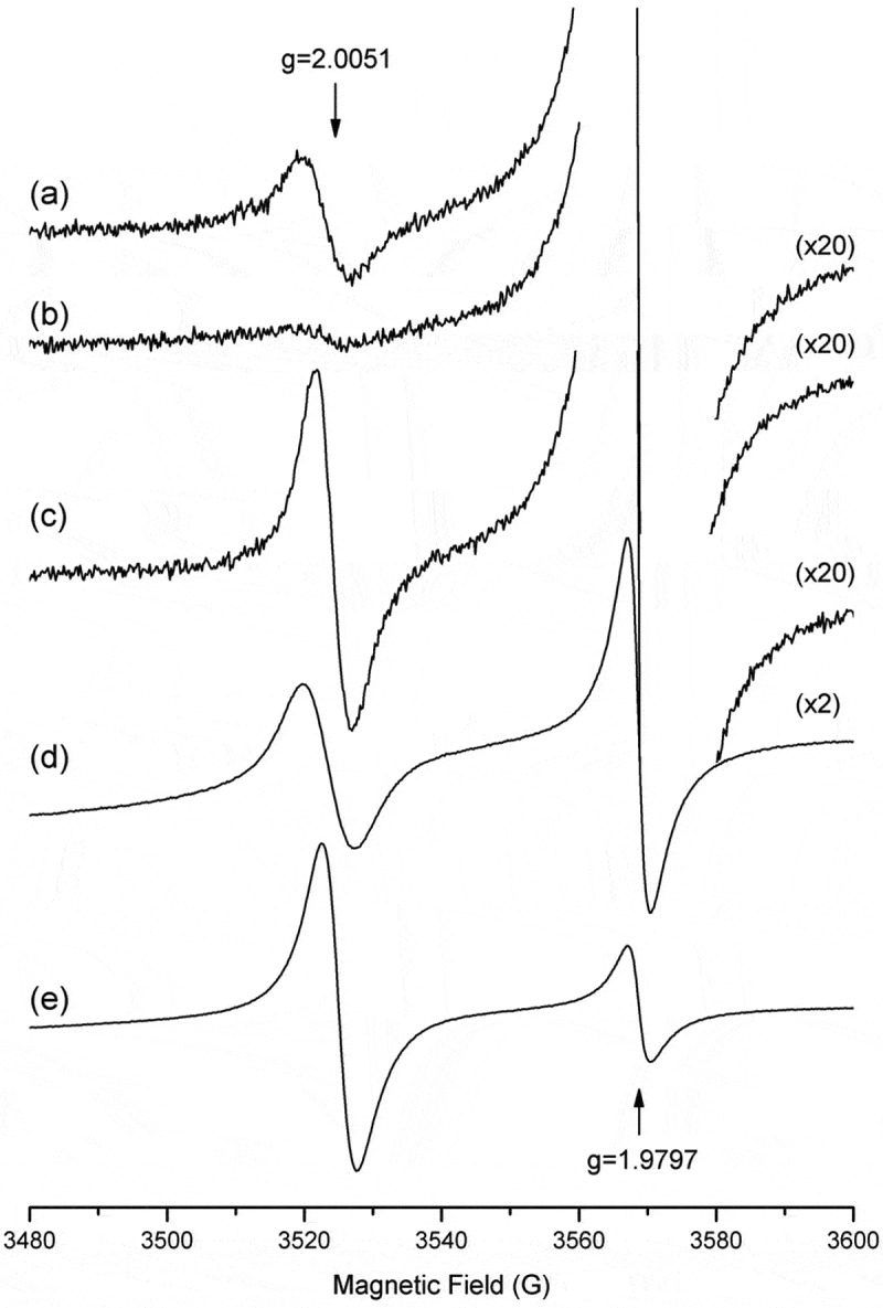
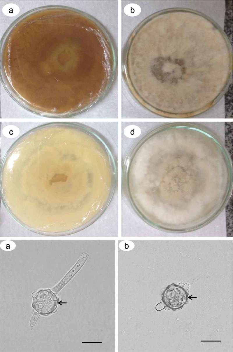
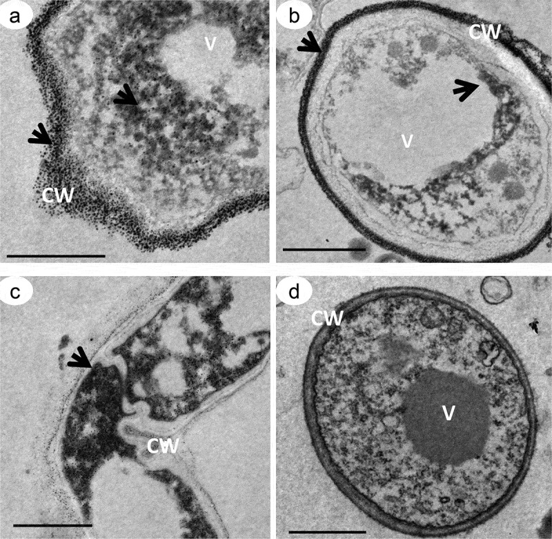
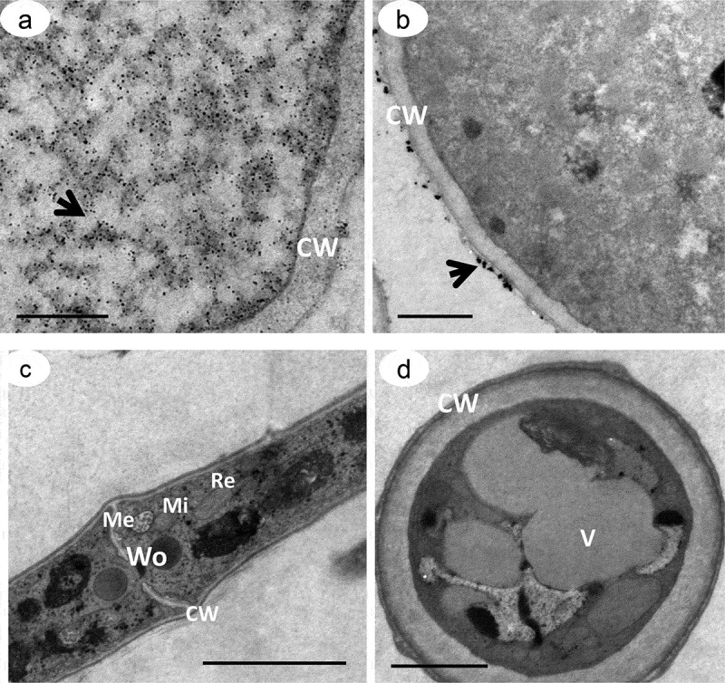
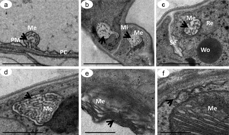
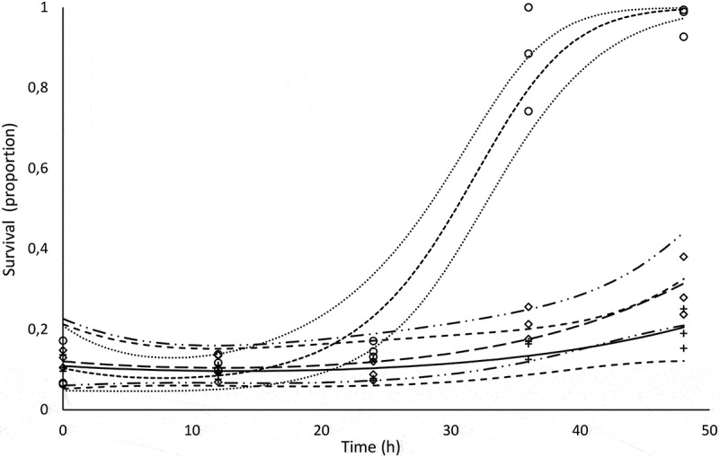
We describe the synthesis and a function of melanin in Duddingtonia flagrans, a nematode-trapping fungus. We tested various culture media treated with L-DOPA, glucose and tricyclazole on fungal growth and melanin distribution using infrared spectroscopy (IS), electron paramagnetic resonance (EPR) and transmission electron microscopy (TEM). In vitro rumen digestion was used to test the environmental stress and then to evaluate the capacity of this fungus to trap nematode larvae. The growth and melanization of the fungus after 21 days of incubation at 30°C were best in Sabouraud dextrose medium. IS indicated the presence of melanin in D. flagrans, with similar bands for commercial melanin used as a control, and assigned the values obtained by EPR (g of 2.0051 ± 0.0001) to the production of melanin by the fungus. TEM indicated that melanin was produced in melanosomes but was not totally inhibited by tricyclazole. Within the limits of experimental error, the predatory activity of fungus treated with tricyclazole was drastically affected after 27 h of in vitro anaerobic stress with rumen inoculum. The deposition of melanin particles on the fungal cell wall contributed to the maintenance of D. flagrans predatory abilities after in vitro anaerobic ruminal stress.

Keywords: Duddingtonia flagrans; L-DOPA; melanin; nematode-trapping fungi; tricyclazole.


Figures

Figure 1.
Figure 1.
Pigmentation of D. flagrans colonies on Sabouraud dextrose agar. Colony view and culture medium with (a) and without (b) addition of L-DOPA and ventral view of a Petri dish with culture medium with (c) and without (d) addition of L-DOPA. e) Melanin particles extracted from five colonies grown in Petri dishes.
Figure 2.
Figure 2.
Infrared spectroscopic analysis. a) One milligram of melanin particles extracted from five colonies of D. flagrans and b) 1 mg of commercial melanin. Note that the amplitude of the signals demonstrates the similarity between the commercial and the D. flagrans melanin. The other wave spectra were due to the presence of radicals in the melanin particles extracted from the fungus.
Figure 3.
Figure 3.
EPR spectra. a) Colony of D. flagrans in SDA medium, b) D. flagrans treated with 160 μg of tricyclazole, c) D. flagrans colony supplemented with 1 mM L-DOPA, (d) melanin particles extracted from D. flagrans and (e) commercial melanin. The 84% lower free-radical concentration can be seen in the tricyclazole-treated sample (b), and an increase of up to three-fold can be seen in the sample supplemented with L-DOPA(c), relative to no treatment (a). A sample MgO:Cr3+ (g of 1.9797) was used as a reference to obtain g for the sample studied.
Figure 4.
Figure 4.
Inhibition of melanization by tricyclazole in the D. flagrans colonies. Colony view and culture medium without (a, b) and with (c, d) treatment with 160 μg/mL of tricyclazole. Chamydospores from plates with (e) and without (f) treatment with 160 μg/mL of tricyclazole. Cell wall (arrow). Bars: 50 μm.
Figure 5.
Figure 5.
Transmission electron microscopy of D. flagrans demonstrating the location of the electron-dense particles of melanin. Culture medium with addition of L-DOPA. Fungal hypha (a, c), chlamydospores (b, d), cell wall (CW) and vacuoles (V). Samples contrasted with 10% silver nitrate (a, b), and samples contrasted with 1% osmium tetroxide and 0.8% potassium ferricyanide (c, d). The arrows indicate the electron-dense particles. Scale bars: 5 μm (a-c) and 2 μm (d).
Figure 6.
Figure 6.
Transmission electron microscopy of D. flagrans treated with 160 μg/mL of tricyclazole, an inhibitor of melanin synthesis. Culture medium without addition of L-DOPA. Note the reduced distribution of the electron-dense particles throughout the cell (a-d) compared to Figure 5. The arrows indicate the melanin particles. Note the absence of melanin granules for the samples stained with 1% osmium tetroxide and 0.8% potassium ferricyanide (c, d). Cell wall (CW), melanosome (Me), mitochondria (Mi), vacuoles (V), endoplasmic reticulum (Re) and Woronin body (Wo). Scale bars: 5 μm (a) and 2 μm (b-d).
Figure 7.
Figure 7.
Transmission electron microscopy of D. flagrans melanosomes. Ultrathin slices demonstrating the formation of melanosomes in stages II to III (a-d) in cells treated with 160μg/mL of tricyclazole and in stage III in cells treated with 1mM L-DOPA (e-f). Melanosome (Me), plasma membrane (PM), mitochondria (Mi), Woronin body (Wo) and septum between hyphae (Se). Arrows show the arrangements of internal matrices in the melanosomes. Scale bars: 500 nm (a, d, e, f) and 1 μm (b, c).
Figure 8.
Figure 8.
Observed and predicted survival proportions of the L3 larvae inoculated with chlamydospores of Duddingtonia flagrans. Diamonds, circles, and crosses are observed survivals for L-DOPA, tricyclazole and control, respectively. The small-spaced dashed line is the predicted survival of L3 with fungal melanin inhibitor (tricyclazole), and the dotted lines above and below it are 0.95 confidence limits (0.95CI). The solid line is the predicted L3 survival in the control medium with the fungus, and the large-spaced dashed lines form the 0.95CI. The long-dashed line is the predicted survival of L3 with L-DOPA melanin stimulant, and the dashed-double-dotted lines correspond to 0.95CI.

References

    1. Allam NG, El-Zaher EHF.. 2014. Protective role of Aspergillus fumigatus melanin against ultraviolet (UV) irradiation and Bjerkandera adusta melanin as a candidate vaccine against systemic candidiasis. Afr J Biotechnol. 24:6566–6577.
    1. Almeida-Paes R, Frases S, Monteiro PCF, Gutierrez-Galhardo MC, Zancopé-Oliveira RM, Nosanchuk JD. 2009. Growth conditions influence melanization of Brazilian clinical Sporothrix schenckii isolates. Microbes Infect. 5:554–562. - PMC - PubMed
    1. Alviano DS, Franzen AJ, Travassos LR, Holandino C, Rozental S, Ejzemberg R, Alviano CS, Rodrigues ML. 2004. Melanin from Fonsecaea pedrosoi induces production of human antifungal antibodies and enhances the antimicrobial efficacy of phagocytes. Infect Immun. 2:229–237. - PMC - PubMed
    1. Andersson KM, Meerupati T, Levander F, Friman E, Ahrén D, Tunlid A. 2013. Characterization of the proteome of the nematode-trapping cells of the fungus Monacrosporium haptotylum. Appl Environ Microbiol. 16:4993–5004. - PMC - PubMed
    1. Andrade MC, Hiura E, Fonseca LA, Magri C, Ferraz CCS, Sena FP, Giradi FM. 2016. Utilization of the Fungus Duddingtonia flagrans in Control of Nematode Larvae Development in Equine Stool. Curr Microbiol. 3:829–835.

LinkOut - more resources