Review
. 2019 Sep 26:9.
doi: 10.7916/tohm.v0.682. eCollection 2019.

Neuroradiological Findings in the Spinocerebellar Ataxias

Affiliations
Review

Neuroradiological Findings in the Spinocerebellar Ataxias

Alex Tiburtino Meira et al. Tremor Other Hyperkinet Mov (N Y). .

Abstract

Background: The spinocerebellar ataxias (SCAs) are a group of autosomal dominant degenerative diseases characterized by cerebellar ataxia. Classified according to gene discovery, specific features of the SCAs - clinical, laboratorial, and neuroradiological (NR) - can facilitate establishing the diagnosis. The purpose of this study was to review the particular NR abnormalities in the main SCAs.

Methods: We conducted a literature search on this topic.

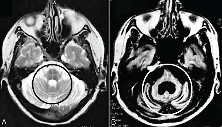
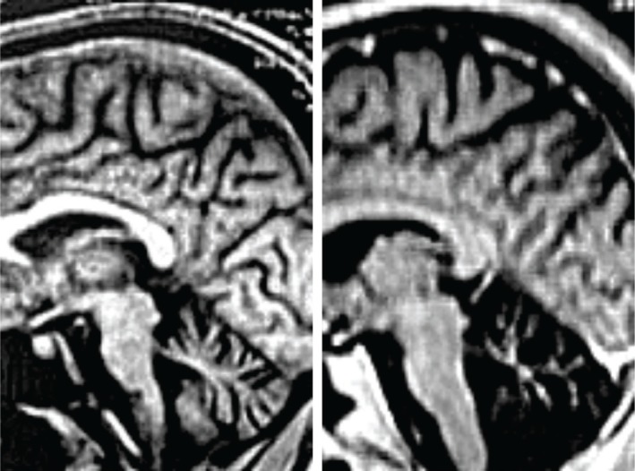
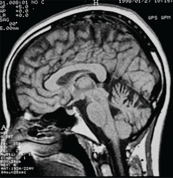
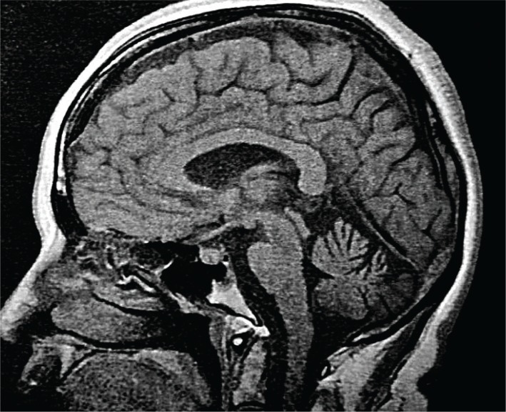
Results: The main NR characteristics of brain imaging (magnetic resonance imaging or computerized tomography) in SCAs were: (1) pure cerebellar atrophy; (2) cerebellar atrophy with other findings (e.g., pontine, olivopontocerebellar, spinal, cortical, or subcortical atrophy; "hot cross bun sign", and demyelinating lesions); (3) selective cerebellar atrophy; (4) no cerebellar atrophy.

Discussion: The main NR abnormalities in the commonest SCAs, are not pathognomonic of any specific genotype, but can be helpful in limiting the diagnostic options. We are progressing to a better understanding of the SCAs, not only genetically, but also pathologically; NR is helpful in the challenge of diagnosing the specific genotype of SCA.

Keywords: Spinocerebellar ataxia; ataxia; brain imaging; cerebellar diseases; gait ataxia; magnetic resonance imaging.


Conflict of interest statement

Funding: None. Conflicts of Interest: The authors report no conflicts of interest. Ethics Statement: Not applicable for this category of article.

Figures

Figure 1
Figure 1
The “Hot Cross Bun Sign” or “Cross Sign”, Typical but Not Pathognomonic Sign of Multiple System Atrophy (MSA), Can be Found in the Spinocerebellar Ataxias (SCAs): (A) a case of MSA and (B) SCA 2. Circles – T2 hyperintensity forms a cross on axial images through the pons, representing selective degeneration of pontocerebellar tracts. (A) Axial T2 image shows atrophy in the pons, bilateral middle cerebellar peduncle, and cerebellum. (B) Axial T2 FLAIR image shows severe atrophy in pons and cerebellum. A larger dilatation of the fourth ventricle is also noted.
Figure 2
Figure 2
Spinocerebellar Ataxia 3 (SCA 3): Initial And Advanced Stages of the Disease. Sagittal T1-weighted images of the brain in patient with SCA 3. Brain stem and cerebellum atrophy are clearly visible from the onset of clinical manifestations. With the evolution of the disease, there is an increase mainly in cortical and cerebellar atrophy.
Figure 3
Figure 3
Spinocerebellar Ataxia 10 (SCA 10) Image. Sagittal T1-weighted image of the brain in patient with SCA 10 showing cerebellar atrophy.
Figure 4
Figure 4
Dentatorubral-pallidoluysian atrophy image. A T1-weighted midsagittal image of a patient with DRPLA shows atrophy of the brain stem and cerebellum.

References

    1. Arruda WO, Teive HA. Ataxias cerebelares hereditárias. Do martelo ao gen. Arq Neuropsiquiatr 1997;55(3 B):666–676. doi: 10.1590/S0004-282X1997000400027 - DOI - PubMed
    1. Döhlinger S, Hauser T, Borkert J, Luft AR, Schulz JB. Magnetic resonance imaging in spinocerebellar ataxias. Cerebellum 2008;7(2):204–214. doi: 10.1007/s12311-008-0025-0 - DOI - PubMed
    1. Deistung A, Stefanescu MR, Ernst TM, et al. . Structural and functional magnetic resonance imaging of the cerebellum: considerations for assessing cerebellar ataxias. Cerebellum 2016;15(1):21–25. doi: 10.1007/s12311-015-0738-9 - DOI - PubMed
    1. Teive HAG, Ashizawa T. Primary and secondary ataxias. Curr Opin Neurol 2015;28:413–422. doi: 10.1097/WCO.0000000000000227 - DOI - PMC - PubMed
    1. Schöls L, Bauer P, Schmidt T, Schulte T, Riess O. Autosomal dominant cerebellar ataxias: clinical features, genetics, and pathogenesis. Lancet Neurol 2004;3:291–304. doi: 10.1016/S1474-4422(04)00737-9 - DOI - PubMed

LinkOut - more resources