

Radiomics Analysis of Dynamic Contrast-Enhanced Magnetic Resonance Imaging for the Prediction of Sentinel Lymph Node Metastasis in Breast Cancer
- PMID: 31632912
- PMCID: PMC6778833
- DOI: 10.3389/fonc.2019.00980
Radiomics Analysis of Dynamic Contrast-Enhanced Magnetic Resonance Imaging for the Prediction of Sentinel Lymph Node Metastasis in Breast Cancer
Abstract
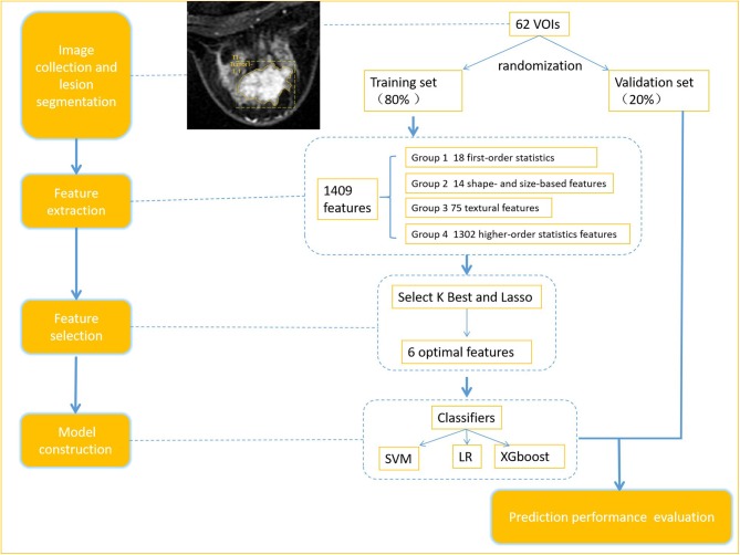
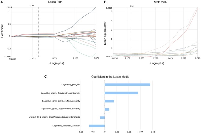
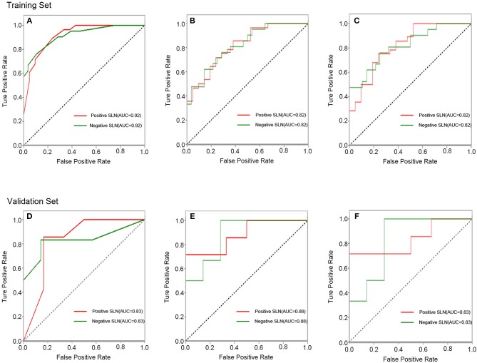
Purpose: To investigate whether a combination of radiomics and automatic machine learning applied to dynamic contrast-enhanced magnetic resonance imaging (DCE-MRI) of primary breast cancer can non-invasively predict axillary sentinel lymph node (SLN) metastasis. Methods: 62 patients who received a DCE-MRI breast scan were enrolled. Tumor resection and sentinel lymph node (SLN) biopsy were performed within 1 week after the DCE-MRI examination. According to the time signal intensity curve, the volumes of interest (VOIs) were delineated on the whole tumor in the images with the strongest enhanced phase. Datasets were randomly divided into two sets including a training set (~80%) and a validation set (~20%). A total of 1,409 quantitative imaging features were extracted from each VOI. The select K best and least absolute shrinkage and selection operator (Lasso) were used to obtain the optimal features. Three classification models based on the logistic regression (LR), XGboost, and support vector machine (SVM) classifiers were constructed. Receiver Operating Curve (ROC) analysis was used to analyze the prediction performance of the models. Both feature selection and models construction were firstly performed in the training set, then were further tested in the validation set by the same thresholds. Results: There is no significant difference between all clinical and pathological variables in breast cancer patients with and without SLN metastasis (P > 0.05), except histological grade (P = 0.03). Six features were obtained as optimal features for models construction. In the validation set, with respect to the accuracy and MSE, the SVM demonstrated the highest performance, with an accuracy, AUC, sensitivity (for positive SLN), specificity (for positive SLN) and Mean Squared Error (MSE) of 0.85, 0.83, 0.71, 1, 0.26, respectively. Conclusions: We demonstrated the feasibility of combining artificial intelligence and radiomics from DCE-MRI of primary tumors to predict axillary SLN metastasis in breast cancer. This non-invasive approach could be very promising in application.
Keywords: DCE-MRI; automatic machine learning; breast cancer; radiomics; sentinel lymph node metastasis.
Copyright © 2019 Liu, Sun, Chen, Fang, Song, Guo, Ni, Liu, Feng, Xia, Zhang and Li.
Figures
Similar articles
-
Pharmacokinetic parameters and radiomics model based on dynamic contrast enhanced MRI for the preoperative prediction of sentinel lymph node metastasis in breast cancer.Cancer Imaging. 2020 Sep 15;20(1):65. doi: 10.1186/s40644-020-00342-x. Cancer Imaging. 2020. PMID: 32933585 Free PMC article.
-
Value of the Application of CE-MRI Radiomics and Machine Learning in Preoperative Prediction of Sentinel Lymph Node Metastasis in Breast Cancer.Front Oncol. 2021 Nov 19;11:757111. doi: 10.3389/fonc.2021.757111. eCollection 2021. Front Oncol. 2021. PMID: 34868967 Free PMC article.
-
Preoperative prediction of sentinel lymph node metastasis in breast cancer by radiomic signatures from dynamic contrast-enhanced MRI.J Magn Reson Imaging. 2019 Jan;49(1):131-140. doi: 10.1002/jmri.26224. Epub 2018 Sep 1. J Magn Reson Imaging. 2019. PMID: 30171822 Free PMC article.
-
The Diagnostic Performance of Machine Learning-Based Radiomics of DCE-MRI in Predicting Axillary Lymph Node Metastasis in Breast Cancer: A Meta-Analysis.Front Oncol. 2022 Feb 4;12:799209. doi: 10.3389/fonc.2022.799209. eCollection 2022. Front Oncol. 2022. PMID: 35186739 Free PMC article.
-
A meta-analysis of the diagnostic performance of machine learning-based MRI in the prediction of axillary lymph node metastasis in breast cancer patients.Insights Imaging. 2021 Nov 3;12(1):156. doi: 10.1186/s13244-021-01034-1. Insights Imaging. 2021. PMID: 34731343 Free PMC article. Review.
Cited by
-
A Nomogram Based on Radiomics with Mammography Texture Analysis for the Prognostic Prediction in Patients with Triple-Negative Breast Cancer.Contrast Media Mol Imaging. 2020 Aug 25;2020:5418364. doi: 10.1155/2020/5418364. eCollection 2020. Contrast Media Mol Imaging. 2020. PMID: 32922222 Free PMC article.
-
Pharmacokinetic parameters and radiomics model based on dynamic contrast enhanced MRI for the preoperative prediction of sentinel lymph node metastasis in breast cancer.Cancer Imaging. 2020 Sep 15;20(1):65. doi: 10.1186/s40644-020-00342-x. Cancer Imaging. 2020. PMID: 32933585 Free PMC article.
-
Application of CT and MRI images based on an artificial intelligence algorithm for predicting lymph node metastasis in breast cancer patients: a meta-analysis.BMC Cancer. 2023 Nov 22;23(1):1134. doi: 10.1186/s12885-023-11638-z. BMC Cancer. 2023. PMID: 37993845 Free PMC article.
-
Identification of ipsilateral supraclavicular lymph node metastasis in breast cancer based on LASSO regression with a high penalty factor.Front Oncol. 2024 Feb 2;14:1349315. doi: 10.3389/fonc.2024.1349315. eCollection 2024. Front Oncol. 2024. PMID: 38371618 Free PMC article.
-
Value of the Application of CE-MRI Radiomics and Machine Learning in Preoperative Prediction of Sentinel Lymph Node Metastasis in Breast Cancer.Front Oncol. 2021 Nov 19;11:757111. doi: 10.3389/fonc.2021.757111. eCollection 2021. Front Oncol. 2021. PMID: 34868967 Free PMC article.
References
-
- Coutant C, Olivier C, Lambaudie E, Fondrinier E, Marchal F, Guillemin F, et al. . Comparison of models to predict nonsentinel lymph node status in breast cancer patients with metastatic sentinel lymph nodes: a prospective multicenter study. J Clin Oncol. (2009) 27:2800–8. 10.1200/JCO.2008.19.7418 - DOI - PubMed
-
- Kootstra J, Hoekstra-Weebers JEHM, Rietman H, Vries JD, Baas P, Geertzen JHB, et al. . Quality of life after sentinel lymph node biopsy or axillary lymph node dissection in stage I/II breast cancer patients: a prospective longitudinal study. Ann Surg Oncol. (2008) 15:2533–41. 10.1245/s10434-008-9996-9 - DOI - PMC - PubMed
LinkOut - more resources
Full Text Sources