Review
. 2020 Jan 1;44(1):54-72.
doi: 10.1093/femsre/fuz026.

Protein aggregation in bacteria

Affiliations
Review

Protein aggregation in bacteria

Frederic D Schramm et al. FEMS Microbiol Rev. .

Abstract

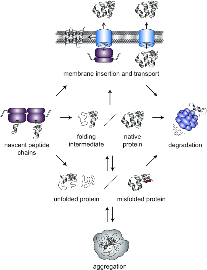
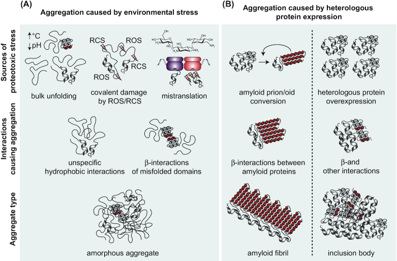
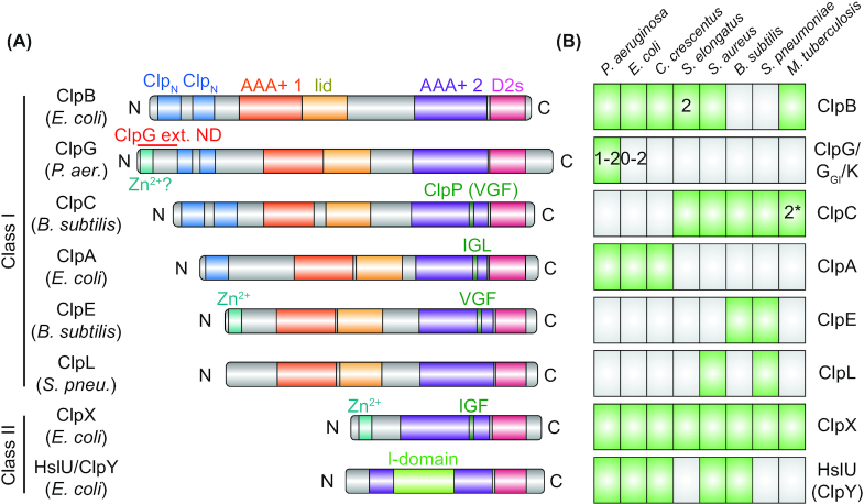
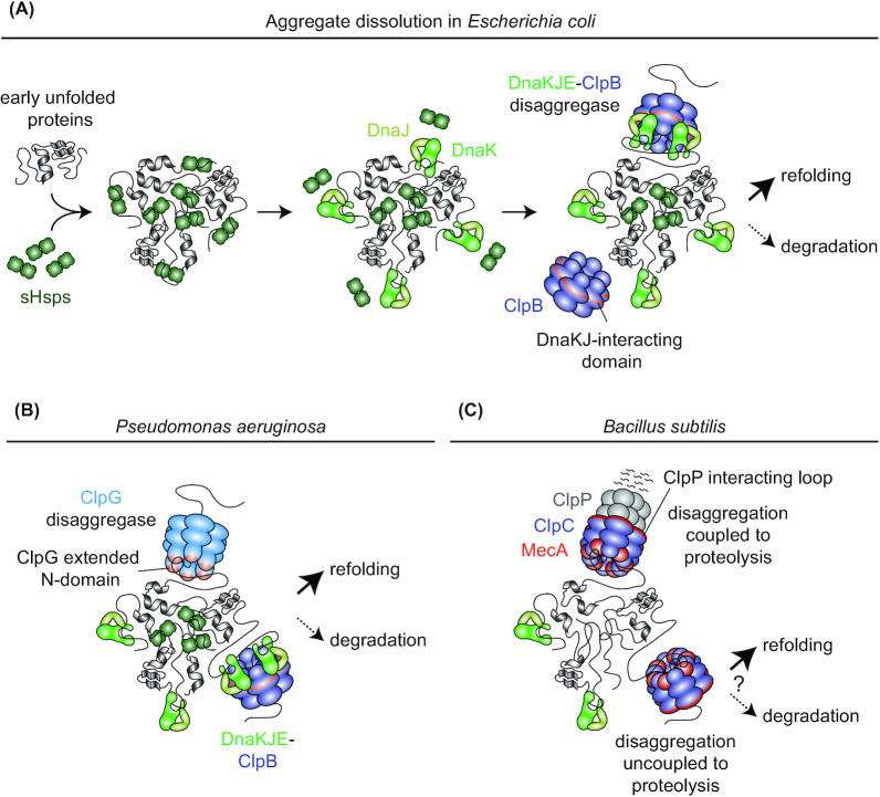
Protein aggregation occurs as a consequence of perturbations in protein homeostasis that can be triggered by environmental and cellular stresses. The accumulation of protein aggregates has been associated with aging and other pathologies in eukaryotes, and in bacteria with changes in growth rate, stress resistance and virulence. Numerous past studies, mostly performed in Escherichia coli, have led to a detailed understanding of the functions of the bacterial protein quality control machinery in preventing and reversing protein aggregation. However, more recent research points toward unexpected diversity in how phylogenetically different bacteria utilize components of this machinery to cope with protein aggregation. Furthermore, how persistent protein aggregates localize and are passed on to progeny during cell division and how their presence impacts reproduction and the fitness of bacterial populations remains a controversial field of research. Finally, although protein aggregation is generally seen as a symptom of stress, recent work suggests that aggregation of specific proteins under certain conditions can regulate gene expression and cellular resource allocation. This review discusses recent advances in understanding the consequences of protein aggregation and how this process is dealt with in bacteria, with focus on highlighting the differences and similarities observed between phylogenetically different groups of bacteria.

Keywords: aggregate inheritance; cellular aging; disaggregases; molecular chaperones; protein aggregation; stress adaptation.


Figures

Figure 1.
Figure 1.
Overview of the major protein homeostatic processes in bacteria. As they exit the ribosome, most peptide chains reach their functional or native state by folding into a specific three-dimensional structure. In the case of larger proteins, this can entail the preceding formation of folding intermediates along the folding pathway. In addition to cytosolic proteins, a fraction of the proteome is inserted into or transported through the membrane. Protein function is threatened by stress conditions that affect protein folding. During stress, noncovalent interactions within the protein can be disrupted, leading to local or global loss of secondary and tertiary structure and the unfolding of a protein (represented as unstructured threads). Through the formation of non-native intramolecular interactions a protein can misfold and assume a structure deviating from its functional state (represented as a red fold). When folding intermediates, unfolded and misfolded proteins are abundant, they can associate with one another through non-native intermolecular interactions and co-aggregate to form larger aggregate structures. In order to maintain a functional proteome, natively folded as well as un/misfolded proteins can be degraded.
Figure 2.
Figure 2.
Summary of proteotoxic stresses, aggregation causing interactions and protein aggregate types. Bacterial protein aggregates can be broadly categorized as those caused by environmental stress and those caused by heterologous protein expression. (A), Different types of environmental stresses lead to protein aggregation. Changes in temperature, osmolarity, ionic strength, pH and macromolecular crowding cause global protein un- and misfolding in a dose-dependent manner. Exposure to substances like hydrogen peroxide and hypochlorous acid cause a surge of reactive oxygen (ROS) and chlorine species (RCS), covalently damaging proteins and irreversibly changing their folding properties. Aminoglycoside antibiotics like kanamycin and streptomycin cause mRNA mistranslation. Incorporation of the wrong amino acids into nascent chains results in aberrant peptides with altered folding properties. Different species of un- and misfolded proteins can interact with one another through unspecific hydrophobic interactions or in a sequence-specific manner through contacting β-strands (β-interactions). Co-aggregation of many protein species results in the formation of globular amorphous aggregates that lack ordered intermolecular interactions. (B), Heterologous protein expression in bacteria can result in the formation of amyloid fibrils and inclusion bodies. Heterologously expressed prion/oid proteins switch between native and misfolded prion conformations, with the prion conformation driving conversion of the same protein species also into prion conformation. Prion/oid proteins can form highly structured amyloid aggregates where monomers of the same protein contact each other through β-sheet interactions running perpendicular to the long axis of the aggregate fibril. In heterologous overexpression for protein production, highly abundant protein can aggregate into globular inclusion bodies containing both amyloid and natively folded structure.
Figure 3.
Figure 3.
Distribution of Hsp100 proteins among bacteria and Hsp100 domain structure. (A), Domain organization of bacterial Hsp100 homologs from different bacterial species. Domains annotated using InterPro (http://www.ebi.ac.uk/interpro/) show the respective identifiers. Hsp100 proteins can be categorized as two classes according to the number of AAA+ domains they contain: Class I Hsp100s ClpA, ClpB, ClpC, ClpE, GlpG (or ClpK) and ClpL have two while class II Hsp100s ClpX and HslU (also called ClpY) have only one. The AAA+ core domains (AAA+ 1/2; IPR003959) are C-terminally bordered by the AAA+ lid domain (lid; IPR041546) or the D2 small domain (D2s; IPR019489), respectively. HslU (ClpY) has an intermediate domain (I-domain, residues 108–243) inserted into its AAA+ core domain. The remaining domain organization of Hsp100s is protein specific, with differences in the length of the region between the AAA+ domains as well as in the N-termini, which may contain one or several conserved Clp N-terminal domains (ClpN; IPR004176) or a zinc-binding motif (Zn2+; IPR010603 in ClpX). The stand-alone disaggregases ClpG and ClpGGI possess an N-terminal extension with a putative zinc-binding motif (Zn2+?), which is involved in protein aggregate interaction (ClpG), or ATPase activity regulation (ClpGGI). HslU (ClpY) associates with the peptidase HslV (ClpQ) while ClpA, ClpC, ClpE and ClpX associate with the peptidase ClpP. ClpP interaction involves a tripeptide motif interaction loop in an AAA+ domain of the Hsp100s (tripeptide sequence indicated). ClpL lacks a known ClpP interacting motif and it is unknown if it interacts with a peptidase subunit. (B), Distribution of Hsp100 proteins in selected bacterial species belonging to the Proteobacteria (Pseudomonas aeruginosa, Escherichia coli and Caulobacter crescentus), Cyanobacteria (Synechococcus elongatus), Firmicutes (Staphylococcus aureus, Bacillus subtilis and Streptococcus pneumoniae) and Actinobacteria (Mycobacterium tuberculosis). ClpG belongs to the Pseudomonas aeruginosa core genome but a second homolog (ClpGGI) can be found on genomic island 1 of P. aeruginosa clone C. Most E. coli species lack ClpG, however, some E. coli strains harbor a ClpG or ClpGGIhomolog on a genomic island or on mobile genetic elements. Synechococcus elongatus possesses two ClpB homologs (ClpB1 and ClpB2) and two potential ClpC homologs (ClpC1 and the unusual truncated ClpC2/ClpX’) can be found in Mycobacterium tuberculosis.
Figure 4.
Figure 4.
Protein disaggregation in different bacterial species. (A), Protein disaggregation via the DnaKJE-ClpB bichaperone machinery in E. coli (GrpE not shown). sHSPs (IbpA and IbpB in E. coli) bind recently unfolded proteins and are incorporated into aggregates while maintaining proteins in a near-native state that facilitates eventual disaggregation by DnaKJE-ClpB. Together with the co-chaperone DnaJ, DnaK binds to aggregated protein and displaces sHSPs from the aggregate surface. DnaK recruits the ClpB disaggregase to the aggregate and this interaction stimulates high ClpB ATPase activity. Substrates are threaded through the central pore of ClpB and, after extraction from the aggregate, can be refolded (preferred) or degraded. (B), Protein disaggregation machineries in Pseudomonas aeruginosa. In addition to the DnaKJE-ClpB disaggregation machinery, P. aeruginosa employs ClpG which does not require stimulating factors for high disaggregation activity. Proteins extracted by both machineries can subsequently be refolded or degraded. (C), Potential protein disaggregation mechanisms in B. subtilis. B. subtilis lacks ClpB or another stand-alone disaggregase. Instead, the ClpP interacting loop containing Hsp100 ClpC interacts with the adaptor MecA and binds to aggregated proteins to drive their disaggregation in vitro. Although protein degradation-coupled disaggregation by MecA-ClpCP has been shown to be more efficient, MecA-ClpC can also disaggregate proteins without associating with ClpP. Thus, potential disaggregation through MecA-ClpC not associated with ClpP could allow for the refolding (or the downstream degradation by another protease) of extracted proteins in vivo.
Figure 5.
Figure 5.
Aggregate formation and inheritance in unicellular organisms. (A), In E. coli, cells are born with an old pole (labeled in red) present in the progenitor cell and a new pole (labeled in blue) built during division. The cell inheriting the old pole of the progenitor cell is termed the old pole daughter cell and the one inheriting the new pole the new pole daughter cell. An E. coli population consists of cell lineages with many different pole inheritance histories (red areas connecting cells indicate old pole inheritance, blue areas new pole inheritance). In E. coli, aggregating proteins (green particles) are occluded from the central nucleoid (violet region), enforcing aggregate localization at the nucleoid-free poles from which they rarely move. This pattern creates a strong asymmetry of aggregate inheritance as aggregates are specifically retained in the cell lineages consecutively inheriting the progenitor cell's old pole. (B), During asymmetric cell division of S. cerevisiae a smaller cell buds off from a larger mother cell. Heat shock-induced aggregates are collected and deposited at different sites in the cell and are actively and passively retained in the mother cell, creating strong asymmetry of aggregate distribution. (C), In the symmetrically dividing S. pombe stress-induced aggregates form and fuse together either in the space between old pole and the nucleus (violet circle) or new pole and the nucleus. Aggregates are mobile however the nucleus impedes frequent movement to the opposite pole half. This leads to preferential aggregate retention in cell lineages consecutively inheriting the progenitor cell's old pole. (D), Schematic showing how growth along the length of the cell outside the pole regions determines aggregate localization and inheritance in C. crescentus. In this organism, cell division is asymmetric and yields a larger stalked cell (the old pole cell) and a smaller swarmer cell (the new pole cell). The C. crescentus nucleoid expands through the entire cell and aggregates form as distributed foci throughout the length of the cell. Numbers represent relative cell positions between the old (0) and new pole (1). Lines depict these relative positions as the cell elongates and divides. With each division event, aggregates will gradually assume a position closer to the new pole until they are inherited by the other cell type. A minority of aggregates remain trapped after forming at the pole. (E), C. crescentus aggregate inheritance pattern resulting from the process described in (D). Aggregates are not retained in the lineage consecutively inheriting the old pole, but instead are constantly distributed between old and new pole cells (with the exception of the minor pole aggregate fraction).
Figure 6.
Figure 6.
Protein aggregation in stress adaptation. (A), Possible role of σ70 aggregation in the regulation of the E. coli heat shock response. In the absence of stress, free DnaKJ (GrpE not shown) will bind to the heat shock sigma factor σ32 and facilitate its degradation by the membrane-bound protease FtsH (contribution of SRP to σ32 regulation not shown). High levels of the housekeeping sigma factor σ70 could outcompete residual free σ32 for binding to the RNA polymerase (RNAP), further inhibiting inappropriate heat shock response induction. During heat shock, DnaKJ will largely relocalize to aggregating protein and liberate σ32. The thermosensitive σ70 will aggregate, potentially reducing the levels of soluble molecules capable of competing with σ32 and further enhancing heat shock response induction. (B), Protein aggregation in the regulation of the B. subtilis heat and oxidative stress response. In the absence of stress, levels of the heat and oxidative stress master regulator Spx are kept low through YjbH adaptor-mediated degradation by the protease ClpXP. Aggregation induced through heat and oxidative stress induces co-aggregation of YjbH. Liberated Spx can induce the expression of stress-adaptive genes and the repression of proliferative genes.
Figure 7.
Figure 7.
The potential impact of protein aggregation on bacterial antibiotic tolerance. After proteotoxic stress exposure, a population of E. coli cells is heterogeneous in its capability to resume growth. The aggregation of a large fraction of the proteome (green signal localized in foci instead of distributed throughout the cell as in growing cells) can render a cell inactive, or dormant, without causing its death. Dormant cells survive antibiotic treatment that otherwise kills metabolically active and growing cells. The exit from dormancy and resumption of the growth program is correlated with ATP-dependent protein disaggregation.

References

    1. Acebrón SP, Martín I, del Castillo Uet al.. DnaK-mediated association of ClpB to protein aggregates. A bichaperone network at the aggregate surface. FEBS Lett. 2009;583:2991–6. - PubMed
    1. Ackermann M, Chao L, Bergstrom CTet al.. On the evolutionary origin of aging. Aging Cell. 2007;6:235–44. - PMC - PubMed
    1. Ackermann M, Stephen CS, Jenal U. Senescence in a bacterium with asymmetric division. Science. 2003;300:1920. - PubMed
    1. Aguilera P, Marcoleta A, Lobo-Ruiz Pet al.. Identification of key amino acid residues modulating intracellular and in vitro microcin E492 amyloid formation. Front Microbiol. 2016;7:35. - PMC - PubMed
    1. Aguzzi A, Lakkaraju AKK, Frontzek K. Toward therapy of human prion diseases. Annu Rev Pharmacol Toxicol. 2018;58:331–51. - PubMed

Publication types

MeSH terms