. 2019 Oct;49(10):1457-1973.
doi: 10.1002/eji.201970107.

Guidelines for the use of flow cytometry and cell sorting in immunological studies (second edition)

Andrea Cossarizza  1 Hyun-Dong Chang  2 Andreas Radbruch  2 Andreas Acs  3 Dieter Adam  4 Sabine Adam-Klages  5 William W Agace  6   7 Nima Aghaeepour  8 Mübeccel Akdis  9 Matthieu Allez  10 Larissa Nogueira Almeida  11 Giorgia Alvisi  12 Graham Anderson  13 Immanuel Andrä  14 Francesco Annunziato  15 Achille Anselmo  16 Petra Bacher  4   17 Cosima T Baldari  18 Sudipto Bari  19   20 Vincenzo Barnaba  21   22   23 Joana Barros-Martins  24 Luca Battistini  25 Wolfgang Bauer  26 Sabine Baumgart  2 Nicole Baumgarth  27 Dirk Baumjohann  28 Bianka Baying  29 Mary Bebawy  30 Burkhard Becher  31   32 Wolfgang Beisker  33 Vladimir Benes  29 Rudi Beyaert  34 Alfonso Blanco  35 Dominic A Boardman  36   37 Christian Bogdan  38   39 Jessica G Borger  40 Giovanna Borsellino  41 Philip E Boulais  42   43 Jolene A Bradford  44 Dirk Brenner  45   46   47 Ryan R Brinkman  48   49 Anna E S Brooks  50 Dirk H Busch  14   51   52 Martin Büscher  53 Timothy P Bushnell  54 Federica Calzetti  55 Garth Cameron  56 Ilenia Cammarata  21 Xuetao Cao  57 Susanna L Cardell  58 Stefano Casola  59 Marco A Cassatella  55 Andrea Cavani  60 Antonio Celada  61 Lucienne Chatenoud  62 Pratip K Chattopadhyay  63 Sue Chow  64 Eleni Christakou  65   66 Luka Čičin-Šain  67 Mario Clerici  68   69   70 Federico S Colombo  16 Laura Cook  37   71 Anne Cooke  72 Andrea M Cooper  73 Alexandra J Corbett  56 Antonio Cosma  74 Lorenzo Cosmi  15 Pierre G Coulie  75 Ana Cumano  76 Ljiljana Cvetkovic  77 Van Duc Dang  2 Chantip Dang-Heine  78 Martin S Davey  79   80 Derek Davies  81 Sara De Biasi  82 Genny Del Zotto  83 Gelo Victoriano Dela Cruz  84 Michael Delacher  85   86 Silvia Della Bella  87 Paolo Dellabona  88 Günnur Deniz  89 Mark Dessing  90 James P Di Santo  91   92 Andreas Diefenbach  2   93   94 Francesco Dieli  95 Andreas Dolf  96 Thomas Dörner  2   97 Regine J Dress  98 Diana Dudziak  99 Michael Dustin  100 Charles-Antoine Dutertre  101   98 Friederike Ebner  102 Sidonia B G Eckle  56 Matthias Edinger  85   103 Pascale Eede  104 Götz R A Ehrhardt  105 Marcus Eich  106 Pablo Engel  107 Britta Engelhardt  108 Anna Erdei  109 Charlotte Esser  110 Bart Everts  111 Maximilien Evrard  98 Christine S Falk  112 Todd A Fehniger  113 Mar Felipo-Benavent  114 Helen Ferry  115 Markus Feuerer  85   86 Andrew Filby  116 Kata Filkor  117 Simon Fillatreau  118 Marie Follo  119   120 Irmgard Förster  121 John Foster  122 Gemma A Foulds  123 Britta Frehse  11 Paul S Frenette  42   43   124 Stefan Frischbutter  2   125 Wolfgang Fritzsche  126 David W Galbraith  127   128 Anastasia Gangaev  129 Natalio Garbi  130 Brice Gaudilliere  131 Ricardo T Gazzinelli  132   133 Jens Geginat  134 Wilhelm Gerner  135   136 Nicholas A Gherardin  56 Kamran Ghoreschi  137 Lara Gibellini  82 Florent Ginhoux  98   138   139 Keisuke Goda  140   141   142 Dale I Godfrey  56 Christoph Goettlinger  143 Jose M González-Navajas  144   145 Carl S Goodyear  146 Andrea Gori  147 Jane L Grogan  148 Daryl Grummitt  122 Andreas Grützkau  2 Claudia Haftmann  31 Jonas Hahn  149 Hamida Hammad  150 Günter Hämmerling  151 Leo Hansmann  94   152   153 Goran Hansson  154 Christopher M Harpur  155 Susanne Hartmann  102 Andrea Hauser  103 Anja E Hauser  2   156   157 David L Haviland  158 David Hedley  64 Daniela C Hernández  2   159 Guadalupe Herrera  160 Martin Herrmann  149 Christoph Hess  161   162 Thomas Höfer  163 Petra Hoffmann  85   103 Kristin Hogquist  164 Tristan Holland  130 Thomas Höllt  165   166 Rikard Holmdahl  167 Pleun Hombrink  168   169 Jessica P Houston  170 Bimba F Hoyer  171 Bo Huang  172 Fang-Ping Huang  173 Johanna E Huber  28 Jochen Huehn  174 Michael Hundemer  175 Christopher A Hunter  176 William Y K Hwang  177   20   178 Anna Iannone  179 Florian Ingelfinger  31 Sabine M Ivison  36   37 Hans-Martin Jäck  77 Peter K Jani  2   180 Beatriz Jávega  181 Stipan Jonjic  182 Toralf Kaiser  2 Tomas Kalina  183 Thomas Kamradt  184 Stefan H E Kaufmann  180 Baerbel Keller  185   186 Steven L C Ketelaars  129 Ahad Khalilnezhad  98   187 Srijit Khan  105 Jan Kisielow  188 Paul Klenerman  115 Jasmin Knopf  149 Hui-Fern Koay  56 Katja Kobow  189 Jay K Kolls  190 Wan Ting Kong  98 Manfred Kopf  188 Thomas Korn  191 Katharina Kriegsmann  175 Hendy Kristyanto  192 Thomas Kroneis  193 Andreas Krueger  194 Jenny Kühne  112 Christian Kukat  195 Désirée Kunkel  196   197 Heike Kunze-Schumacher  194 Tomohiro Kurosaki  198 Christian Kurts  130 Pia Kvistborg  129 Immanuel Kwok  98   199 Jonathan Landry  29 Olivier Lantz  200 Paola Lanuti  201 Francesca LaRosa  68   70 Agnès Lehuen  202 Salomé LeibundGut-Landmann  203 Michael D Leipold  204 Leslie Y T Leung  105 Megan K Levings  36   37   205 Andreia C Lino  2   97 Francesco Liotta  15 Virginia Litwin  206 Yanling Liu  105 Hans-Gustaf Ljunggren  207 Michael Lohoff  208 Giovanna Lombardi  209 Lilly Lopez  210 Miguel López-Botet  211 Amy E Lovett-Racke  212 Erik Lubberts  213 Herve Luche  214 Burkhard Ludewig  215 Enrico Lugli  12   16 Sebastian Lunemann  216 Holden T Maecker  217 Laura Maggi  15 Orla Maguire  218 Florian Mair  219 Kerstin H Mair  135   136 Alberto Mantovani  220   221 Rudolf A Manz  11 Aaron J Marshall  222 Alicia Martínez-Romero  223 Glòria Martrus  216 Ivana Marventano  68   70 Wlodzimierz Maslinski  224 Giuseppe Matarese  225 Anna Vittoria Mattioli  82   226 Christian Maueröder  227   228 Alessio Mazzoni  15 James McCluskey  56 Mairi McGrath  2 Helen M McGuire  229 Iain B McInnes  146 Henrik E Mei  2 Fritz Melchers  2   180 Susanne Melzer  230 Dirk Mielenz  77 Stephen D Miller  231 Kingston H G Mills  232 Hans Minderman  218 Jenny Mjösberg  207   233 Jonni Moore  234 Barry Moran  232 Lorenzo Moretta  235 Tim R Mosmann  236 Susann Müller  237 Gabriele Multhoff  238   239 Luis Enrique Muñoz  149 Christian Münz  31   32 Toshinori Nakayama  240 Milena Nasi  82 Katrin Neumann  241 Lai Guan Ng  98   187   199   242   243 Antonia Niedobitek  2 Sussan Nourshargh  244 Gabriel Núñez  245 José-Enrique O'Connor  181 Aaron Ochel  241 Anna Oja  169 Diana Ordonez  246 Alberto Orfao  247 Eva Orlowski-Oliver  248 Wenjun Ouyang  249 Annette Oxenius  250 Raghavendra Palankar  251 Isabel Panse  2 Kovit Pattanapanyasat  252 Malte Paulsen  246 Dinko Pavlinic  29 Livius Penter  153 Pärt Peterson  253 Christian Peth  53 Jordi Petriz  254 Federica Piancone  68   70 Winfried F Pickl  255 Silvia Piconese  21   23 Marcello Pinti  256 A Graham Pockley  123   257 Malgorzata Justyna Podolska  149   258 Zhiyong Poon  177 Katharina Pracht  77 Immo Prinz  24 Carlo E M Pucillo  259 Sally A Quataert  236 Linda Quatrini  235 Kylie M Quinn  260   261 Helena Radbruch  104 Tim R D J Radstake  262 Susann Rahmig  263 Hans-Peter Rahn  264 Bartek Rajwa  265 Gevitha Ravichandran  241 Yotam Raz  266 Jonathan A Rebhahn  236 Diether Recktenwald  267 Dorothea Reimer  77 Caetano Reis e Sousa  268 Ester B M Remmerswaal  168   269 Lisa Richter  270 Laura G Rico  254 Andy Riddell  81 Aja M Rieger  271 J Paul Robinson  272 Chiara Romagnani  2   159 Anna Rubartelli  273 Jürgen Ruland  274 Armin Saalmüller  135 Yvan Saeys  275   276 Takashi Saito  277 Shimon Sakaguchi  198 Francisco Sala-de-Oyanguren  278 Yvonne Samstag  279 Sharon Sanderson  280 Inga Sandrock  24 Angela Santoni  281 Ramon Bellmàs Sanz  112 Marina Saresella  68   70 Catherine Sautes-Fridman  282 Birgit Sawitzki  283 Linda Schadt  31   32 Alexander Scheffold  4 Hans U Scherer  192 Matthias Schiemann  14 Frank A Schildberg  284 Esther Schimisky  285 Andreas Schlitzer  286 Josephine Schlosser  102 Stephan Schmid  287 Steffen Schmitt  288 Kilian Schober  14 Daniel Schraivogel  289 Wolfgang Schuh  77 Thomas Schüler  290 Reiner Schulte  291 Axel Ronald Schulz  2 Sebastian R Schulz  77 Cristiano Scottá  209 Daniel Scott-Algara  292 David P Sester  293 T Vincent Shankey  294 Bruno Silva-Santos  295 Anna Katharina Simon  100 Katarzyna M Sitnik  67 Silvano Sozzani  296 Daniel E Speiser  297 Josef Spidlen  298 Anders Stahlberg  299 Alan M Stall  300 Natalie Stanley  8 Regina Stark  168   169 Christina Stehle  2   159 Tobit Steinmetz  77 Hannes Stockinger  301 Yousuke Takahama  302 Kiyoshi Takeda  198 Leonard Tan  98   187 Attila Tárnok  303   304   305 Gisa Tiegs  241 Gergely Toldi  117 Julia Tornack  2   306 Elisabetta Traggiai  307 Mohamed Trebak  308 Timothy I M Tree  65   66 Joe Trotter  300 John Trowsdale  72 Maria Tsoumakidou  309 Henning Ulrich  310 Sophia Urbanczyk  77 Willem van de Veen  9   311 Maries van den Broek  31   32 Edwin van der Pol  312 Sofie Van Gassen  275   276 Gert Van Isterdael  313 René A W van Lier  169 Marc Veldhoen  295 Salvador Vento-Asturias  130 Paulo Vieira  76 David Voehringer  314 Hans-Dieter Volk  315 Anouk von Borstel  79   80 Konrad von Volkmann  316 Ari Waisman  317 Rachael V Walker  318 Paul K Wallace  319 Sa A Wang  320 Xin M Wang  321 Michael D Ward  44 Kirsten A Ward-Hartstonge  36 Klaus Warnatz  185   186 Gary Warnes  322 Sarah Warth  197 Claudia Waskow  263   323 James V Watson  324 Carsten Watzl  325 Leonie Wegener  53 Thomas Weisenburger  3 Annika Wiedemann  2   97 Jürgen Wienands  326 Anneke Wilharm  24 Robert John Wilkinson  327   328   329 Gerald Willimsky  330 James B Wing  198 Rieke Winkelmann  4 Thomas H Winkler  3 Oliver F Wirz  9 Alicia Wong  98 Peter Wurst  331 Jennie H M Yang  65   66 Juhao Yang  174 Maria Yazdanbakhsh  111 Liping Yu  332 Alice Yue  333 Hanlin Zhang  100 Yi Zhao  334 Susanne Maria Ziegler  216 Christina Zielinski  51   335   336 Jakob Zimmermann  337 Arturo Zychlinsky  180
Affiliations

Guidelines for the use of flow cytometry and cell sorting in immunological studies (second edition)

Andrea Cossarizza et al. Eur J Immunol. 2019 Oct.

Abstract

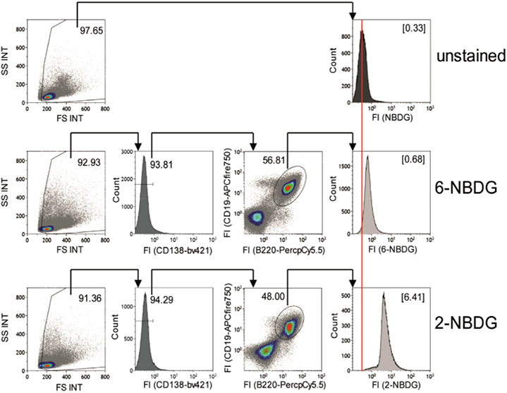
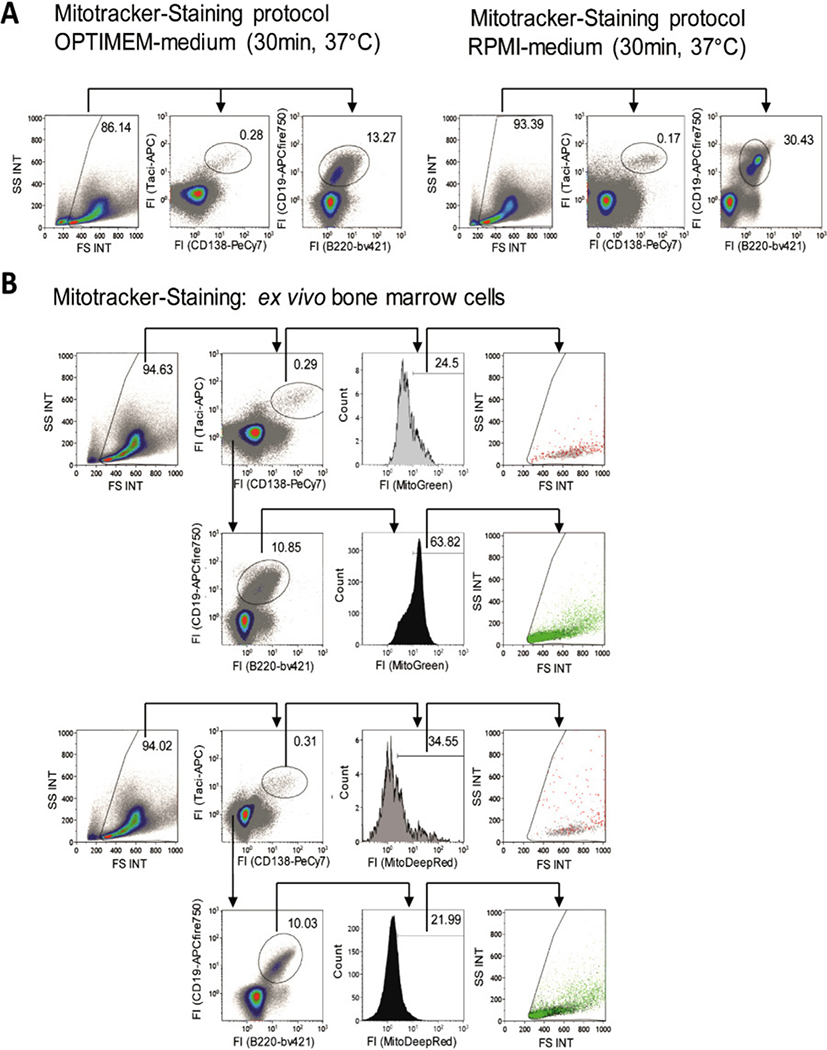
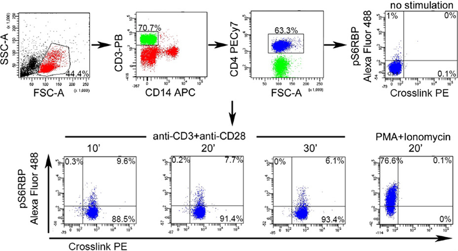
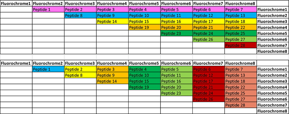
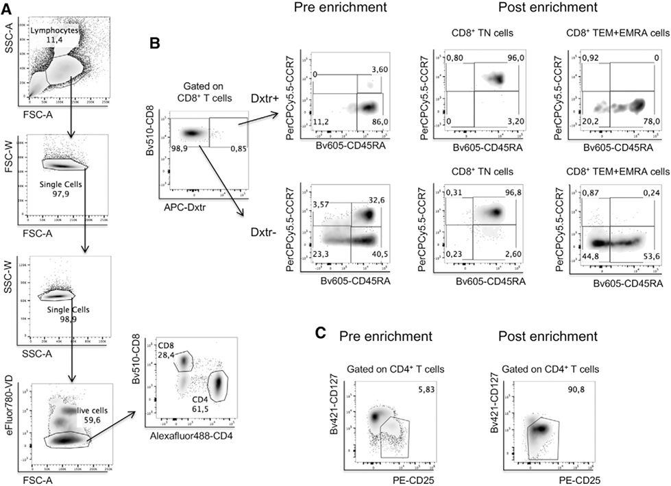
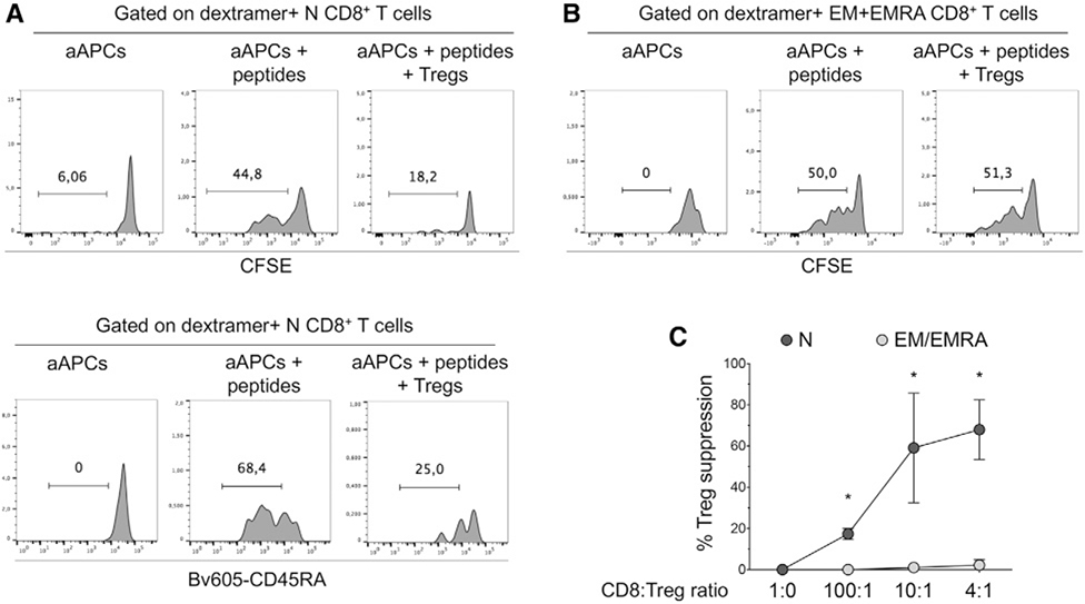
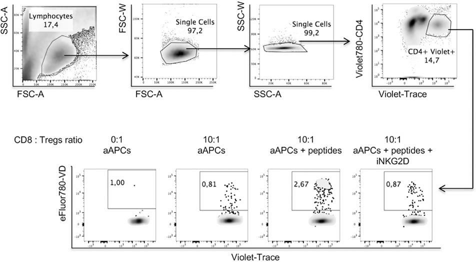
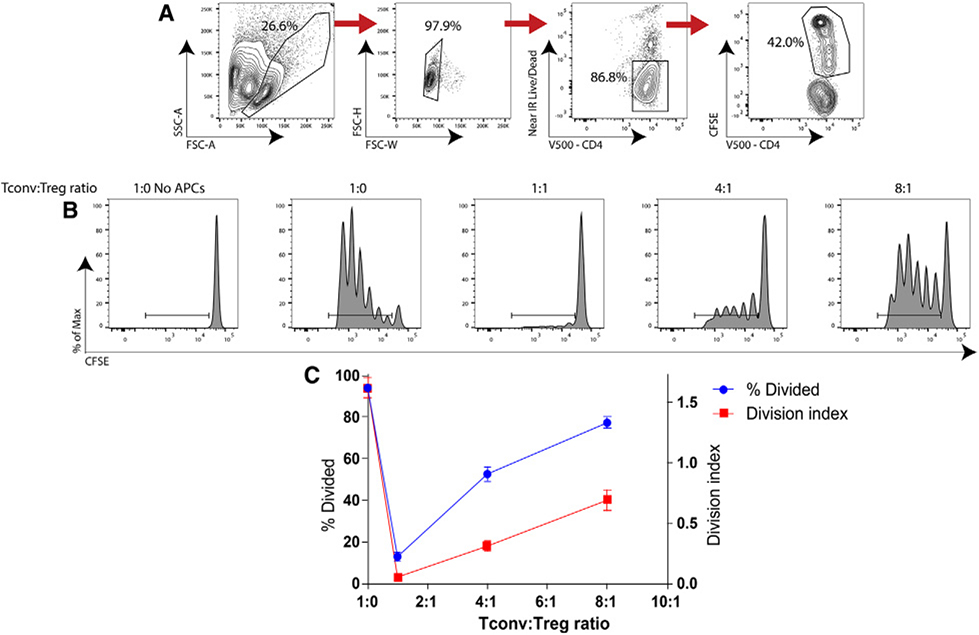
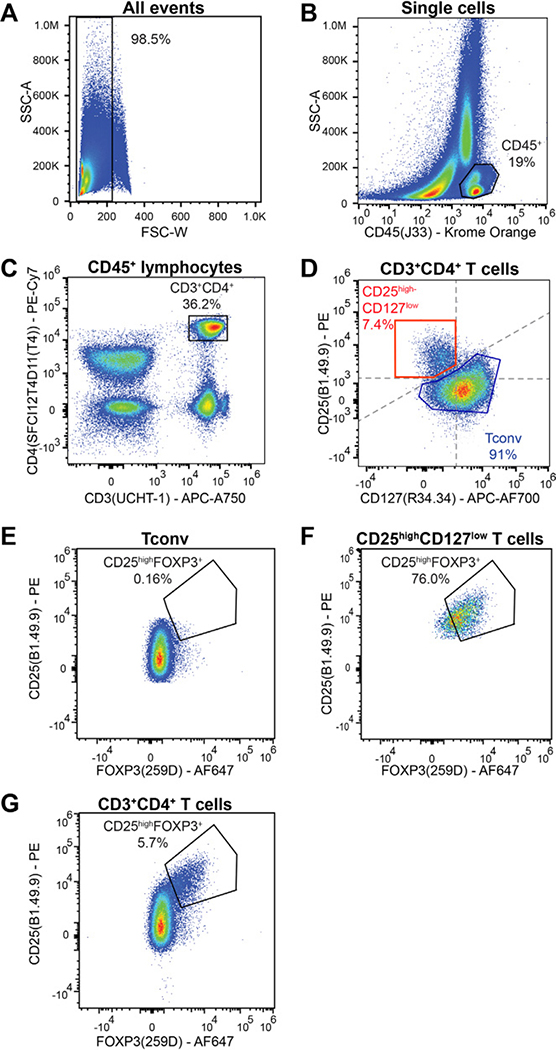
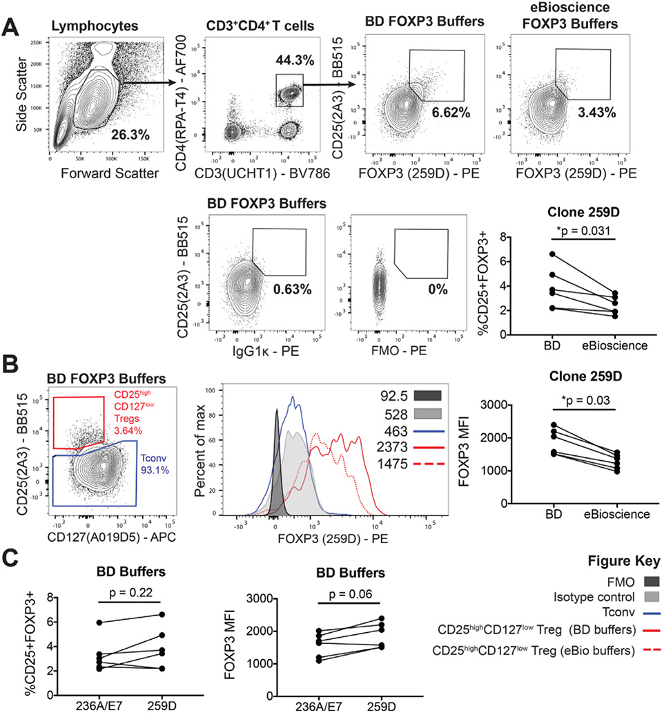
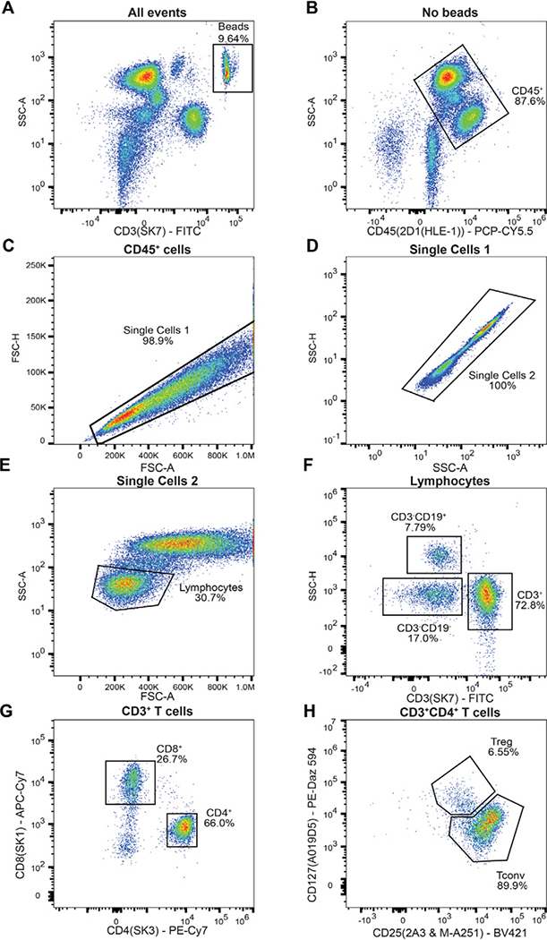
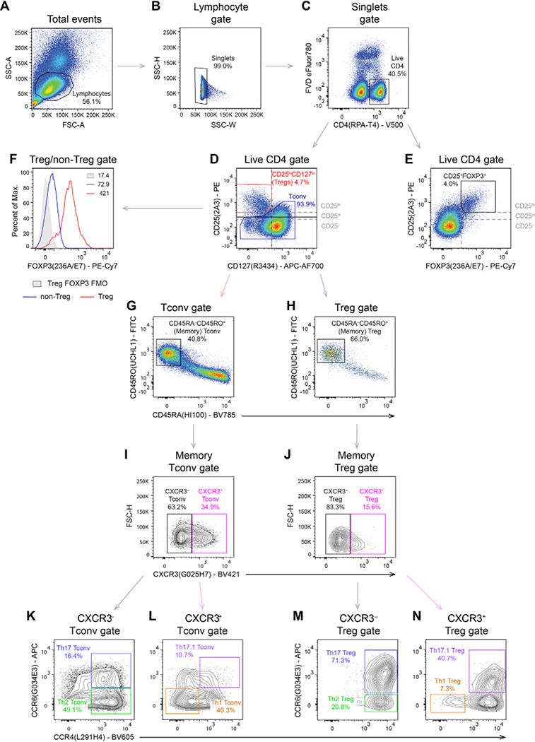
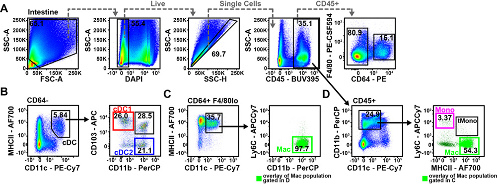
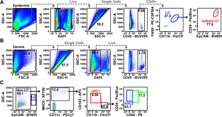
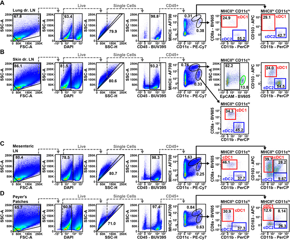
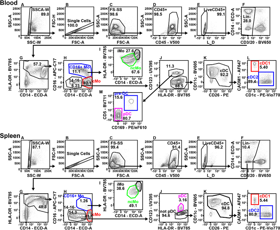
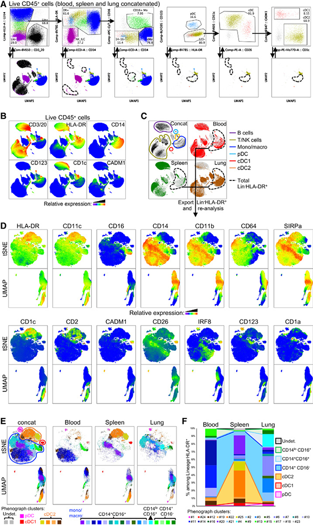
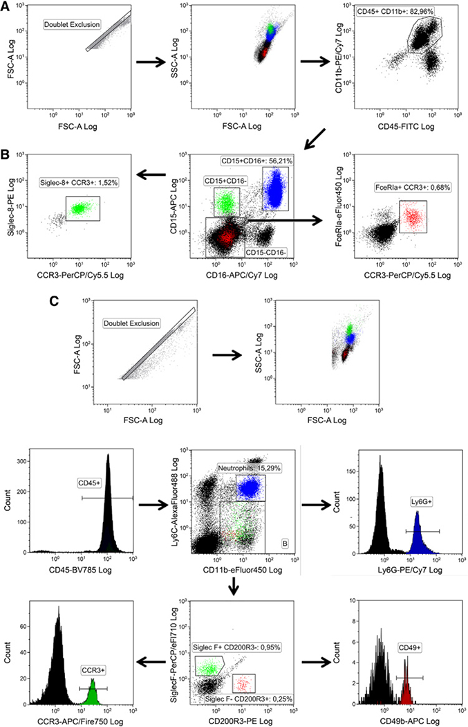
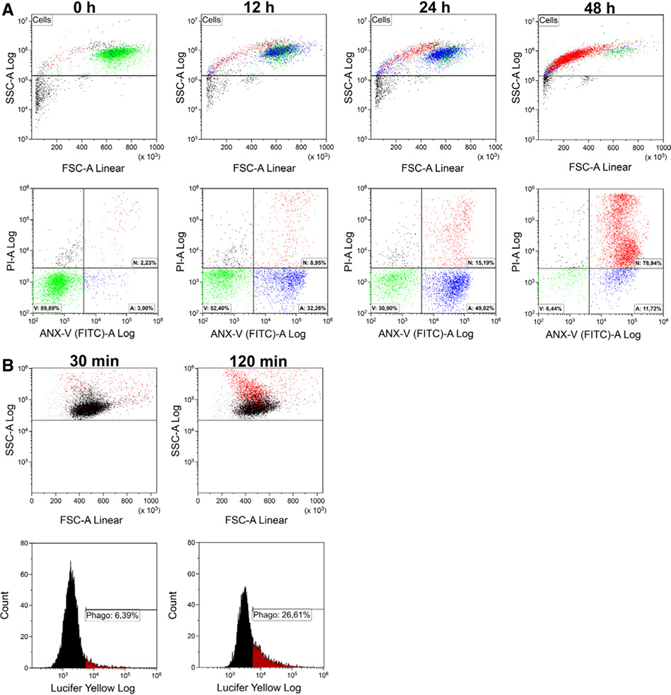
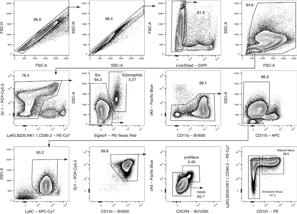
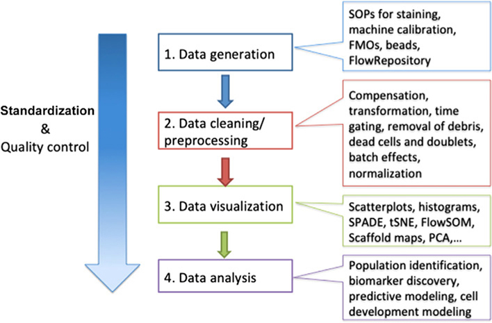
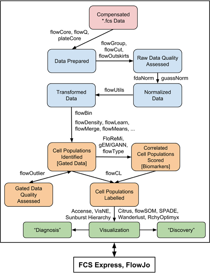
These guidelines are a consensus work of a considerable number of members of the immunology and flow cytometry community. They provide the theory and key practical aspects of flow cytometry enabling immunologists to avoid the common errors that often undermine immunological data. Notably, there are comprehensive sections of all major immune cell types with helpful Tables detailing phenotypes in murine and human cells. The latest flow cytometry techniques and applications are also described, featuring examples of the data that can be generated and, importantly, how the data can be analysed. Furthermore, there are sections detailing tips, tricks and pitfalls to avoid, all written and peer-reviewed by leading experts in the field, making this an essential research companion.


Conflict of interest statement

Conflict of interest

Some of the authors of these guidelines work for companies manufacturing FCM equipment and reagents. The mention of a particular company's equipment or reagents does not imply endorsement of these products but are included as examples. The content of these guidelines is editorially independent of any company and has been peer-reviewed.

Figures

Figure 1.
Figure 1.
Sample core after hydrodynamic focussing by laminar sheath flow in a flow chamber.
Figure 2.
Figure 2.
Intensity profile of a focus spot of a gaussian laser beam. Note: if a cell is out of the center of the laser focus by 10 μm (20 μm sample core), laser intensity goes down about 5% with a 60 μm diame ter laser focus.
Figure 3.
Figure 3.
Liquid stream of a jet in air sensing cell sorter. Depending of abort settings of the cell sorter, cells that are too close together are aborted from sorting. Reproduced with permission from ref. [16].
Figure 4.
Figure 4.
Schematics of fluidics of a jet in air sensing cell sorter.
Figure 5.
Figure 5.
Typical electronic signal processing of a flow cytometer. The signal coming from a PMT or photo diode is amplified by a preamp and a main amp. The analogue signals are then digitized by an ADC board. A personal computer (PC) is used for further data processing and HV controlling.
Figure 6.
Figure 6.
Principle of a spectral flow cytometer. (A) Excitation light source (laser), (B) labeled cell, (C) dispersing element, (D) multichannel light detector (CCD or multichannel PMT).
Figure 7.
Figure 7.
Spillover and compensation: (A) the emission spectra of PerCP-Cy5.5 and PE-Cy7. (B) Peripheral blood lymphocytes stained with PerCP-Cy5.5 CD4 mAb. The MdFI is shown for the PerCP-Cy5.5 and PE-Cy7 detectors without (left) and with (right) compensation.
Figure 8.
Figure 8.
Brightness of positive population.
Figure 9.
Figure 9.
Accuracy for SOV: The figure shows two different assays in which lysed whole blood was stained with the same fluorochromes: BD HorizonTM Brilliant Violet 510 (BV510) and BD HorizonTM Brilliant Violet 605 (BV605). Both assays used the same BV605 reagent. In the top panels, the BV510 positive population was dim while in the bottom panels the BV510 positive population is very bright. For each assay, the SOVs were determined and the correct spillover was applied (Middle panels). For the left panels, the BV510→BV605 SOV was increased by 1% (overcompensated) and compensation applied. For the right panels, the BV510→BV605 SOV was decreased by 2% (undercompensated) and compensation applied.
Figure 10.
Figure 10.
Examples for performance tracking with and without a CS&T module [130]. (A) A Levey-Jennings chart of a weekly measured performance for one parameter (out of 10) is shown. The cross in red indicates a failure in the performance check (a higher PMT-Voltage is needed to reach the target values of the beads, which corresponds to a loss of sensitivity). After checking and changing the band-pass filter in front of the corresponding PMT, the performance is measured again and is compared to the previous situation (blue dots). With the correct band-pass filter installed, the performance of the PMT is back to the previous level. The graph is taken from a CS&T-Cytometer Performance Report of a BD FAC-SCanto II equipped with 3 lasers.(B) The histogram of channel A of the violet 405 nm laser shows the corresponding measurement to the situation described above in (a) and is taken from a self-defined, instrument-specific calibration worksheet. The blue population represents the “standard” setup (with a 510/50 band-pass filter in front of the PMT of channel A, where the beads are reaching the respective target values (brackets). The red curve shows a measurement with a 610/20 nm band-pass filter instead. The beads are clearly outside the target values and the positive and negative populations are barely separated from each other. This is an example, how one can easily track basic instrument performance without having a separate software module available.
Figure 11.
Figure 11.
How one can detect suboptimal alignment of lasers. Both histograms display a negative and positive bead population in the 450/50 channel of the UV-Laser of a BD FAC-SAria SORP cell sorter. Although the positive peak in (A) still falls into the defined target area (brackets = P2), the shape and %CV of the peak suggest a suboptimal alignment of the UV-Laser. After realignment the shape of the positive peak become narrower with only the half of the %CV. (B) Laser-alignment is optimal, when the lowest %CV values are reached.
Figure 12.
Figure 12.
Voltage walk displaying rCV and rSD of the second peak of sperotech 8 peak rainbow calibration particles. The arrow indicates the point of minimal PMT voltage.
Figure 13.
Figure 13.
Titration of a CD4 mAb (clone GK1.5) conjugated to FITC and titrated on murine splenocytes. The antibody was titrated in 1:2 dilution steps starting from a 1:100 dilution (5.4 μg/mL) up to 1:12 800 (0.04 μg/mL). (A) Histograms of the stained samples are shown. (B) MFI of the positive and negative populations (left axis) and SNR between the positive and negative populations (right axis) are plotted. Best separating titer for this particular antibody was determined to be 0.7 μg/mL (1:800 dilution).
Figure 14.
Figure 14.
Human whole blood fixed with formaldehyde and permeabilized with TX-100. White blood cell populations were identified using CD14-PE-Cy7 and CD45-Krome Orange. Debris (red) is identified using CD45 versus SS (top panel—region C). Identification of peripheral blood monocytes (shown in blue in both panels) was accomplished using CD-14-PE-Cy7 (not shown).
Figure 15.
Figure 15.
Impact of methanol concentration on P-STAT5 immunoreactivity in peripheral blood monocytes activated in vitro using GM-CSF. Whole blood from a normal donor was treated with GM-CSF for up to 20 min in vitro at 37°C. One part of the fixed and permeabilized samples was treated with 50% methanol (A) and the other with 80% methanol (B) at 4°C. After washing, all samples were stained with (-◊-) P-STAT5, (-□-) P-ERK, and (-Δ-) P-S6.
Figure 16.
Figure 16.
Effect of methanol treatment on CD14 staining of human peripheral blood monocytes. Whole blood samples from one individual were stained with CD-14-PE-Cy7 before (left panels) or after (right panels) fixation and permeabilization. Samples were treated with either 50% (top panels) or 80% (lower panels) methanol. See text for details.
Figure 17.
Figure 17.
Effect of formaldehyde concentration on P-STAT5 immunoreactivity in K562 cells (reproduced from ref. [75] with permission). Cells were fixed at 37°C for 10 min using increasing final concentrations of formaldehyde, permeabilized, and stained with anti-P-STAT5-PE as described.
Figure 18.
Figure 18.
Structural characteristics of Igs. Ribbon diagram of a mouse monoclonal IgG anti-body consisting of two identical heavy and light chain proteins, respectively. Antibody heavy chain residues are indicated in blue and light chain residues in green. Amino acid residues encoding the CDR1, 2, and 3 regions are shown in red. (Image was generated using the Swiss PDB viewer and PDB accession number 1IGT).
Figure 19.
Figure 19.
Structural characteristics of VLR antibodies. (A) Ribbon diagram of the antigen-binding units of a monoclonal VLR antibody. Parallel β-sheets lining the concave antigen-binding surface are shown in blue and a variable loop structure involved in antigen binding is depicted in red. The invariant stalk region necessary for multimerization of the secreted VLR antibody was omitted (Model was generated using the Protein Model Portal Algorithm [84]). (B) Structural characteristics of VLR antibodies. Individual VLRB units consist of a signal peptide, N-terminal LRR (LRR-NT), LRR-1, up to nine variable LRRv units, a connecting peptide, C-terminal capping LRR (LRR-CT) and the invariable stalk region and can be modified by inclusion of engineered 6xHis and HA-epitope tags or Fc-fusion sequences.
Figure 20.
Figure 20.
In cell sorting experiments one often needs to find a compromise between purity, yield, and time, which cannot be optimized all at the same time.
Figure 21.
Figure 21.
This figure shows a summary of discussed enrichment methods. (A) The separation of different cell types with a Ficoll® density gradient is shown. (B) Once one applies a centrifugal force in an elutriation chamber, cells will stop passing through and start separating along a density gradient built inside the chamber. The equilibrium formed depends on the speed of the cellular flow, the amount of applied centrifugal force, and the viscosity of the medium used. This is the reason why elutriation is compatible with a wide range of cell types and carrier media. (C) Target cells (black) can form aggregates with antibody-labeled beads and If you add beads that are coated with specific antibodies against your target cells (black) to the cell suspension, the target cells will form aggregates with the beads. These aggregates are held back on the top of the mesh while the rest of the cell suspension is passing through. With this method one can either deplete or enrich for a specific cell population. Combining different mesh and bead sizes is allowing for a serial enrichment of target cells.
Figure 22.
Figure 22.
Enrichment of human B cells out of whole blood stained for CD45 BV421 and CD19 APC. (A) Staining prior enrichment. (B) Staining after lysis of erythrocytes with Lysing-buffer. (C) Staining after CD19+ MACS® enrichment. (D) Staining of the CD19 fraction (MACS® flow through).
Figure 23.
Figure 23.
Check-list: Parameters for selecting a sorting method. The parameters that affect cell sorting and therefore must be prioritized when choosing a sorting strategy are shown. Starting from the available material (amount, fragility), they range from the mundane cost aspect to practical and methodological concerns such as the available time, to the important experimental approaches regarding what yield, purity, or versatility is needed for down-stream applications. Optimization of one parameter may downgrade another parameter, e.g., a high purity may be at the expense of a high yield or speed, or unchanged functionality of the cells may not allow direct positive selection.
Figure 24.
Figure 24.
Improvement of population discrimination after pre-enrichment. Cytometer histograms of unwanted (gray lines) and wanted (solid green) populations. (A) A large excess of an unwanted population may create substantial overlap with the target population, making it impossible to achieve a good single cell sort. (B) After a pre-enrichment bulk sort, which removes most of the unwanted population a good discrimination between the two populations can be achieved.
Figure 25.
Figure 25.
PBMC Sort. A PBMC sort on a BD FACSAriaTM where by adding both FSC and SSC Height versus Width plots and carefully gating on singlets an additional 9% of likely doublets are removed (reproduced with permission from ref. [142]).
Figure 26.
Figure 26.
The threshold value defines a signal intensity, in one or more parameters, above which the cytometer starts to recognize an event. All other events will be invisible to the instrument’s electronics. A particle passing the laser beam emits a certain amount of light over time. The threshold is set on the height of the signal that is emitted by each particle. On the left-hand side a dotplot with the forward scatter as the trigger parameter is shown. Only particles with a signal higher than this threshold value are recognized by the software as an event and shown in the dotplot (black and orange dots). The dots in on the left side of the threshold value (grey and blue dots) are not included in the data file.
Figure 27.
Figure 27.
Staining pattern and gating strategy for a CD34+ enrichment. The cells are stained with CD34-Pe and CD45-APC. For analysis purposes only, PI was added for post analysis only.
Figure 28.
Figure 28.
Result of a sequential sorting process. 109 total cells have been processed sequentially in 5 h to a final purity greater than 99%. Overall 280 000 target cells have been harvested from 800 000 target cells starting material, resulting in an overall yield of approximately 35%.
Figure 29.
Figure 29.
Custom-made GMP-compliant flow cytometric cell sorter (A) with auxiliary equipment cabinet (left) and laminar air flow hood (right) containing the sort chamber and (B) the sort unit in operation (safety-cabinet-enclosed sort unit seen between the two operators).
Figure 30.
Figure 30.
An example of a gating stategy for rare cells. Gating stategy used to identify circulating endothelial cells (CECs) and their precursors (EPCs) among peripheral blood leukocytes. (A) Debris and aggregates were eliminated using FSC-Area versus FSC-Height, (B) possible clogs were removed using the parameter Time versus SSC. (C) A DUMP channel was used to remove CD45+ cells and dead cells from the analysis. (D) Nucleated cells were identified based on Syto16 positivity. (E) Stem cells were identified according to CD34 positivity, (F) EPCs (CD133+, CD31+) and CECs (CD133, CD31+) were identified. The expression of CD276, also named B7-H3 (G, I), and CD309 (H, J), also named VEGFR-2 or KDR, was evaluated in each subpopulation. In this example, more than 10 million events were initially acquired in order to enumerate a population that, according to the literature, is always represented less than 0.1%.
Figure 31.
Figure 31.
TMRM and JC-1 staining of CD4+ T cells. The K+ ionophore valinomycin depolarizes mitochondria of CD4+ T cells, as revealed by the decrease in TMRM fluorescence, and by the decreased fluorescence of JC-1 aggregates and increased fluorescence of JC-1 monomers. Untreated cells (CTRL) are shown in left panels. For TMRM, unstained sample is also shown in right panel. Dot plot combining untreated sample and valinomycin-treated sample is also reported (lower right panel).
Figure 32.
Figure 32.
MitoTracker Green staining of different subsets of CD8+ T cells. Different CD8+ T-cell subsets, i.e., central memory (CM), naïve (N), effector memory (EM), and terminally differentiated effector memory (EMRA) were identified according to the expression of CD45RA and CD197. Among them, the use of MitoTracker Green (MT Green) allows to determine mt mass, which is clearly different among cell subsets.
Figure 33.
Figure 33.
MitoSOX Mitochondrial Red superoxide indicator and Mitochondria Peroxy Yellow-1 staining of different subsets of CD8+ T cells. Doublets were excluded from the analysis of PBMCs by using FCS-A and FSC-H (upper left panel); viable cells were selected according to negativity for annexin-V (ANX-V) Pacific Blue conjugate and TO-PRO-3 iodide (upper right panel). Then, CD4+ or CD8+ T lymphocytes were selected on the basis of positivity for a CD4APC-H7 mAb or a CD8-PO mAb respectively. Among these, fluorescence intensity of MitoSOX Mitochondrial Red Superoxide Indicator (MitoSOX) and Mitochondria Peroxy Yellow-1 (mitoPY) was analyzed.
Figure 34.
Figure 34.
Flow cytometry (FCM) detection of extracellular vesicles (EVs). (A) Transmission electron microscopy (TEM) image of EVs from human urine after one freeze-thaw cycle [251]. (B) Size distribution of EVs from human urine by TEM [251]. Please note that all graphs have logarithmically scaled vertical axes. The size distribution has a maximum and follows a power-law distribution for larger EVs. For comparison, λ1 and λ2 (dashed lines) indicate illumination wavelengths typically used in FCM. (C) Forward scatter (FSC) versus diameter for EVs and platelets exposing integrin β3 (CD61+) from human blood plasma and polystyrene particles measured (symbols) by FCM (A60-Micro, Apogee Flow Systems, UK) and calculated (lines) with Mie theory (Rosetta Calibration, Exometry, the Netherlands) [252]. The diameters of EVs, platelets, and polystyrene particles were obtained from the FCM scatter ratio [253], literature values [254], and specifications of the manufacturer, respectively. Polystyrene particles were modeled as solid spheres with a refractive index (RI) of 1.633, whereas EVs and platelets were modeled as concentric spheres having a shell with a thickness of 4 nm and an RI of 1.48 and a core with an RI of 1.38. FSC increases with increasing diameter and refractive index and scatter of polystyrene particles cannot be directly related to the scatter and diameter of EVs. (D) CD61-APC versus diameter for EVs and platelets exposing CD61+. The parabolic fit describes CD61-APC expression of EVs and platelets (R2 = 0.59), meaning that EVs and platelets have a similar density of CD61. In (C) and (D), the EV diameter was determined by the FCM scatter ratio [253], whereas the platelet diameter was obtained from the literature [254].
Figure 35.
Figure 35.
Representative DNA fluorescence histogram of PI-stained cells. Isolated cells are fixed and stained as described above, and their fluorescence determined on a linear fluorescence scale. The presence of a sub-G1 peak can be used to indicate the presence of cells undergoing apoptosis (See Apoptosis: Measurement of apoptosis).
Figure 36.
Figure 36.
Identification of single cell populations for analysis using FCM. Cultured tumor cells were harvested, washed, and stained [316]. (A) Tumor cells are identified on an FSC versus SSC plot and gated to exclude debris, which is found in the lower left corner. (B) Single cells can be separated from cell aggregates by analyzing cell height and area (upper right)—single cells will show as a correlated line, with any clumped cells below. (C) Viable cell populations can be identified using viability stains such as the LIVE/DEAD® fixable range of products from Life Technologies, the eFluor® fixable dyes from eBioscience, BioLegend’s Zombie range of fixable dyes, Tonbo biosciences’ Ghost Dyes™, and the Fixation and Dead Cell Discrimination Kit from Miltenyi Biotec, as described in Chapter III Section 4 Dead cell exclusion, cell viability, and sample freezing. Reproduced with permission from ref. [316].
Figure 37.
Figure 37.
Schematic representation of fluorescent dot plot for the flow cytometric analysis of cell proliferation based on BrDU incorporation. Human PBMCs have been labeled with BrDU and a phenotypic marker, with unlabeled cells acting as the control. The total viable cell population was used for the analysis.
Figure 38.
Figure 38.
Schematic fluorescence histogram depicting a progressive decline in the fluorescent intensity of proliferating cells stained with CFSE. For the assays, 106 isolated cells (e.g., human PBMCs) are incubated with CFSE (~5 mM final concentration) at room temperature for 8 min, at which time the reaction is blocked by the addition of FBS (2% v/v final concentration). Cells are washed in PBS containing 2% v/v FBS, after which they are stimulated. The fluorescence of the stimulated cells is then measured at appropriate time-points using FCM. (A) The bright/strong, undiluted fluorescent signal of nonproliferating/arrested cells. (B) The (serially) diluted fluorescence intensity of cell populations from successive generations of proliferated cells.
Figure 39.
Figure 39.
Identifying healthy and apoptotic cells based on Annexin V staining. The human prostate cancer cell line LNCap was seeded into six-well plates and allowed to adhere overnight. The following day, cells were left untreated (A) or incubated for 6 h with 4 μg/mL human recombinant granzyme B [323, 324] (B). After the incubation period, cells were harvested and processed as described above, with 105 cells being stained with Alexa-Fluor® 647 Annexin V (following the manufacturer's instructions) and PI (final concentration 1 μg/mL). Cells were analyzed on a Beckman Coulter Gallios™ flow cytometer. Plotting Annexin V binding on the x-axis of a 2D dot/density plot and PI/7-AAD on the y-axis enables the identification of healthy (Annexin VnegativePI/7-AADnegative, bottom left quadrant), apoptotic (Annexin VpositivePI/7-AADnegative, bottom right quadrant) and late apoptotic/dead (Annexin VpositivePI/7-AADpositive, top right quadrant) cells. The cells incubated in the presence of granzyme B showed induction of apoptosis and increased cell death.
Figure 40.
Figure 40.
Identifying healthy and apoptotic cells based on the expression of activated caspase-3. The human breast cancer cell line MDA-MB-231 was seeded into six-well plates and allowed to adhere overnight. The next day, cells were left untreated or incubated for 24 h with the topoisomerase I inhibitor camptothecin (4 μg/mL, induces apoptosis). After the incubation period, cells were harvested and stained using the FITC active caspase-3 apoptosis kit (BD Biosciences) following the manufacturer's instructions and analyzed on a BD Biosciences LSRII flow cytometer. Cells were identified using FSc and SSc measurements (A) and the expression of active caspase-3 determined on the basis of FITC fluorescence (B; control sample shown on open histogram and camptothecin treated shown on grey histogram).The cells incubated in the presence of camptothecin showed activation of caspase-3
Figure 41.
Figure 41.
(A) BxPC-3 pancreatic adenocarcinoma cells were left untreated (top) or stimulated to elicit apoptosis (middle) or necroptosis (bottom). The left panels show dot plots of cell size (FSC-H) versus granularity (SSC-H) (relevant cell populations gated as gate A). The middle panels show dot plots of Pulse Width versus Pulse Area of the PI fluorescence channel to gate for singlets (gate B). The right panels depict the respective cell cycle profiles, with percentages of hypodiploid (sub-G1) cells indicated. (B) BxPC-3 cells were treated as in A and analyzed for loss of membrane integrity by PI staining. The percentages of PI-negative and PI-positive cells are indicated in the lower and upper right corners of the dot plots.
Figure 42.
Figure 42.
(A) Single-parameter analysis of BxPC-3 cells left untreated or treated with nigericin for 48 h and analyzed for caspase-1 activity by FAM-FLICA staining (48 h were determined in previous experiments as optimal to achieve the best separation between the FAM-FLICA-positive and the FAM-FLICA-negative population). FSC-H versus SSC-H dot plot is depicted on top, the respective histogram is presented at the bottom. The percentages of FAM-FLICA-positive cells in untreated and nigericin-stimulated cells are indicated in the upper right corner of the histogram. (B) Dual parameter analysis of BxPC-3 pancreatic adenocarcinoma cells either untreated for 48 h or stimulated with nigericin for 48 h to induce pyroptosis (cells are from the same experiment as shown in (A) but from a parallel stimulation). For each sample (untreated and treated cells) FSC-H versus SSC-H dot plots are shown on the left and PI versus FAM-FLICA on the right. The compensation gating is not shown. This flow cytometric analysis also needs pyroptosis to be validated by methods such as Western blot.
Figure 43.
Figure 43.
Representative examples of strategies to differentiate between attached and internalized fluorescent bacteria in whole-blood phagocytosis assays by conventional flow cytometry (A–C) and imaging flow cytometry (D–F). In both assays, whole-blood samples anticoagulated with heparin were stained with CD45-APC (A) or CD45-PE (D) Ab and incubated for 30 min at 37°C (B) or at 4°C (C) with a suspension of Escherichia coli (ATCC 11775) transformed by electroporation with a plasmid containing the GFP gene (pMEK91 GFP). The ratio of bacteria to leukocytes was 1:4. Then, samples were lysed with BD FACS Lysing Solution, put on ice and analyzed immediately in a BD Accuri C6 conventional flow cytometer (A–C) or in an Amnis ImageStream 100 multispectral imaging flow cytometer (D–F), both using a 488 nm blue laser. Graphs B and C show the intensity of GFP fluorescence emission in granulocytes distinguished by higher granularity (SSC) and lower CD45 expression (purple-colored events in graph A) after incubation of whole blood with GFP-expressing E. coli at 37°C (graph B) or at 4°C (graph C). Comparison of B and C shows the difference between granulocytes with adherent and/or internalized bacteria (74.5% of the population after incubation at 37°C) and granulocytes with only adherent bacteria (3.8% of the population after incubation at 4°C). Graph (D) shows the features of the main leukocyte populations identified on an imaging flow cytometer by their light scatter under darkfield illumination (intensity SSC) and the expression CD45 (intensity CD45-PE). Composite graphs (E) and (F) show the intracellular localization of GFP bacteria in single cells of the granulocyte subpopulation (gated on NEUTRO, graph D) after incubation of whole blood with GFP-expressing E. coli at 37°C. Merged images (BF/GFP) from the brightfield illumination channel (BF) and the green fluorescence channel (GFP) allow distinguishing cells with external bacteria (graph E) from cells with internalized bacteria (graph F). Numbers on the BF image in (E) and (F) composites indicate the sequential number of the event in the sample run.
Figure 44.
Figure 44.
A representative example of flow cytometric assay of phagocytosis in whole blood of Bottlenose dolphin (Tursiops truncatus) using pHrodo Red E. coli BioParticles. The assay was performed following the protocol described in the text. Data were obtained in a CytoFlex flow cytometer (Beckman Coulter). Graph (A): Doublet exclusion based on FSC-A versus FSC-H features. Graph (B): Selection of live cells based on exclusion of DAPI dye. Graph (C): Identification of leukocytes based on staining with CD11a-PECy5 Ab. Graph (D): Morphological features of live blood cells, showing the populations of phagocytic cells (monocytes and polymorphomu- clear neutrophils) where ingestion of pHrodo Red E. coli BioParticles should be expected. Graph (E): Fluorescence intensity of pHrodo Red E. coli BioParticles in the negative control tube, showing the low level of E. coli ingestion in the sample treated with Cytochalasin A. Graph (F): Fluorescence intensity of pHrodo Red E. coli BioParticles in the assay tube, showing the high level of E. coli BioParticles ingestion by phagocytic cells from the blood of a healthy dolphin.
Figure 45.
Figure 45.
Autophagy pathway (modified and reproduced with permission from ref. [423]). In the first step of autophagy, a double-membraned vesicle is formed, which elongates to form an autophagosome (left). During the elongation step, the cytosolic protein LC3-I is lipidated to become LC3-II, which is inserted into the membrane of the growing autophagosome. The autophagosome closes, engulfing bulk cytoplasmic material to be degraded (middle). The autophagosome then fuses with a lysosome to breakdown the autophagy vesicle and its contents (right). Autophagy inducers are shown in RED, and autophagic flux inhibitors are shown in BLUE.
Figure 46.
Figure 46.
An example of autophagy induction and flux measurement with the FlowCellect LC3 kit in murine B cells. (A) Unstimulated murine splenocytes were cultured with 10 nM Bafilomycin A1 (BafA) or Vehicle (Basal) for 2 h. LC3-II expression was quantified by FCM. (B) Purified murine B cells were stimulated with 10 μg/mL LPS for 3 days and treated with BafA as in (A) during the last 2 h before harvest. LC3-II expression was measured by FCM. (C–E) The quantification of basal LC3-II levels (MFI Basal LC3-II) (C), net autophagic flux (MFI (BafA-Basal) LC3-II) (D), and normalized autophagic flux (MFI (BafA-Basal)/Basal LC3-II) (E) are shown. Data are represented as mean ± SEM. Studenťs t-test. *P ≤ 0.05, **P ≤ 0.01, ****P ≤ 0.0001.
Figure 47.
Figure 47.
Identification of leukocytes in human whole blood using violet laser and Vybrant DyeCycle Violet stain on the Attune NxT Flow Cytometer. Leukocytes are outnumbered by red blood cells 700-fold in whole blood and generally require enrichment by RBC lysis or gradient centrifugation prior to analysis. This strategy exploits the use of Vybrant DyeCycle Violet stain (DCV), a low cytotoxicity permeable DNA-specific dye that can be used for DNA content cell cycle analysis and stem cell side population by FCM. DCV threshold levels were set empirically to exclude the large amounts of red blood cells that are found in unlysed whole blood. A proper threshold is shown in a SSC-H versus DCV-H dotplot (left). DCV can be excited with violet lasers and can be used for simultaneous staining with Abs. This protocol is ideally suited to study the numbers of nucleated cells in unlysed whole blood. Using a gate in this figure as the parent gate (R1), the three main leukocyte cell populations (R2) in human blood are identified using classic forward and side scatter plots (right).
Figure 48.
Figure 48.
Identification of leukocytes in human whole blood using violet side scatter on the flow cytometer. Resolution of leukocytes from red blood cells in whole blood is improved by incorporating violet 405 nm side scatter. Using both violet and blue side scatter (left) or both violet and red side scatter (right), allows identification of leukocytes in whole blood. Using a gate in this figure as the parent gate, the three main leukocyte cell populations in human blood can be identified using classic forward and side scatter plots (PLTs, platelets; Ly,: lymphocytes; Mo, monocytes; Gr, granulocytes; RBCs, red blood cells).
Figure 49.
Figure 49.
Use of the Attune NxT No-Wash No- Lyse Filter Kit. The standard configuration for the 405 nm violet laser optical filter block is shown in (A) and the same optical filter block using the No-Wash No-Lyse Filter Kit shown in (B), with changes outlined in red. To use the filter kit, remove the 440/50 BP filter in VL1 slot 1 and place the 405/10 BP filter that is placed in the VL1 slot 1in slot 1. Remove the 495 Dichroic Longpass (DLP) filter in a lot A the 415DLP. The Blank filter in slot 1A is switched with the 417LP filter in slot 0.
Figure 50.
Figure 50.
Reactive oxygen species production. Representative experiment of activated leukocytes in unlysed whole blood. Cells were stained with Vybrant DyeCycle Violet stain to discriminate nucleated cells from erythrocytes (excitation/emission (nm): 405/437), in combination with dihydrorhodamine (DHR) 123 or Total ROS 520 nm (excitation/emission (nm): 488/530) PE-CD14 (excitation/emission (nm): 561/578), APC-CD33 (Excitation/Emission (nm): 637/660), and 7-ADD (Excitation/Emission (nm): 488/647). A polygonal gate was drawn to enclose the DCV-positive cells, and subsequent bivariate plots were generated based on this gate. Cells were stimulated with PMA dissolved with DMSO and incubated in presence of DHR (upper row) or Total ROS (lower row) for 30 min at 37°C. Subsequently, cells were stained with DCV and PE-CD14, PECy7-CD11b, APC-CD33, and APCCy7-CD3 Abs for 20 min at room temperature. Following incubation, blood was diluted in HBSS and immediately analyzed on the flow cytometer. DRAQ7™ was added to discriminate necrotic cells (data not shown).
Figure 51.
Figure 51.
Measuring intracellular Ca2+ mobilization in human B cells in response to anti-IgM stimulation after labeling with Indo-1 AM. (A) The shift in Indo-1 bound to Indo-1 unbound at low intracellular Ca2+ concentrations (grey) and high intracellular Ca2+ concentrations (black). Ca2+ increase was induced in Indo-1-labeled PBMCs by addition of iono. (B) Setting of Indo-1 AM bound versus Indo-1 AM unbound on x-axis and y-axis, respectively. The PMTs should be adjusted so that unstimulated cells occur on a line about 45° to the y-axis. (C) Gating strategy for the analysis of Ca2+ mobilization in naïve (na), IgM memory (M Mem), and switched (sw) memory B cells after stimulation with anti-IgM. PBMCs were labeled with Indo-1 AM and stained with CD27, CD19, IgG, and IgA After gating on living Indo-1 bound cells, lymphocytes were determined. Gating of CD19+ B cells is followed by differentiation of IgG/IgA/CD27 naive (na) B cells, IgG/IgA/CD27+ IgM Memory B cells (M Mem), and IgG/IgA+/CD27+ class switched memory B cells (sw). Dot plots of time versus ratio of Indo-1 bound/unbound (middle panels) and kinetics (lower panels) are shown for the three subpopulations. After baseline acquisition anti-IgM (arrow) was added inducing a shift of Indo-1 AM bound/unbound in IgM-expressing naive and IgM Memory B cells whereas this ratio is at baseline levels in IgM- negative class switched memory B cells. After addition of iono the ratio of Indo-1 AM bound/unbound is rapidly increasing in all subsets. Data were acquired with a BD LSR Fortessa™ and analyzed by FlowJo™.
Figure 52.
Figure 52.
Schematic workflow for the quantification of transcripts by FCM using the PrimeFlowTM RNA Assay. Step 1: Single-cell suspensions are stained for surface and intracellular proteins according to standard protocols for FCM followed by a second fixation step. Step 2: Cells are incubated with mRNA-specific probe sets. Step 3: The signal of the mRNA-specific probes is amplified using different sets of amplifiers and probes. Step 4: The fluorescence of stained cells is measured using a flow cytometer and analyzed by suitable software after gating on live singlets. The example shows the expression of intracellular gagpol mRNA and surface p24 Gag protein of J89 cells that were latently infected with HIV-1 and activated with hTNF-α (10 ng/mL) for 40 h [495]. Steps 1–3 reproduced with permission from ThermoFisher Scientific © 2019.
Figure 53.
Figure 53.
An example of intracellular cytokine detection. Shown are viable, single, CD3+ CD4+ C57BL/6 WT Th cells from the inflamed colon of T-cell transfer-induced colitis. (A) Cells were restimulated for 4 h with PMA/iono (and Brefeldin A added after 1 h) in RPMI, IMDM, or CaCl2- supplemented RPMI and stained for intracellular cytokine expression. (B) Frequency of IL-17+ cells among colonic Th cells restimulated with PMA/iono at the indicated densities for n = 7 mice. (C) Frequency of IL-17+ cells among colonic Th cells restimulated with PMA/iono for the indicated amount of time for n = 4 mice per group. *p < 0.05, **p < 0.01, and ***p < 0.001 by one-way ANOVA for repeated measurements and Tukey's post hoc test. Reproduced with permission from ref. [514].
Figure 54.
Figure 54.
An example of intranuclear transcription factor detection. (A–D) Shown are viable, single, CD3+CD4+ C57BL/6 WT Th cells from the inflamed colon or the spleen of T cell transfer-induced colitis. (A) Transcription factor expression can depend on activation state of the cell: Interferon regulatory factor 4 (Irf4) and T-box expressed in T cells (T-bet) were stained directly ex vivo (grey shaded) or after 4 h restimulation with PMA/iono (black line). (B) Fixation time can positively or negatively influence staining quality of transcription factors: Eomesodermin (Eomes) and retinoic acid receptor-related orphan receptor gamma t (ROR-γt) were stained after 1 h or after overnight (o/n) fixation with the eBioscience Foxp3/transcription factor staining buffer set. (C–F) Transcription factor staining can be combined with cytokine staining or fluorescent reporter genes. (C and D) ROR-γt, T-bet, IFN-γ, and IL-17 were stained simultaneously with the eBioscience Foxp3 staining buffer set. (D) Black indicates the full staining and grey the FMO control for the T-bet antibody (ab). (E and F) Depicted are viable, single, CD45+B220CD11bF4/80Gr-1CD90+, TCRβ+, TCRγδ cells from the small intestine of C57BL/6 RorcGFP/+ reporter mice. (E) IL-22 was stained after 4 h of restimulation with PMA/iono and 5 μg/mL IL-23 with the Miltenyi Biotec inside stain kit. (F) ROR-γt stained directly ex vivo with the Miltenyi inside stain kit is depicted for ROR-γtGFP (grey shaded) and ROR-γtGFP+ cells (black line).
Figure 55.
Figure 55.
“Canonical” pathways for LPS activation of multiple signaling pathways in peripheral blood monocytes via TLR-4 (adapted from Guha and Mackman [524] and reproduced with permission). Inhibition of PI3K (right) by Ly294002 or GDC-0941) or of MEK 1/2 (left) by U0126 is also illustrated here. Also shown, in monocytes, activation of the ribosomal S6 protein is predominantly through activated ERK.
Figure 56.
Figure 56.
LPS activation of the ERK pathway in human peripheral blood monocytes. Samples were pre-incubated with the indicated inhibitors for 60 min at 37°C before the addition of LPS to all samples. After 4 min incubation with LPS, all samples were fixed using formaldehyde and permeabilized using Triton X-100 (see Chapter III Section 5: Cell fixation and permeabilization for flow cytometric analyses, for details on fixation and permeabilization steps). Only monocyte responses are shown here, based on CD45 and CD14 gating (not shown here).
Figure 57.
Figure 57.
Simultaneous measurement of four different signaling targets. Human peripheral blood was incubated with LPS for 10 min at 37°C. Here, each of the measured phospho-epitopes is shown versus SSC, with the CD-14pos monocytes in red.
Figure 58.
Figure 58.
Kinetics of LPS activation of the AKT and ERK pathways in peripheral blood monocytes. Whole blood samples were pretreated with the PI3K inhibitor GDC-0941 (right panel), or vehicle controls (left panel), followed by activation with LPS for 0 to 15 min at 37oC. P-AKT (orange, lower line in both panels) and P-ERK (red, upper line in both panels). Note that in the GDC-0941 treated sample (right), the P-ERK peak seen in the untreated sample is missing (arrow, right panel).
Figure 59.
Figure 59.
Glucose uptake activity of murine splenocytes. Splenic single cell suspensions from naïve C57BL/6-mice were incubated with or without (control) the glucose-derivates 2-NBDG or 6-NBDG in glucose-free RPMI1640 medium for 45 min (37°C, 5% CO2). Then, cells were stained with fluorochrome-coupled antibodies directed against CD138 (Brilliant violet 421), B220 (PerCP/Cy5.5), and CD19 (APCFire) (in the dark; 20 min, 4°C). Viable cells were defined by FSC/SSC. B cells were defined as CD138B220+CD19+ cells. Numbers indicate percentage gated cells, numbers in brackets indicate MFI. The red line indicates the main peak of the unstained, NBDG, negative fraction.
Figure 60.
Figure 60.
MitoTracker FM-staining of plasma cells in different media and buffers. Single cell bone marrow suspensions from femur and tibia of naïve C57BL/6 mice were prepared and incubated in serum-free medium with MitoTracker Green FM or MitoTracker Deep Red FM (37°C, 30 min, 5% CO2). Suspensions were then stained with fluorochrome-coupled Abs against the surface markers CD138 (PE/Cy7), TACI (APC), B220 (Brilliant Violet 421), and CD19 (APCFire) [547] in the dark (20 min, 4°C) after incubation with MitoTracker FM. Viable cells were defined by FSC/SSC. Plasma cells were defined as CD138+TACI+-cells and B cells as B220+CD19+CD138TACI [547]. (A) Influence of medium (OPTIMEM or RPMI1640) on plasma cell staining. (B) Analysis of MitoTracker Green FM and MitoTracker Deep Red FM uptake in bone marrow plasma cells and B cells. Cells were re-gated on FSc and SSc. Plasma cells and B cells exhibiting a high MFI for MitoTracker Green FM or MitoTracker DeepRed FM are depicted in red or black whereas plasma cells and B cells exhibiting a low MFI for MitoTracker Green FM or MitoTracker DeepRed FM are depicted in grey or green, respectively. Numbers indicate percentage gated cells, numbers in brackets indicate MFI.
Figure 61.
Figure 61.
pS6ribo evaluation on PBMNC following TCR stimulation. Lymphocytes were gated based on physical parameters, then T cells were identified as CD3+CD14. T helper cells were gated as CD4+. pS6ribo was evaluated either on unstimulated cells or upon CD3 mAb and CD28 mAb stimulation for 10, 20, or 30 min. As a positive control of the procedure PBMNC were stimulated with PMA and Iono for 20 min. In this experiment CD3 mAb and CD28 mAb capping was performed by the addition of PE-conjugated anti-isotype mAbs (anti-mouse IgG1 and anti-mouse IgG2a). Thus, cells with an efficient crosslink can be detected as PE-positive.
Figure 62.
Figure 62.
An example of a combinatorial staircase giving 28 unique dual fluorochrome codes to 28 different peptides, allowing the detection of 28 different T cell responses in parallel.
Figure 63.
Figure 63.
FCM analysis of PBMCs from a patient suffering from advanced melanoma. Dot plots show the gating strategy (A) used to identify live single CD8+, PE+, and BV421+ cells. Fluorochrome+ dot plots are made for all fluorochromes used, however to reduce the image size only gating for PE+ and BV421+ CD8+ cells is shown here. The “Dump channel” consists of CD4+, CD14+, CD16+, CD19+ and dead cells. Dual (AND) boolean gating of fluorochrome+ channels, combined with all the NOT gates of the other channels to get rid of the background, is done to identify neoantigen-specific CD8 T cell populations (B). The neoantigen-specific T cells are positioned in the diagonal of the upper right corner of the plot as they are positive for the two fluorochromes. The gating strategy shown in the upper panel is the one performed for the identification of the CMTR2 neoantigen-specific T cell population in the lower panel.
Figure 64.
Figure 64.
Production and usage of pMHC multimers. (A) pMHC monomer generation through folding and functionalization; (B) Epitope exchange technologies enable high-throughput generation of pMHC complexes for different antigen-specificities; (C) Different usage of nonreversible, reversible, and dye-conjugated reversible pMHC multimers.
Figure 65.
Figure 65.
Versatile analysis of a murine H2-kb/SIINFEKL-specific T cell population. Double staining with nonreversible pMHC multimerized with streptavidin-PE (“Tetramer”) and reversible pMHC multimerized with streptactin-APC (“Streptamer”) before (red) and after (blue) addition of D-biotin; dissociation of Alexa488-conjugated monomeric SIINFEKL-pMHC molecules over time after addition of D-biotin (outside red box); pregating on lymphocytes, singlets, living CD19, CD8+ CD45-color coded T cells; gate of SIINFEKL-MHC-A488 additionally pregated on streptavidin-PE+ T cells; CD45 color coding enables simultaneous analysis of multiple samples.
Figure 66.
Figure 66.
Principal of antigen-specific stimulation assays. (A) PBMCs or single cell suspensions from tissues are incubated with the antigen of interest or without antigen as negative control to determine background levels of the assay. If whole proteins are used for stimulation, the antigen has to be taken up by the autologous APCs of the cell source, processed and presented on MHC molecules. Peptides of a certain length can bind externally to MHC molecules. (B) The antigen-specific T-cells will start to secrete cytokines and/or cytotoxic molecules (5–12 h), express activation markers (5–16 h) and at later time points start to proliferate (3–5 days). For the different functions of T-cells, such as cytokine release, cytotoxicity, expression of activation markers, and proliferation single-cell flow-cytometric assays are available and for most technologies also selection markers on the cell surface are available allowing additional isolation of the specific cells.
Figure 67.
Figure 67.
Enrichment of antigen-specific T-cells increases sensitivity for the detection of rare cells. (A) CD154 and TNF-α expression was analyzed on human CD4+ T-cells without addition of an antigen and following stimulation wit the neo-antigen keyhole limpet hemocyanin (KLH). Cells are gated on CD4+ T-cells and percentage and absolute numbers of CD154+ cells after acquiring 5 × 10⁵ PBMCs (upper plots) or obtained from 1 × 10⁸ PBMCs after enrichment of CD154+ cells (lower plots). (B) Phenotypic characterization of the enriched CD154+CD4+ T-cells to discriminated between CD45RO+ memory cells and CD45ROCCR7+ naïve T-cells, following stimulation with a peptide pool of C. albicans MP65 as recall antigen or KLH as neoantigen. (C) Parallel detection of antigen-specific Tcons (CD154+) and Tregs (CD137+) following stimulation with birch pollen lysate and magnetic enrichment for CD154+ and CD137+ cells from 2 × 10⁷ stimulated PBMC. Upper plots: cells are gated on CD4+ T-cells and absolute cell counts of CD154+ and CD137+ cells with and without stimulation are indicated. Lower plots: Overlayed flow-cytometric analysis of birch-specific CD154+ and CD137+ cells. Numbers indicate percentages among CD137+CD154 CD4+ T-cells and absolute numbers of CD137+CD25+FOXP3+ Treg. (D) To describe the precision of flow cytometry data, the CV can be calculated from the variance and the SD) For rare cell analysis, the approximations SD = √r and CV [%] = 100/√r can be used, where r is the number of positive events [635]. From CV [%] = 100/√r follows r = [100/CV]2. Using this approximation the number of total required events is illustrated depending on the frequency of target cells for different CVs.
Figure 68.
Figure 68.
Cytokine secretion assay performed on PBMNC for the detection of IFN-γ and IL-17 producing T helper cells. Cells were stimulated with PMA/Iono. Lymphocytes were gated based on physical parameters, then doublets removed using FSC Height and Area (FSC-H and FSC-A, respectively). Dead cells were excluded as PerCP-positive and PE-positive following PI addition. T helper cells were then identified as CD3+ positive, CD4+, CD8. IFN-γ and IL-17 expression were subsequently analyzed.
Figure 69.
Figure 69.
Flow cytometer setup for multiplex-bead based array. (A) FSC–SSC plot for the identification of beads based on their physical parameters. Histogram plots of APC-Cy7 (B) and ACP (C) channels showing that PMT voltages are optimally set to the highest visible MFI. By this way, it is possible to properly distinguish different types of beads used. Panel (D) represents histogram plot of PE channel (the fluorochrome bound to the secondary antibody) measured on unstained beads.
Figure 70.
Figure 70.
Quantification of human soluble cytokines with cytometric bead array (CBA) (A) Representative flow cytometry analysis of an experimental setting for evaluation of four different cytokines from culture supernatants of polyclonally stimulated human CD4+ T cells. The FSC/SSC plot allows identification of the total beads population; the APC-APC-Cy7 plot allows the identification of each bead corresponding to a specific analyte. Single beads are clustered based on the conjugation with different quantities of two different flurochromes. (B) Representative flow cytometric plots of an experiment for evaluation of six different cytokines from culture supernatants of polyclonally stimulated human CD4+ T cells. The FSC/SSC plot allows identification of the total bead population; the APC-APCCy7 plot allows identification of each bead corresponding to a specific analyte. Single beads are clustered based on their fluorescence intensity; in this case each bead population is conjugated with the same quantity of two different flurochromes used for its identification. (C and D) Representative flow cytometric plots of a standard curve from an experiment for measurement of six different cytokines, as reported in panel B: the “zero” tube in panel C (0 pg/mL) and the “top” tube in panel D (2500 pg/mL). Beads clusters are identified in APC (or APC-Cy7) channel and the different quantities of each analyte are defined by PE MFI.
Figure 71.
Figure 71.
Quantification of ex vivo cytotoxicity by influenza-specific CTLs. Seven days after pulmonary infection with influenza A/WSN/33, untouched flu-specific murine CTLs in unfractionated bronchoalveolar lavage (Effectors, E) were incubated in vitro with a titrated number of target cells (T). Targets consisted of an equal mixture of spleen cells loaded with the MHC-I-binding influenza peptide NP366–374 (flu) or an irrelevant MHC-I ligand (control). Flu peptide-loaded spleen cells were labeled with a higher concentration of Cell Proliferation Dye eFluor 670 than their control counterparts. Five hours later, the relative frequency of the remaining target cells was quantified by FCM. The exact frequency of flu-specific CTLs can be determined in parallel by staining with the corresponding MHC-I multimer. (A) Flow cytometric gating strategy to identify target cells. Shown are results for the Effector:Target ratio of 2. (B) Histograms showing the percentage for each target cell population at the end of the assay. (C) Quantification of technical duplicates shown in (B). The percentage of flu-specific kill was calculated as: 100 − [100 × (Tflu / Tcontrol)with E / (Tflu / Tcontrol)without E].
Figure 72.
Figure 72.
Quantification of in vivo cytotoxicity by influenza-specific CTLs. Seven days after pulmonary infection with influenza A/WSN/33, infected and naive mice received target cells intravenously. Targets consisted of an equal mixture of spleen cells loaded with an MHC-I-binding influenza peptide (flu) or an irrelevant MHC-I ligand (control). Flu peptide-loaded spleen cells were labeled with a higher concentration of Cell Proliferation Dye eFluor 670 than their control counterparts. Four hours later, target cells in lung-draining mediastinal LNs and non-draining inguinal (distal) LNs were quantified by flow cytometry. (A) Flow cytometric gating strategy to identify target cells in the indicated organs. (B) Representative histograms indicating the percentage of eqch target cell population at the end of the assay in the indicated organs.
Figure 73.
Figure 73.
(A) Gating example of murine CD3+CD4+B220CD25+Foxp3-GFP+ Treg cells. (B) Alternative strategy if Foxp3 reporter is not available. (C) Gating example of human CD4+CD25+CD127loFOXP3+ cells and further sub gating into fractions I (Naïve Tregs), II (effector Tregs), and III (Non-Tregs/Tfr). In this example, CD4 APC-Cy7 was used to avoid clash with CXCR5 BV421 but we would recommend CD4 V500 and IR live/dead when this is not the case.
Figure 74.
Figure 74.
Flow chart illustrating steps of cell subsets isolation. A portion of PBMCs is used for enrichment of CD8+ T cells, another portion is used for enrichment of Tregs. The enriched CD8+ T cells fraction (untouched) is used for isolation of Naïve (negative fraction) and EM+EMRA (positive fraction) CD8+ T cells, with Naïve CD8+ T cell isolation kit.
Figure 75.
Figure 75.
(A) Representative flow-cytometry (FC) analysis of the gating strategy applied for the identification of CD8+ and CD4+ T cells. Briefly, lymphocytes were first gated by the physical parameter Forward and Side scatter area (FSC-A and SSC-A) and doublets and debris were eliminated by plotting the width against the area of FSC and SSC parameters (FSC-W and SSC-W). Dead cells were excluded using viability dye (VD), and gating into live cells we identified CD8+ and CD4+ T cells. (B) Representative FC analysis of pre-and post-enrichment of naïve (N) or effector memory plus effector memory RA+ (EM+EMRA) CD8+ T cells, gated on (dextamer+)-CD8+ T cells (upper) or (dextramer)-CD8+ T cells (lower). (C) Representative FC analysis of pre- and post-enrichment of Treg cells with magnetic beads.
Figure 76.
Figure 76.
Representative histograms of purified CFSE-stained CD8+ T(N) cells (A) or effector memory plus effector memory RA+ (EM+EMRA) CD8+ T cells (B) stimulated with autologous (a)APCs pulsed (or not) with 20 μg/mL of peptides (aAPCs + peptides) and co-cultured (or not) with Treg cells at a CD8:Treg ratio of 10:1 for 7 days. Histograms indicate the percentage of cell proliferation (as detected by CFSE dilution) and differentiation (as detected by CD45RA downregulation) in (dextramer+)-CD8+ T cells. (C) Mean values of four independent suppression assays at different CD8:Treg ratios. %Treg suppression = (MFI CFSE-stained dextramer+ CD8+ T cells with Treg cells – MFI CFSE-stained dextramer+ CD8+ T cells without Treg cells) / (MFI CFSE-stained dextramer+ CD8+ T cells unstimulated – MFI CFSE-stained dextramer+ CD8+ T cells without Treg cells) × 100. *P < 0.05 one-way ANOVA with Tukey's multiple comparison test.
Figure 77.
Figure 77.
Representative FC analysis of dead Tregs, as detected by the percentage of VD+ cells in Tregs, alone (0:1) or co-cultured with purified CD8+ TEM + EMRA cells (10:1) and aAPCs stimulated or not with peptides in the presence or absence of iNKG2D.
Figure 78.
Figure 78.
(A) Gating strategy for identification of responder cells in human polyclonal suppression assay. (B) Proliferation histograms of human Tconv cells cultured with various ratios of Treg cells, irradiated CD4 splenocytes, and CD3 mAb for 3 days. (C) Summary data comparing percentage divided and division index of Tconv cells performed in duplicate. Division index is the average number of divisions by each cell as calculated in Flowjo Software.
Figure 79.
Figure 79.
Comparison between different methods to analyze proliferation in suppression assay of antigen-specific T cells (as described in Figure 76); mean values of four independent experiments are reported. (A) Left panel shows % divided of N or EM+EMRA (dextramer+)-CD8+ T cells, at different CD8:Treg ratio. Right panel shows %Treg suppression calculated using % divided T cells (see formula reported in Fig. 76C). (B) Left panel shows MFI of CFSE of N or EM+EMRA (dextramer+)-CD8+ T cells, at different CD8:Tregratio. Right panel shows %Treg suppression calculated using MFI of CFSE (see formula reported in Figure 76 C). *P < 0.05 one-way ANOVA with Tukey's multiple comparison test.
Figure 80.
Figure 80.
Gating strategy for the identification of CTV-labeled, Thy1.1+ OT-II cells by FCM. Wild-type C57BL/6 mice were injected i.v. with 5x10⁵ naïve OT-II TCRtg CD4+ T cells. In this setting, such high numbers of naïve OT-II TCRtg CD4+ T cells (in contrast to classical adoptive transfer experiments with typically less than 1–5 × 10⁴ naïve OT-II cells per mouse) are required for recovering enough events for proper cell division analyses. One day later, recipient mice were immunized with 5 μg OVA and 2 μg LPS in the hock. Three and a half days later, draining popliteal lymph nodes were dissected, single-cell suspensions were prepared and the cell surface was stained with appropriate combinations of fluorescently labeled mAbs. Thereafter, samples were fixed and stained with the Foxp3 transcription factor staining set and samples were then acquired on a BD LSRFortessa. Single lymphocytes were first gated based on FSC/SSC characteristics. CD4+ T cells were further gated to exclude dead cells and B cells, and finally with the congenic marker Thy1.1 and CTV to differentiate transferred OT-II cells from endogenous (Thy1.1) T cells of the recipient. The CTV profile of the identified OT-II cells is shown in the histogram. To reduce the overall size of the acquisition data file, 50 000 lymphocytes were acquired first and then only TCRtg Thy1.1+ CD4+ T cells were appended to the file.
Figure 81.
Figure 81.
Murine CD4 and CD8 T cells. Sample gating tree for the identification of CD4 and CD8 T cell subsets from the spleen. Conventional CD4 and CD8 T cells can be identified by gating on time, lymphocytes according to FSC and SSC (R1, R2), exclusion of doublets (R3) and dead cells (R4) and gating on CD3ε+ or TCRβ+ cells (R4) and CD4+CD8α cells (R5).
Figure 82.
Figure 82.
Schematic of murine CD4 T cell differentiation. An array of CD4 helper T cell subsets differentiate from CD4 Tn cells, including Th1, Th2, Th9, Th17, Th22, Tfh, Treg, and cytotoxic CD4 T cells. Molecules under each CD4 helper T cell subsets heading indicate the key effector cytokine/molecules, key transcription factor/s, and key chemokine receptors.
Figure 83.
Figure 83.
Chemokine receptors for identification of murine CD4 subsets. Subsets of CD4 T cells can be identified based on the expression of chemokine receptors. CD4 T cells were gated on lymphocytes according to scatter parameters, live cells, dump negative (CD25, NKT tetramer, B220), and CD3+/CD4+. Examples shown include Th1 cells that express the chemokine receptor CXCR3 and CD4+ Tfh cells that express PD1 and CXCR5.
Figure 84.
Figure 84.
Transcription factors for identification of murine CD4 subsets. Subsets of CD4 T cells can be identified based on their expression of master transcription factors (TFs). Examples shown include Th1 cells identified by expression of T-bet, Th17 cells by RORγt, and CD4+ Tfh cells by Bcl6 expression. Live CD3+ cells are displayed for Th1 and Th17 cells both gated as shown in Fig. 81. For analysis of Bcl2 expression, within live CD3+/ CD4+ cells Tfh cells (CXCR5+/CD44 high) and naïve T cells (TN CXCR5/CD44 low) are displayed.
Figure 85.
Figure 85.
Effector molecules produced by murine CD4 T cells. CD4 helper T cell subsets produce distinct sets of cytokines. To analyze production of cytokines, in vitro generated Th-subsets were restimulated with PMA and Iono in the presence of BrefA. Examples shown include Th1 cells that produce IFN-γ, Th2 cells that produce IL-4 and Th17 cells that produce IL-17. All dot plots are gated on live CD4+ T cells as shown in Fig. 81.
Figure 86.
Figure 86.
Discriminating murine CD8 T cell subsets. The expression of CD44, CD62L, and CD69 can be used to identify CD8 T cell populations in the different phases of the immune response. CD8 T cells displayed in the top row were gated as shown in Fig. 81. Naïve mice mainly contain naïve CD8 T cells. Pathogen-specific T cells can be identified using tetramer staining, here GP33-specific CD8 T cells after LCMV infection. During the effector phase (d8 post infection), the majority of LCMV-specific CD8 T cells upregulate CD44 and downregulate CD62L. In the memory phase (day 30+ post infection), T cells retain high expression of CD44 and can be divided in Tcm, Tem, and Trm using expression of CD62L and CD69, with distinct contribution of Tcm, Tem, and Trm in different tissues.
Figure 87.
Figure 87.
Delineating murine SLEC and MPEC populations. The expression of KLRG1 and CD127 can be used to differentiate SLECs (KLRG1+CD127) from MPECs (KLRG1CD127+). Plots are gated on CD8α+ T cells as in Fig. 81 (total CD8 T cells, top two rows) and additionally on tetramer+ cells as in Fig. 86 (bottom row). Cells are derived from peripheral blood at the peak (day 27, left) or memory timepoint (day 230, right) post-vaccination with recombinant adenoviral vector expressing SIV-Gag as a target antigen.
Figure 88.
Figure 88.
Discriminating murine Tcm cells from Tvm cells. The expression of CD44, CD62L, and CD49d can be used to differentiate antigen-experienced Tcm cells from antigen-inexperienced Tvm cells. Tcm cells are CD44hiCD62LhiCD49dhi while Tvm cells are CD44hiCD62LhiCD49dlo. Shown are splenocytes from 3-month-old naïve or flu-infected (day 60 post-infection) C57BL/6 mice. Cells in contour plots are gated on singlets, lymphocytes, live, dump (B220, NK1.1, CD4, CD11c, CD11b, F4/80)-, CD8α+ T cells as given in Quinn et al. [739].
Figure 89.
Figure 89.
Effector molecules produced by murine CD8 T cells (A) Splenocytes of virus-immune mice were stimulated with peptide for 6 h in the presence of BrefA to identify virus-specific CD8 T cells based on their cytokine expression and degranulation using CD107a (gated on live CD8α+ T cells). (B) Virus-specific CD8 T cells from different tissues were stained Granzyme B (gated on CD8α+ T cells as in Figure 81, with identification of transferred OT1 CD8 T cells using congenic markers CD45.1 /CD45.2).
Figure 90.
Figure 90.
Unconventional and conventional murine T cells can have overlapping phenotypes. Splenocytes were gated on scatter parameters (see Figure 81), live, CD3+/CD4+ T cells. Staining with CD1d PBS-57 tetramer identifies NKT cells that mainly express CD69.
Figure 91.
Figure 91.
Identifying Trm cells. Expression of CD69, CD103, and CXCR6 can be used to identify Trm. Lymphocytes from different tissues of LCMV-immune mice (d30+ post infection) were isolated and LCMV-specific memory T cells identified as live, CD8α+, tetramer+ (see Figs. 81 and 84).
Figure 92.
Figure 92.
Gating strategy used to define TN, TVM, TCM, and TEM CD8 T cell subsets in naïve mice, using splenocytes from naïve SPF 3 month old and 18 month old C57BL/6J mice. (A) Gating strategy, where cells are gated on singlets, lymphocytes, live, dump-, CD8+ T cells, and then (B) CD44 versus CD62L then CD49d or (C) CD44 versus CD49d to define the populations indicated in the key. Frequencies indicate the frequency of indicated subsets within the CD8 T cell population.
Figure 93.
Figure 93.
Gating strategy used to define naïve, memory and TTDE CD8 T cell subsets in aged chronically infected mice (applies also to Figs. 95 and 94). FCM analysis of the peripheral blood of 8-month-old C57BL/6J mouse experimentally infected for 6 months with 106 PFU of a chronically persistent β-herpesvirus, murine cytomegalovirus (MCMV).
Figure 94.
Figure 94.
FCM analysis of CD122 and CD62L expression in naïve, memory, and TTDE CD8 T cell subsets (pre-gated according to the gating strategy shown in Fig. 93) in the peripheral blood of 8 month old C57BL/6J mouse experimentally infected for 6 months with 106 PFU of MCMV.
Figure 95.
Figure 95.
FCM analysis of KLRG1 and CD27 expression on total CD44hiCD11ahi CD8 T cells (pregated according to the gating strategy shown in Fig. 93) in the peripheral blood of 15-month-old (BALB/c × DBA/2) F1 mice experimentally infected for 9 months with 106 PFU of a nonpersistent virus, Western Reserve vaccinia virus (VACV), or 105 PFU of a chronically persistent β-herpesvirus, murine cytomegalovirus (MCMV) compared to uninfected littermate mice (MOCK).
Figure 96.
Figure 96.
Phenotyping of Treg cells from the murine thymus. Gating strategy to identify Treg cells in the thymus. From all events, lymphocytes can be distinguished by their FSC/SSC properties (gate G0). After lymphocyte gating, doublets are excluded twice (gates G1 and G2), followed by exclusion of dead or autofluorescent cells (gate G3). From G3, CD4SP thymocytes (gate G4) are gated, from which two Treg cell precursors (G5 and G6) and thymic Treg cells (G7) can be identified. Thymic Treg cells (G7) and CD4SP thymocytes (G4) can be subdivided into two subsets of CD24highCD69+ immature (G8 and G10) and C D24dim/low C D69 (G9 and G11) mature cells. Figures are based on thymocyte isolations from Foxp3EGFPCreERT2ROSA26YFP mice.
Figure 97.
Figure 97.
Phenotyping of Treg cells from murine spleen and lymph nodes. (A) Gating strategy to identify Treg cells in the spleen. From all events, lymphocytes can be distinguished by their FSC/SSC properties (gate G0). Based on G0, doublets are excluded twice (gates G1 and G2) followed by exclusion of dead or autofluorescent cells (gate G3). From G3, CD4+CD3ε+ T cells (gate G4) are gated, from which Foxp3+ Treg cells (gate G6) and Foxp3 Tcon cells (gate G5) can be further identified. From G6, Helios+ tTreg (gate G7) and Helios pTreg cells (gate G8) are gated. Finally, a staining for CD62L and CD44 on Treg cells (gate G6, blue) and Tcon cells (gate G5, orange) are shown together, with CD62LCD44+ effector/memory cells being gated (gate G9). (B and C) Gating strategy to identify Treg cells in skin-draining lymph nodes (B) and mesenteric lymph nodes (C). Gates as described in panel A. Figures are based on spleen and lymph node isolations from wild type mice.
Figure 98.
Figure 98.
Isolation and analysis of Treg cells from murine liver and spleen. (A) Image of liver tissue pre-cut (left) and after cutting (right) in a metal sieve. After cutting, a syringe plunger can be used to disseminate the tissue. (B) Image of the preparation of a liver suspension in the Percoll gradient (left). The bottom phase consists of 80 % Percoll-PBS, the top phase of 40 % Percoll-PBS and the digested liver cells. On the right, a representative image of a sample after centrifugation is shown. Three layers can be discriminated: a top layer consisting mainly of hepatocytes, the middle layer with target cells, and a bottom layer with unwanted cells. (C) Gating strategy to identify tisTregST2 cells in liver. From all events, a CD4-gate to identify T cells can be drawn (gate G0). In the plot, the smaller color-coded plots indicate expression of CD3ε in the same SSC-A vs CD4 plot. Presence of CD3ε+ cells in the G0 gate can be appreciated. Based on G0, lymphocytes can be identified by their FSC/SSC properties (gate G1). Next, doublets are excluded (gate G2) as well as unwanted, dead or autofluorescent cells (gate G3). From G3, CD4+CD3ε+ T cells (gate G4) are gated, from which Treg cells (gate G6) and Tcon cells (gate G5) can be identified. Finally, Klrg1+ST2+ tisTregST2 (gate G7) are gated from Treg cells (gate G6). A staining of Gata-3, shown in the histogram, exemplifies the expression of this marker in liver Tcon cells (gate G5, orange, dotted line), liver Klrg1+ST2+ tisTregST2 cells (gate G7, red), and liver Klrg1ST2 Treg cells (gate G8, blue). In (D), the same gating strategy as described for liver is applied to a spleen sample. In both tissues, CD4+Foxp3+Klrg1+ST2+Gata-3high tisTregST2 cells can be identified with the proposed gating strategy. CD3ε or TCRβ antibodies can be used. Figures are based on liver digestions and spleen isolations from Foxp3DTR, GFP animals.
Figure 99.
Figure 99.
Isolation and analysis of Treg cells from murine skin. (A) Representative image of skin tissue in digestion buffer after cutting with scissors. Cutting can be performed directly in the GentleMACS® C tube. (B) Image of the skin tissue after digestion. The sample is poured onto a metal mesh and can be dissociated manually using a syringe plunger. (C) Sequential filtration workflow for skin samples. (D) Gating strategy to identify tisTregST2 cells in skin tissue. From all events, a CD4-gate to identify T cells can be drawn (gate G0). In the plot, the smaller color-coded plots indicate expression of Foxp3 in the same SSC-A vs CD4 plot. Presence of Foxp3+ cells in the G0 gate can be appreciated. Based on G0, lymphocytes can be identified by their FSC/SSC properties (gate G1). Smaller plot shows FCS/SSC of all events without CD4 pre-gating. Next, doublets are excluded (gate G2) as well as unwanted, dead or autofluorescent cells (gate G3). From G3, CD4+TCRΒ+ T cells (gate G4) are gated, from which Treg cells (gate G6) and Tcon cells (gate G5) can be identified. Finally, Klrg1+ST2+ tisTregST2 (gate G7) are gated from Treg cells (gate G6). A staining of Gata-3, shown in the histogram, exemplifies the expression of this marker in skin Tcon cells (gate G5, orange, dotted line) and skin Klrg1+ST2+ tisTregST2 cells (gate G7, red). Figures are based on skin digestions from Foxp3DTR, GFP animals.
Figure 100.
Figure 100.
Isolation and analysis of T cells from the murine fat and lung tissue. Gating strategy to identify Treg cells in fat (A) and lung (B) tissue. From all events, a CD4-gate to identify T cells can be drawn (gate G0). Based on G0, lymphocytes can be identified by their FSC/SSC properties (gate G1). Next, doublets are excluded (gate G2) as well as unwanted, dead or autofluorescent cells (gate G3). From G3, CD4+TCRβ+ T cells (gate G4) are gated, from which Treg cells (gate G6) and Tcon cells (gate G5) can be identified. Finally, Klrg1+ST2+ tisTregST2 (gate G7) are gated from Treg cells (gate G6). A staining of Gata-3, shown in the histogram, exemplifies the expression of this marker in Tcon cells (gate G5, orange, dotted line), Klrg1+ST2+ tisTregST2 cells (gate G7, red), and Klrg1ST2 Treg cells (gate G8, blue). Figures are based on lung and fat digestions from Foxp3DTR, GFP animals.
Figure 101.
Figure 101.
Isolation and analysis of Treg cells from the murine colon tissue. (A) Image of colon tissue after excision. The appendix is still attached (left image) and should be removed (right image). (B) Image of the colon tissue after cleanup (left). Feces have been removed and the colon has been cut longitudinally. The colon is then cut into 1 cm pieces (right) and can be washed. (C) Gating strategy to identify Treg cells in colon tissue. From all events, a CD4-gate to identify T cells can be drawn (gate G0). Based on G0, lymphocytes can be identified by their FSC/SSC properties (gate G1). Next, doublets are excluded (gate G2) as well as unwanted, dead or autofluorescent cells (gate G3). From G3, CD4+TCRβ+ T cells (gate G4) are gated, from which Treg cells (gate G6) and Tcon cells (gate G5) can be identified. Finally, Klrg1+ST2+ tisTregST2 (gate G7) are gated from Treg cells (gate G6). A staining of Gata-3, shown in the histogram, exemplifies the expression of this marker in Tcon cells (gate G5, orange, dotted line), Klrg1+ST2+ tisTregST2 cells (gate G7, red), and Klrg1ST2 Treg cells (gate G8, blue). Figures are based on colon digestions from Foxp3DTR, GFP animals.
Figure 102.
Figure 102.
Representative gating strategy for γδ T cells among live peripheral lymph node cells. (A) Representative contour plot for direct gating of γδ T cells. (B) Representative contour plots for exclusion of αβ T cells before gating γδ T cells.
Figure 103.
Figure 103.
Representative gating strategy for the identification of genuine γδ T cells in pLN based on the H2BeGFP fluorescence in Tcrd-H2BeGFP mice and counterstaining with anti-TCRβ.
Figure 104.
Figure 104.
Dot plots show strategies to identify IL-17- versus IFN-γ-producing γδ T cells. γδ T cells from pLN of Tcrd-H2BeGFP mice were gated as in Fig. 103 above. (A and B) Intracellular cytokine staining in correlation to CD44 (A) and CD27 (B) surface marker expression. (C and D) Representative analyses of γδ T cells from pLN of Tcrd-H2BeGFP mice correlate CD27, CD44, and Ly6C surface staining to expression of Vγ4 and Vγ6.
Figure 105.
Figure 105.
Preparation of ear skin. Schematic illustration of preparing ear skin for subsequent isolation of lymphocytes after enzymatic digestion of the tissue.
Figure 106.
Figure 106.
Representative gating strategy of murine ear skin lymphocytes stained with DAPI, anti-CD45, TCRαβ (H57), γδ TCR (GL3), and CD3 to detect dermal γδ T cells (CD3+ and GL3+) and epidermal γδ T cells (DETC, CD3hi, and GL3hi).
Figure 107.
Figure 107.
Representative gating strategies of Vγ4+ (red) and Vγ6+ (green) γδ T cells in peripheral lymph nodes (pLN) (A) as well as epidermal Vγ5+ γδ T cells (red) and dermal Vγ4+ and Vγ6+ γδ T cells (blue) in ear skin (B) GL3+CD3+ γδ T cells among TCRß cells were separated into different γδ subsets by staining with anti-Vγ4 as well as 17D1 followed by conjugated anti-IgM to detect Vγ6+/Vγ5+ γδ T cells.
Figure 108.
Figure 108.
(A) Basic gating strategy for murine thymic iNKT cells. (B) Basic gating strategy for thymic iNKT cells following magnetic-bead enrichment. Sample was additionally stained with Zombie Aqua viability dye and Abs against lineage markers. Numbers adjacent to gates indicate frequency of parent population.
Figure 109.
Figure 109.
Murine thymic iNKT cell populations. (A) Magnetic-bead enriched iNKT cells from C57BL/6 mice were additionally stained with antibodies against CD44, NK1.1, and CD24. The upstream gating strategy is shown in Fig. 108. (B) Magnetic-bead enriched iNKT cells from C57BL/6 mice were additionally stained intracellulary with Abs against PLZF, T-bet, and RORγt. The upstream gating strategy is shown in Fig. 108. (C) Magnetic-bead enriched iNKT cells from C57BL/6 mice were additionally stained with antibodies against CD122 and CD4. Numbers adjacent to gates indicate frequency of parent population. The upstream gating strategy is shown in Fig. 108. Boldface S0, S1, S0/1, S2, S3 adjacent to gates indicate developmental stages. Boldface p, 1, 2, and 17 adjacent to gates indicate NKTp, NKT1, NKT2, and NKT17 subsets, respectively.
Figure 110.
Figure 110.
Murine peripheral iNKT cell populations. (A) Magnetic-bead enriched iNKT cells from C57BL/6 mice were additionally stained intracellulary with Abs against PLZF, T-bet, and RORγt. The upstream gating strategy is analogous to that shown in Fig. 108. (B) Magnetic-bead enriched iNKT cells from C57BL/6 mice were additionally stained with Abs against CXCR3 and CD4. Numbers adjacent to gates indicate frequency of parent population. The upstream gating strategy is analogous to that shown in Fig. 108. Boldface 1, 2, and 17 adjacent to gates indicate NKT1, NKT2, and NKT17 subsets, respectively.
Figure 111.
Figure 111.
A) Basic gating strategy for thymic MAIT cells. B) Basic gating strategy for thymic MAIT cells following magnetic-bead enrichment. Numbers adjacent to gates indicate frequency of parent population. Stainings with control tetramer MR1–6-FP-APC are displayed as well.
Figure 112.
Figure 112.
Thymic MAIT cell populations. A) Magnetic-bead enriched MAIT cells from C57BL/6 mice were additionally stained with antibodies against CD44 and CD24. Upstream gating was performed as shown in Fig. 111. B) Magnetic-bead enriched MAIT cells from C57BL/6 mice were additionally stained intracellulary with antibodies against PLZF, T-bet and RORγt. Numbers adjacent to gates indicate frequency of parent population. Upstream gating was performed as shown in Fig. 111. Boldface S1, S2, S3 adjacent to gates indicate developmental stages. Boldface 1 and 17 adjacent to gates indicate MAIT1 and MAIT17 subsets, respectively.
Figure 113.
Figure 113.
Representative gating strategy for TCRγδ+ population analysis of (A) murine small intestine intraepithelial lymphocytes (IEL) and (B) lamina propria (LPL). After isolation, lymphocytes were stained with Zombie (Live/Dead-Biolegend), CD45 (104 -Biolegend), Tcrß (REA318 -Miltenyi), TCRγ/δ (GL3 -Biolegend), CD8α (53–6.7 -Biolegend), Vγ7 (F2.67 - provided by P. Pereira: Institut Pasteur, Paris, France), Vδ6.3 (C504.17C -eBioscience), Vδ4 (GL2 -Biolegend) for the IEL cell suspension A and Vγ1 (2.11 -Biolegend) and Vγ4 (UC3–10A6 -Biolegend) for the LPL cell suspension.
Figure 114.
Figure 114.
Representative gating strategy and analysis of A TCRαβ+ murine small intestine intraepithelial lymphocytes (IEL) and B lamina propria (LPL). After isolation, lymphocytes were stained with Zombie (Live/Dead - Biolegend), CD45 (104- Biolegend), Tcrß (REA318 - Miltenyi), TCRγ/δ (GL3 - Biolegend), CD8α (53–6.7- Biolegend), CD8β (YTS156.7.7 - Biolegend) and CD4 (GK1.5 - Biolegend).
Figure 115.
Figure 115.
Gating of CD4+ and CD8+ T cells in peripheral blood. Lymphocytes are identified on based of the FSC and SSC. Single cells are discriminated from doublets by plotting the pulse width and height against each other for both the SSC and FSC. CD3+ T cells are gated and excluded from apoptotic cells by viability dye. Including dead cells can result in large errors because of their property to bind nonspecifically to antibody conjugates. Although not applied here, in the same channel other cell types may be excluded by using a DUMP channel, meaning a channel that contains all cellular markers in one color that should be excluded e.g. Abs against CD14 (monocytes), CD19, and CD21 (B cells). Peripheral blood ratios of CD4+ and CD8+ T cells vary from donor to donor. A normal CD4:CD8 ratio is between 1 and 2. Low frequencies of double negative CD3+CD4CD8 cells are common and contain populations of NKT cells.
Figure 116.
Figure 116.
A 4D model to address T-cell differentiation stages. At least seven stages of T-cell differentiation can be distinguished for peripheral blood derived CD8+ T cells by using the markers; CD45RA, CD27, CD28, and CCR7. Phenotyping and gating of T cells in peripheral blood according to backbone described in Figure 115.
Figure 117.
Figure 117.
Adhesion, chemokine and cytokine receptor expression identify up to eight functional subsets within the human CD4+ memory pool. Peripheral blood derived CD4+ T cells can be divided between naïve, cytotoxic, and eight different T-helper subsets based on the surface expression of (A) CCR4, CCR6, CXCR3, CXCR5, CX3CR1, CD28, and CD161 and (B) production of IFN-γ, IL-4, IL-10, IL-17, IL-21, and IL-22. For detection cells were stimulated with Ionomycin and PMA in the presence of BFA and MN. Phenotyping and gating of T cells in peripheral blood according to backbone described in Fig. 115.
Figure 118.
Figure 118.
Effector CD8+ T-cell differentiation during acute infection using KLRG1 and CD127. In humans four different effector populations can be identified during acute infection based on the expression of KLRG1, CD127, CD45RA, and CD27. Phenotyping and gating of T cells in peripheral blood according to backbone described in Fig. 115
Figure 119.
Figure 119.
T-cell subsets as identified by intracellular staining of transcription factors and cytolytic molecules. Peripheral blood derived CD3+ T cells are divided between CTL and TH cells. (A) CTL can be identified by the mutual expression of GZMB and Perforin. (B) CTL but also TH 1 cells can be identified within the CD8 and CD4 lineage by the expression of T-bet and further divided by the expression of Eomes. (C) Hobit expression strongly correlates with T-bet expression in CD8+ T cells. (D) Treg cells can be identified among the CD25+CD4+ T cells by the expression of FoxP3 and Helios. Phenotyping and gating of T cells in peripheral blood according to backbone described in Figure 115.
Figure 120.
Figure 120.
Detection of cytokine production and degranulation after stimulation of T cells. Peripheral blood T cells were stimulated for 4 h with Iono and PMA or medium control in the presence of BFA and MN. (A) Stimulated CD8+ and CD4+ T cells were stained for expression of IFN-γ and IL-2. (B) TNF-α production was captured in combination with degranulation of stimulated CD8+ T cells as detected by capture of CD107. Phenotyping and gating of T cells in peripheral blood according to backbone described in Fig. 115.
Figure 121.
Figure 121.
Gating strategy for human TRM. The above gating strategy is shown for human lung tissue TRM, but similar gating strategies apply for other tissues. Side (SSC) and forward (FSC) scatter are used to gate on lymphocytes, followed by gating out doublets also using SSC and FSC. Live/dead marker Near-IR is used to gate out dead cells and CD3 eVolve605 to gate on T cells. To gate out aggregates, CD45RA QDot655 and CD27 PE-CF594 are used. Whether or not this aggregate gating is necessary depends on your antibodies and cells used. To distinguish between CD4+ and CD8+ T cells, CD4 BUV737 and CD8 BUV805 are used. The most widely used markers of TRM are CD69 and CD103, which can be stained for on both CD4+ and CD8+ T cells
Figure 122.
Figure 122.
Gating strategy to quantify CD25highCD127lowFOXP3+ Tregs using whole blood and DuraClone tubes. (A–C) From total events, single cells were selected and CD45+ lymphocytes were gated based on SSC properties and CD45 expression. (D) From CD4+CD3+ T cells the CD25highCD127low gate was identified. If the CD25 resolution is adequate then typically there is a clear separation of this population on a diagonal axis indicated by the grey dashed line. (E and F) show the expression of FOXP3+ within the indicated CD25highCD127low or Tconv cells gates. (G) Identification of CD25highFOXP3+ Tregs from total CD3+CD4+ T cells (panel C).
Figure 123.
Figure 123.
Phenotyping CD25highCD127lowFOXP3+ Tregs in whole blood. Representative staining of healthy adult peripheral whole blood with the Ab panel listed in Table 32. (A) Gating strategy and representative data for CD25high FOXP 3+ staining following fixation and permeabilisation with either BD or eBioscience FOXP3 buffer sets. Gates were set on the basis of an isotype control (for comparison the lack of utility of an FMO control for setting the FOXP3 gate is shown). (B) Representative data for CD25high CD1 27low staining and FOXP3 MFI with the indicated gated populations of CD25high CD1 27low or Tconv cells. Right graph shows the FOXP3 MFI if samples are processed with BD or eBioscience buffers. (C) CD25high FOXP 3+ frequencies and FOXP3 MFI in CD25high CD1 27low cells if staining is performed with the 236A/E7 or 259D anti-FOXP3 mAbs. All graphs show data from 6 healthy adults. Wilcoxon signed-rank tests were performed on paired samples.
Figure 124.
Figure 124.
Quantification of CD25highCD127low Tregs using whole blood. (A) Count beads were gated based on SSC properties and CD3 expression. (B-E) After the exclusion of the beads, CD45+ whole blood cells were selected, doublet cells were excluded, and total lymphocytes were gated based on SSC and FSC properties. (F–H) From CD3+ T cells, CD4+CD8 T cells were selected. Within the latter gate, CD25highCD127low Tregs and T conventional cells were identified. The Trucount tubes contain a number of beads that is used to calculate the absolute counts of the Tregs per μL based on the equation: (Number of positive Treg events/Number of bead events) × (Number of beads per tube/Test blood volume).
Figure 125.
Figure 125.
Identification of human Treg subsets in PBMCs. (A–C) Lymphocytes were gated according to their size and granularity, doublets excluded and live CD4+ T cells gated. (D) Regulatory T cells (Treg) were identified as CD4+ CD25high CD1 27low (red gate) and the remaining cells were identified as “non-Treg” Tconvs (blue gate). (E & F) If the cells are fixed and permeabilized, FOXP3 staining can be performed. In (E), Dashed lines show how CD25 negative, low and high expression are defined. In (F), FOXP3 expression in the CD4+ CD25high CD1 27low Tregs (red line) and non-Tregs (blue line) is shown, relative to a Treg FOXP3 FMO control (solid grey). Mean fluorescence intensity (MFI) values are provided. (G and H) Memory Tregs and non-Tregs were selected as CD45RA CD45RO+. (I–N) Treg and non-Treg Th subsets were defined according to their expression of CXCR3, CCR4, and CCR6 as follows: Th17 (CXCR3CCR4+CCR6+), Th1 (CXCR3+CCR4 CCR6), Th17.1 (CXCR3+CCR4+CCR6+), and Th2 (CXCR3CCR4+CCR6).
Figure 126.
Figure 126.
Gating strategy to identify CD25highFOXP3+ Tregs in human intestinal biopsies. (A) Representative Tregs staining from PBMCs and (B) LPMCs. From total events, doublets were excluded based on FSC-H and FSC-A. Live cells were selected based on negative expression of FVD and CD4+ T cells were gated based on CD3 and CD4 expression. From CD4+ T cells, Tregs were gated as CD25highFOXP3+ cells. From the Treg gate, the expression of CD161 and Helios are shown. Dashed lines show how CD25 negative, low, and high expressions are defined.
Figure 127.
Figure 127.
Human γδ T cells found in the peripheral blood. Each population is divided based on their Vδ chain usage, primarily due to the availability of TCR Vδ1 and TCR Vδ2 mAbs. Each subset is displayed alongside a set of cell surface markers that accurately define them in the steady state. Vγ9/Vδ2+ and Vδ2 γδ T cells seem to undergo postnatal selection in the periphery from a naive γδ T cell pool. Vδ2+/Vγ9+ T cells are established in the perinatal period and are rapidly matured after birth, resulting in a uniform responsiveness to pAgs. Non-Vδ1 or -Vδ2 T cells express a Vδ3–8 TCR chain pairing and are rare in the peripheral blood but enriched in the tissues, such as the liver. The markers that define them and if they form further subsets is unclear.
Figure 128.
Figure 128.
Representative dot plots of Vδ2+ γδ T cells in human peripheral blood mononuclear cells (hPBMCs). This gating strategy involves gating of Lymphocytes > Single cells (FSC-W/FSC-H) > Single cells (SSC-W/SSC-H) > CD45+DAPI- cells > CD3+ T cells. CD3+ cells were stained with Vδ2 clone 123R3 (top) or Vδ2 clone B6 (bottom) and anti-γδ TCR (clone 11F2). (Figure kindly contributed by Inga Sandrock [Hannover Medical School, Germany]).
Figure 129.
Figure 129.
Gating strategy to define human γδ T cells in the peripheral blood. The gating strategy used to define human γδ T cells involves manual gating of Lymphocytes > Single Cells > Live cells > CD3+ cells > TCRγδ+ T cells. The use of TCRγδ vs TCRαβ mAbs provides the consistent ability to accurately discriminate γδ T cells even in the most challenging samples i.e. where γδ T cells numbers are very low or viability is poor. γδ T cell subsets are then defined based on expression of TCRγδ+ T cells > Vδ1+, Vδ2+, Vδ1/2. Vδ2+ T cells can then be sub-divided in those that express Vγ9+ or not Vγ9 (rare in peripheral blood).
Figure 130.
Figure 130.
Functional sub-populations of human Vδ2+/Vγ9+ T cells. After using the gating strategy to define human γδ T cells described in Fig. 129, human Vδ2+/Vγ9+ T cells can be further split into effector subsets based on CD27, CD28, and CD16 expression. These populations are highly variable between individuals and it is unclear how these populations are derived.
Figure 131.
Figure 131.
Identifying naïve and effector sub-groups of adaptive Vδ2+/Vγ9 and Vδ2 γδ T cells. After application of the gating strategy described in Fig. 129, the distribution of clonally diverse naïve γδ T cells can be identified by the expression of CD27 and CD45RA (CD27hi; marked in red) and clonally expanded effector γδ T cells (CD27lo;marked in blue), see Davey et al. 2017 [1009]. These naïve and effector subsets display very distinct phenotypes and can be further defined by the expression of CX3CR1, Granzyme A/B, or IL7Rα. The data shown here is an example of the expression of these markers in human Vδ1+ γδ T cells.
Figure 132.
Figure 132.
Gating on human blood NKT cells. (A) Lymphocytes are distinguished amongst PBMCs based on their relative FSC-A and SSC-A intensities. Single cells are then isolated by their relationship between FSC-H versus FSC-A, and SSC-W versus SSC-A. To remove any non-specific or TCR-independent CD1d-tetramer staining, dead cells are removed from analysis based on their uptake of LIVE/DEAD™ Fixable Near-IR viability dye. Monocytes and B cells are also excluded based on their CD14 and CD19 expression respectively. (B) The frequency of circulating Type I NKT cells, as determined by co-staining for CD3ε and α-GalCer (PBS-44)-loaded CD1d-tetramer (left) in relation to a vehicle control CD1d-tetramer (right). (C) The frequency of iNKT cells assessed by co-staining with 6B11 and anti-Vβ11. (D) Co-staining with anti-Vα24 and anti-Vβ11, which nonexclusively enriches for iNKT cells.
Figure 133.
Figure 133.
MR1-tetramer staining controls. Representative plots depict MR1–5-OP-RU tetramer staining among CD19 lymphocytes from human PBMCs in comparison to a MR1-Ac-6-FP tetramer control and a fluorescence minus one (FMO) control. Refer to Fig. 134A for gating strategy.
Figure 134.
Figure 134.
(A) Gating on human peripheral blood MAIT cells. Lymphocytes are distinguished from PBMC preparations based on their FSC-A and SSC-A. Single cells are identified by their linear relationship between FSC-H versus FSC-A, enabling doublets to be excluded. To remove any nonspecific or TCR-independent MR1–5-OP-RU tetramer staining, dead cells are excluded with the use of a viability dye (7-AAD), and monocytes and B cells are excluded based on the expression of CD14 and CD19, respectively. MAIT cell frequencies can be presented as a percentage of CD19 lymphocytes, or as a percentage of T cells. (B) MAIT cells can be divided into subsets based on expression of CD4 and CD8 co-receptors and, relative to non-MAIT T cells, are typically enriched for CD8+ and CD4CD8 double negative (DN) subsets, with only minor populations of CD4+ or CD4+CD8+ double positive (DP) cells.
Figure 135.
Figure 135.
Identifying MAIT cells using surrogate markers. Gating strategy utilized similar to Fig. 134A. Plots depict the identification of human MAIT cells among CD19, CD3+ lymphocytes via their expression of TRAV1–2 and CD161 and how this relates to MR1–5-OP-RU tetramer staining from a normal donor (top) and an abnormal donor (bottom).
Figure 136.
Figure 136.
MAIT cell enrichment. Gating strategy utilized similar to Fig. 134A. Top panel depict plots with the percentages of MAIT cells among CD19, CD3+ lymphocytes from PBMCs either prior to (first panel) or following MR1–5-OP-RU tetramer enrichment (second panel). Bottom panel depict plots with the percentages of MAIT cells among CD19, CD3+ thymocytes either prior to (first panel) or following TRAV1–2 Ab enrichment (second panel). Further phenotypic analysis of MAIT cells reveal heterogeneous subpopulations based on CD4, CD8, CD27, and CD161 (third and fourth panel).
Figure 137.
Figure 137.
Discrimination of B cell progenitors in BM. Single cell suspensions from BM were stained for B220, CD43, IgM, and IgD. (A) Left plot: Gating strategy to exclude debris. Middle plot: Gating strategy to exclude doublets. Right plot: Pre-B cells (gate I), pro-B cells (gate II) and pre-pro-B cells (gate III) are identified by their distinct B220/CD43 phenotypes. (B) Cells were gated through the gates I, II or III as indicated. Exclusion of IgDpos and IgMpos cells eliminates contaminating immature and mature B cells.
Figure 138.
Figure 138.
Discrimination of immature and mature B cells in BM. Single cell suspensions from BM were stained for CD19, B220, IgM, and IgD. (A) Gating strategy to exclude doublets and debris. (B) B220high/CD19neg cells (gate I) include pre-pro B cells, while all other B cell subtypes (except plasma cells) are included in the B220 high/CD19pos population (gate II). (C) Cells were gated through gate II. Immature (gate III) and mature B cells (gate IV) were identified according to their IgM/IgD phenotypes. Gate V includes a mixture of pre- and pro B cells.
Figure 139.
Figure 139.
Analysis of follicular and MZ B cells. Single cell suspensions from spleen were stained for B220, CD21, CD23, IgM, and IgD. (A) Gating strategy to exclude doublets and debris. (B) B cells are gated according to B220 expression and follicular and MZ B cells were further discriminated by their CD21intmed/CD23high and CD21high/CD23low/neg phenotype, respectively. (C) Gated follicular and MZ B cells exhibit distinct IgD/IgM expression characteristics.
Figure 140.
Figure 140.
Analysis of B-1 cells. Single cell suspensions from the peritoneal cavity were stained for CD19, CD5, CD23, CD43 and IgM. (A) Gating strategy to exclude doublets and debris. (B) B-1 cells were identified by CD19, CD43 and IgM expression. (C) B-1a and B-1b cells are distinguished according to CD5 expression.
Figure 141.
Figure 141.
Two gating strategies for the identification of murine splenic GC B cells from single cell suspensions by flow cytometry. C57BL/6 mice were immunized with sheep red blood cells (SRBC) and analyzed on d10 post-immunization. In (A) GC B cells were stained as being CD19+ CD38low and GL7+ PNA+. In (B) GC B cells were stained as being CD19+ CD38low and GL7+ Fas+. Both variants unambiguously help to identify GC B cells.
Figure 142.
Figure 142.
Staining of murine splenic GC B cells from single cell suspensions to identify GC subpopulations by flow cytometry. C57BL/6 mice were immunized with sheep red blood cells (SRBC) and analyzed on d10 post-immunization. The GC can be divided into the dark zone (DZ, CXCR4hi CD86low) and the light zone (LZ, CXCR4low CD86hi) and can be distinguished by their respective surface makers.
Figure 143.
Figure 143.
Gating strategy for the identification of human B cells. (A–E) Gating example for peripheral blood: (A) Lymphocytes are identified by their light scattering properties. (B) Exclusion of doublets. (C) Cells positive for CD3 and CD14 and DAPI stained dead cells are excluded. (D) B cells are identified by their expression of CD19 and CD20 including CD20low plasmablasts. (E) B cell subsets are discriminated by CD27 and IgD: CD27IgD+ naïve B cells, CD27+IgD+ pre-switch memory B cells, CD27+IgD switched memory B cells, CD27IgD B cells containing switched memory B cells. (F) B cell subsets discriminated by CD27 and CD20 in peripheral blood, spleen, tonsil, and bone marrow: conventional naïve B cells are CD27 CD20+ (containing CD27 memory B cells) memory B cells CD27+ CD20+ and plasmablasts CD27++ CD20low. Cell subsets defined by CD27 and CD20 expression were color-coded and depicted in a CD27 versus CD38 plot (pink: CD27CD20+ B cells, dark blue: CD27+CD20+ B cells, green (only in tonsil): CD27intCD20high, turquois (only in bone marrow): CD27CD20).
Figure 144.
Figure 144.
Ig isotype expression of B cell subsets in different human tissues. Gating strategy is the same as depicted in Figure 143A–D. B cell subsets discriminated by CD27 and CD20 in peripheral blood, spleen, tonsil and bone marrow: conventional naïve B cells are CD27 CD20+ (containing CD27 memory B cells) memory B cells CD27+ CD20+ and plasmablasts CD27++ and CD20low. Cell subsets defined by CD27 and CD20 expression were color-coded and depicted in a IgD versus IgM and IgA versus IgG plot to show Ig surface expression of each subset (pink: CD27CD20+ B cells, dark blue: CD27+CD20+ B cells, green (only in tonsil): CD27intCD20high, turquois (only in bone marrow): CD27CD20).
Figure 145.
Figure 145.
(A) After gating on lymphocytes, doublets, and in the subsequent gating step, CD3+ T cells, CD14+ monocytes, and dead cells are excluded. B cells including CD20low plasmablasts are gated by their CD19 and CD20 expression. Conventional naïve and memory B cells and plasmablasts are identified by using CD20 and CD27. (B) Identification of TT-specific memory B cells and plasmablasts before (day 0) and after TT vaccination (day 7 and day 14) in peripheral blood. Staining and block with unlabeled TT are shown.
Figure 146.
Figure 146.
Determining optimal concentrations of multimerized antigen-tetramers for staining. (A) titration of CCP2-SA-APC, CArgP2-SA-APC, and of “empty” streptavidin APC tetramers on ACPA-expressing HEK 293T (HEK-ACPATM) and wild-type HEK 293T (HEKWT) cells. Gates are based on unstained controls. The red square marks the optimal concentration of CCP2-SA-APC. (B) Staining of HEK-ACPATM and HEKWT cells with combinatorial CCP2 and CArgP2 tetramers.
Figure 147.
Figure 147.
Gating strategy to identify ACPA-expressing B cells. (A) Setting up a “B cell store gate” that will be used during sample measurement to store data in order to obtain a manageable size of data to be analyzed. (B) Gating strategy to identify ACPA-expressing B cell subsets. The CD20 versus CD27 gates for ACPA-expressing B cells are copies of the same gates from the CCP2−/− population. (C) Back-gating of ACPA-expressing B cells as an additional measure of control to verify cell size and granularity within the large pool of PBMC-derived B cells.
Figure 148.
Figure 148.
Identification of regulatory B cell subsets from CpG-stimulated PBMC. PBMC from healthy female adult subject cultured for 72 h with media alone or media containing 1μM CpG-ODN 2006. Before staining, cells stimulated for 5 h with 25 ng/mL PMA and 1 μg/mL Iono and for the last 2h with 10 μg/mL Brefeldin A. Cells harvested and surface and intracellular antibody stainings performed. Total viable B cells gated from lymphocytes after doublet discrimination (A). Breg subsets gated from viable single CD19+ B cells (B–E). IL-10+ B cells gated from (B) CD19+ CD24high CD38high B cells, (C) B10/pro- B10 cells (CD19+ CD24high CD27+), (D) suppressive plasmablasts (CD19+ CD27int CD38+), and (E) CD19+ CD73 CD25+ CD71+ B cells. Breg subsets gated from IL-10+ CD19+ B cells based on surface markers showing enrichment of IL-10+ B cells (F).
Figure 149.
Figure 149.
IL-10 staining and control stainings. PBMC cultured for 72 h with media alone or media containing 1 μM CpG-ODN 2006. The last 5 h before staining, PBMC additionally stimulated with 25 ng/mL PMA and 1 μg/mL ionomycin and for the last 2 h with 10 μg/mL Brefeldin A (A and C) or medium control (B). IL-10+ B cells gated from single viable CD19+ B cells. Upstream gating was performed as in Fig. 148. Anti-IL-10 antibody staining (A) after stimulation with PMA, Iono, and Brefeldin-A or (B) without stimulation and (C) isotype control staining.
Figure 150.
Figure 150.
Identification of B cells expressing different Immunoglobulin heavy chain isotypes in a human PBMC sample (healthy individual age 47, male). (A) Lymphocytes were identified based on their FSC and SSC, Doublet exclusion was performed on FSC-H vs FSC-A, and B cells were gated as CD19+ and zombie yellow (viability dye) negative. (B) Nonswitched B cells (IgD+) and class-switched (IgMIgD) were gated. (C) Within the IgMIgD population, IgA+ B cells, IgA2+, and IgA cells can be distinguished. IgA1+ B cells were defined as IgA+IgA2. (D and E) IgA B cell were further differentiated based on expression of IgG1, IgG2 (D), IgG3, and IgG4 (E).
Figure 151.
Figure 151.
FMO controls for IgG subclasses. (A) FMO for IgG1-Dylight- 405. (B) FMO for IgG2-PE-Cy5.5. (C) FMO for IgG4-APC. (D) FMO for IgG3- PC7. Upstream gating was performed as in Fig. 150.
Figure 152.
Figure 152.
Comparison of common two-color flow cytometric analyses of plasma cell populations. (A) Exemplary gating strategy for single extended lymphocytes in spleen. (B) Single cell suspensions from bone marrow (BM), spleen and mesenteric lymph nodes (mLN) of Blimp1:GFP-reporter mice were isolated and stained as described with Abs against CD138 and one additional surface marker indicated on the y-axis. The Blimp1:GFPhi/CD138hi gate was used as the reference gate for the plasmablast/plasma cell populations and events in this gate are high-lighted in green in the following plots.
Figure 153.
Figure 153.
Flow cytometric distinction between plasmablasts, early- and late mature plasma cells. Single cell suspensions from the bone marrow (femur and tibia) of Blimp1:GFP-reporter mice were analyzed for their surface expression of CD138, TACI, CD19, and B220. Viable cells were defined using FSc/SSc characteristics. (A) CD138+/TACI+ cells and subpopulations defined on their B220 and CD19 abundance were analyzed for their Blimp1:GFP-expression (MdFI: Median fluorescence intensity); MdFI values are indicated in the depicted histograms. CD19 and B220 surface expression was used to further subdivide the CD138+TACI+ population (P1: CD19+/B220+; P2: CD19+/B220low; P3: CD19low/B220low). CD138/TACI cells were used as a negative control for Blimp1:GFP-expression. (B) Blimp1:GFP+/CD138+ cells were divided based on their fluorescence intensities in high-expressing population (CD138high/Blimp1:GFPhigh) and low- expressing population (CD138+/Blimp1:GFP+). These two subpopulations are further subdivided based on heterogeneous CD19/B220 expression.
Figure 154.
Figure 154.
Representative gating strategy and analysis of human peripheral blood PB/PC. Whole blood from a patient with systemic lupus erythematosus (SLE) was diluted with PBS at room temperature, and subjected to density gradient centrifugation over Ficoll (GE Healthcare, Uppsala, Sweden) to isolate PBMC. Active SLE patients exhibit significant B lymphopenia, with increased frequencies and absolute numbers of PB/PC in peripheral blood [1246, 1315]. PBMC were washed with PBS/0.2% BSA, and stained at 4°C for 15 min with a cocktail of the following mAbs: CD19 (clone SJ25C1, BD), CD27 (clone 2E4; Sanquine), CD20 (L27, BD), CD14 (M5E2, BD), CD3 (UCHT1, BD), CD38 (HIT2, BD), CD138 (B-B4, Miltenyi Biotec). Cells were washed with PBS/0.2% BSA. DAPI was added prior to acquisition of the sample on a BD FacsCANTO II instrument for dead cell labeling. In total, 300 000 events were collected. (A) Gating strategy. Data were analyzed for changes of scatter or fluorescence parameters over the time of data acquisition, and optionally gated to remove parts of the acquisition that show irregular or discontinuous cytometric patterns. Then, a large light scatter parameter gate was used to identify lymphocytes and monocytes. FSChigh cells represent doublets and were excluded. SSChigh cells correspond to remaining granulocytes, likely low density granulocytes described before in SLE [1332] that were co-enriched along with PBMC. Next, cell aggregates were removed by gating on cells showing closely correlating area and height values of the FSC signal. Most cell doublets are characterized by a relatively increased FSC-area vs. FSC-height ratio. Live B cells were detected by staining for CD19, and exclusion of T cells, monocytes and dead cells according to CD3, CD14, and DAPI staining. Note that the B cell gate captures CD19dim cells, which can be strongly enriched for PB/PC. CD19 expression it self is subject to regulation in, e.g., autoimmune conditions [1328, 1333], so that boundaries of the CD19 B cell gate should be carefully validated. CD19+CD3CD14DAPI B cells were then analyzed for CD20 and CD27 expression, revealing CD20+ subsets of naïve and memory B cells besides PB/PC with a CD27highCD20low/− phenotype. In this (SLE) sample, PB/PC are detectable at increased frequencies; normal donors show commonly less than 2% PB/PC among CD19+ B cells. (B) PB/PC were then analyzed for expression of CD38 and CD138. Virtually all CD27highCD20low/− gated PB/PC (red) expressed high levels of CD38, and two thirds expressed CD138. CD3+ T cells and CD14+ monocytes not expressing CD138 and containing very few CD38high cells are shown for comparison (grey). (C) As an alternative to the PB/PC gating shown in (A-B), total PB/PC, or CD138+ PB/PC can be gated in various combinations of the markers CD20, CD38, CD27 and CD138, with consistent results. (D) PB/PC show a unique FSC and SSC profile distinct from that of total lymphocytes, B lymphocytes, and monocytes. (E) Backgating confirms the validity of the gating strategy. In particular, it shows that the entire PB/PC subsets was included during light scatter gating, some PB/PC events were excluded as doublets, and that significant amounts of T cells and/or monocytes share the CD27highCD20−/low phenotype of PB/PC and may contaminate this population unless careful CD19 gating and DUMP channel exclusion is employed. An example for the detection of blood PB/PC by icIg staining is published in ref. [1322].
Figure 155.
Figure 155.
Representative gating strategy and analysis of human bone marrow PC. Bone marrow (BM) cells were flushed from a femoral head piece of a healthy donor using PBS/0.2% BSA/5 mM EDTA buffer and mononuclear cells were isolated via density gradient centrifugation over Ficoll. BM cells were washed with PBS/0.2% BSA/5 mM EDTA and stained at 4°C for 15 min with a cocktail of the following Abs:CD3-VioBlue (BW264/56, Miltenyi), CD16-VioBlue (REA423, Miltenyi), CD38-BV 510 (HIT2, BioLegend), CD138-FITC (44F9, Miltenyi), CD45-PE (HI30, BioLegend), HLA-DR-PerCP (L243, BioLegend), CD19-PE-Cy7 (HIB19, BioLegend), CD10-APC-Fire 750 (HI10a, BioLegend), CD14-APC-Fire 750 (M5E2, BioLegend). Cells were then washed with PBS/0.2% BSA/5 mM EDTA and stained with DAPI for later dead cells exclusion prior to acquisition of the sample on a MACS® Quant Analyzer. (A) Analytical gating strategy. Time/CD45 visualization confirms the stability of the cytometric measurement over time. Time frames showing discontinuous data should be excluded. As PC exhibit particular light scatter and background fluorescence properties, the CD138+CD38high PC population was gated first, followed by cell aggregate exclusion and gating on CD3, CD16, CD10, CD14, and DAPI cells, for exclusion of dead cells and cell types potentially contaminating the gated PC population. Then, the FSC-A/SSC-A plot reveals that PC show a broader light scatter value distribution than typical lymphocytes, which is in agreement with their increased size and ellipsoid shape. Should the FSC-A/SSC-A plot reveal remaining FSClow and/or SSClow cell debris or electronic artifacts, these should be excluded by gating at this step. (B) Human BM PC consistently display distinct populations with either high or low to no expression of CD19 [1214, 1324]. The absence of HLA-DR expression confirms at large the absence of PB [1245, 1322], and remaining HLA-DR+ PB are excluded. (C) Backgating analyses of the procedure shown in (A). (D) Comparison of antibody staining and light scatter properties of total CD138+CD38+ BM PC vs total, ungated BM mononuclear cells. PC exhibit increased background fluorescence signals compared to other cells (possibly integrating cell size effects, autofluorescence, and nonspecific binding of labelled antibodies) stressing that subset gating should be adjusted at the level of PC rather than at global levels. Consistent with their increased size, nonspherical shape, and high organelle content, BM PC show a FSC/SSC pattern distinct from that of other BM cells.
Figure 156.
Figure 156.
Identification of murine SI LP ILCs. Representative gating strategy of ILCs derived from the small intestinal (SI) lamina propria LP of 6-week-old C57BL/6 mice. Mononuclear cells (MCs) were prepared as previously described [1350]. Cells were gated as viable (LD), B220 CD11cGr-1 F4/80 FcεR1α (Lin) CD45+ TCRβ TCRγδ and either as NKp46+ (grey gate) T-bet+ Eomes ILC1, Eomes+ T-bet+ NK cells or as CD127+ (black gate) GATA3+ RORγt ILC2 and RORγt+ GATA3lo ILC3 which can be further separated according to NKp46 and CD4 expression.
Figure 157.
Figure 157.
Identification of human tonsil ILCs. Representative gating strategy (upper panel) and expression of transcription factors (lower panel) of human tonsil ILCs. After magnetic depletion of CD3+ cells, cells were gated as viable (LD), CD3 CD14 CD19 FcεRIα CD123 CD11c BDCA3 (Lin) and either CD94+ CD127−/lo CD56+ NK cells; CD94 CD127hi CD117+ CRTH2 ILC3; CD94 CD127hi CD117+/lo CRTH2+ ILC2; or CD94 CD127hi CD117 CRTH2 NKp44 CD56 ILC1.
Figure 158.
Figure 158.
Identification of NK cells in the blood and spleen of C57BL/6 mice. Whole blood (A) was stained in BD Trucount tubes and analyzed after red blood cell lysis. Lymphocytes were gated among CD45+ leucocytes based on their morphology and, after exclusion of CD3+ T cells and CD19+ B cells, NK cells were gated as NK1.1+NKp46+ cells. For the analysis of spleen NK cells (B), due to extraction techniques, doublets and dead cells need to be gated out. CD3+ T cells and CD19+ B cells were excluded and NK cells were gated as NK1.1+NKp46+ splenocytes.
Figure 159.
Figure 159.
Identification of liver NK cells in C57BL/6 mice. After Percoll density gradient centrifugation of single cell suspension obtained scratching the liver, lymphocytes were analyzed. Doublets, dead cells, CD45 cells, CD3+ T cells, and CD19+ B cells were sequentially excluded. Among NK1.1+NKp46+ cells NK cells were gated as CD49b+CD49a cells, and distinguished from CD49bCD49a+ ILC1s.
Figure 160.
Figure 160.
Identification of small intestine lamina propria NK cells in C57BL/6 mice. After enzymatic digestion and Percoll density gradient centifugation, single cell suspension obtained from the small intestine was analysed. As in figure 159, doublets, dead cells, CD45 and CD19+ B cells were sequentially excluded. T cells were gated out based on their expression of TCRβ. RORγt+ cells represent ILC3s, which can be further distinguished in NCR+ and NCR ILC3s. Among RORγt NKp46+ cells, NK cells are gated as NK1.1+Eomes+ cells.
Figure 161.
Figure 161.
In this PB samples, lymphocytes are first gated based on their physical parameters (upper left grey dot plot) then human NK cells can be identified for their CD56 surface expression and lack of CD3. The CD56bright NK subpopulation (in blue) is positive for NKG2A, negative for KIRs and CD57 while CD16 can be either negative or dimly expressed (as shown). NKG2A and KIR surface expression allows three subpopulations of CD56dim NK cells (in red), namely “maturing” (NKG2A+ KIR in dark red), “double positive” (NKG2A+ KIR+ in dark red) and “mature” (NKG2A KIR+ in light red), to be identified. To discriminate among these CD56dim maturation steps, we used a cocktail of anti-KIR (clones: EB6B, GL183, Z27) that did not include anti-LIR1, for this reason in the dot plot also a double negative population is present. Among the mature population (in light red), CD57 molecule is expressed on the, so called, “terminally differentiated” NK cells. In CMV positive donors, a percentage of this latter population could also express NKG2C representing the so called “memory NK cells.” Recently it has been demonstrated that in some CMV positive individuals a fraction of the NKG2C subset can also express PD1. Percentage of subpopulation are not specified because they are extremely diverse among different individuals and do not give additional information to the gate strategy.
Figure 162.
Figure 162.
Flow cytometric analysis of mouse blood DCs and monocytes. Example for basic gating strategy from FSC-A/SSC-A, over doublet exclusion and gating on Live, CD45+ LIN cells (defined as CD3/CD19/CD49b/Ly6G) on a blood sample (A), as used for all tissues. Conventional DCs are identified as CD11chiMHCII+ cells and can be divided into cDC1 (CD8/CD24+CD11b, red gates) and cDC2 (CD8CD11b+, blue gates) (B). Plasmacytoid DCs are identified as CD11cintSiglecH+ and can be further purified by gating on B220+ mPDCA-1+ cells (pink gates; C). Monocytes are identified as CD115+CD11b+ cells and can be further divided into Ly6Clo and Ly6Chi monocytes (blue and red gates, respectivley; D).
Figure 163.
Figure 163.
Flow cytometric analysis of mouse bone marrow DCs, macrophages, and monocytes. Example for basic gating strategy from FSC-A/SSC-A, over doublet exclusion and gating on Live, CD45+ LIN cells (defined as CD3/CD19/CD49b/Ly6G) on a BM sample (A). Conventional DCs are identified as CD11chiMHCII+ cells and can be divided into cDC1 (CD8/CD24+CD11b, red gates) and cDC2 (CD8CD11b+, blue gates) (B). Plasmacytoid DCs are identified as CD11cintSiglecH+ B220+mPDCA-1+ cells (pink gates; C). Monocytes are identified as CD115+CD11b+ cells and can be further divided into Ly6Clo and Ly6Chi monocytes (blue and red gates, respectivley; D), while macrophages can be gated as CD11b+ F4/80+ (green gate; E). Backgating of monocyte and macrophage populations, that were gated independently of CD115 expression, onto CD115 versus CD11b expression confirms CD115 as a valid marker for these populations (F).
Figure 164.
Figure 164.
Flow cytometric analysis of mouse spleen DCs, macrophages, and monocytes. Example for basic gating strategy from FSC-A/SSC-A, over doublet exclusion and gating on Live, CD45+ LIN cells (defined as CD3/CD19/CD49b/Ly6G) on a spleen sample (A). Conventional DCs are identified as CD11chiMHCII+ cells and can be divided into cDC1 (CD8+/XCR1+CD11b, red gate) and cDC2 (CD8CD11b+, blue gate) (B). Plasmacytoid DCs are identified as CD11cintSiglecH+ but can also be identified by using B220 or mPDCA-1 (pink gates; C). Red pulp macrophages are identified as CD11bloF4/80+ or CD11bloCD64+ (green gate; D). Monocytes are identified as CD115+ CD11b+ cells and can be further divided into Ly6Clo and Ly6Chi monocytes (blue and red gates, respectivley; E). Backgating of Ly6Clo and Ly6Chi monocytes that were gated independently of CD115 expression confirms CD115 as a valid marker for both populations (F).
Figure 165.
Figure 165.
Flow cytometric analysis of mouse lung macrophages and DCs. Example for basic gating strategy from FSC-A/SSC-A, over doublet exclusion and gating on Live, CD45+ cells on a lung sample (A). Conventional DCs are gated as MerTK CD64 MHCII+ CD11c+ before being identified for cDC1 (red) and cDC2 (blue) (A and B). Macrophages are first gated as MerTK+ CD64+ cells, before being separated into SiglecF+ CD11b Alveolar Macrophages (AM, green) and SiglecF CD11b+ Interstitial Macrophages (IM, pink) (A and C).
Figure 166.
Figure 166.
Flow cytometric analysis of mouse small intestine macrophages and DCs. Example for basic gating strategy from FSC-A/SSC-A, over doublet exclusion and gating on Live, CD45+ cells on a small intestine sample (A). Conventional DCs are gated as CD64 MHCII+ CD11c+ before being identified as CD103+ CD11b cDC1 (red), CD103 CD11b+ cDC2 (blue), and CD103+ CD11b+ “double positive” cDC2 (black) (B). Macrophages can be identified as CD64+ F4/80lo MHCII+ CD11c+ CD11b+ (green) (C) or alternatively can be gated from the CD11b+ CD11clo population. From this gate cells can be split into Ly6Chi MHCII monocytes (pink), Ly6C+ MHCII+ transitional monocytes (tMono, black) and Ly6C+ MHCII+ macrophages (green; D).
Figure 167.
Figure 167.
Flow cytometric analysis of mouse skin macrophages and DCs. Example for basic gating strategy from FSC-A/SSC-A, over doublet exclusion and gating on Live, CD45+ cells on an epidermis sample (A). Langerhans cells (LC) are mainly found in the epidermis and are gated as F4/80+ CD11b+ (blue), while mature LC can be further identified by being CD24+ EpCAM+ (red; A). Example for basic gating strategy from FSC-A/SSC-A, over doublet exclusion and gating on Live, CD45+ cells on a dermis sample (B). Conventional DCs are gated as EpCAM MHCII+ CD11c+ before being identified as CD103+ CD11b cDC1 (red) or CD24+ CD11b+ cDC2 (blue; C) Within this last gate macrophages can be identified as CD64+ cells (green; C).
Figure 168.
Figure 168.
Flow cytometric analysis of mouse lymph node macrophages and DCs in steady-state. Example for basic gating strategy from FSC-A/SSC-A, over doublet exclusion and gating on Live, CD45+ cells on lung draining (dr.) LN sample (A), skin draining LN sample (B), mesenteric LN sample (C) and for Peyer's Patches (D). Generally, migratory cDCs express higher levels of MHCII but lower levels of CD11c on their surfaces as compared to lymphoid resident cDCs (MHCIIlo CD11chi). Lymphoid-resident cDC1 are further gated as CD8+ CD11b (red), cDC2 as CD8 CD11b+ (blue) (A–D). Migratory cDC1s (MHCIIhi CD11clo) can be identified by their subsequent expression of CD103 (red) while migratory cDC2s are identified as CD103 CD11b+ (blue; A–D). The skin dr. LN migratory fraction further consists of EpCAM+ Langerhans cells (green; B). In the mesenteric LNs and Peyer's Patches the migratory cDC2 population can be divided into CD103 CD11b+ cDC2 (blue) and CD103+ CD11b+ “double positive” cDC2 (black; C and D).
Figure 169.
Figure 169.
Gating strategies for flow cytometric analysis of human DCs and monocytes in blood and spleen. For blood and spleen: (A) Exclusion of doublets, (B) exclusion of doublets, (C) identification of cells based on their Forward and Side Scatter profile, (D) gating on CD45+ cells, (E) exclusion of Live/Dead+ dead cells, (F) gating on Lin(CD3/CD19/CD20) cells, and (G) gating on HLA-DR+ cells. (H) Gating on CD14−/lo CD16 DCs, CD14−/+ CD16+ monocytes and identification of CD14hi CD16 classical monocytes (cMo). (I) Identification of HLA-DRlo/+ CD14lo/+ nonclassical monocytes (ncMo) and HLA-DRhiCD14hi intermediate monocytes (iMo). For Blood: (J) Gating on HLA-DR+ CD123+ cells for identification of early pre-DCs and pDCs as well as HLA-DR+ CD123 cDCs. (K) Gating on classical DCs (cDC) based on expression of CD11c and CD26 (exclusion of CD11c CD26 non-cDCs). (L) Identification of CD1c CADM1+ cDC1 and CD1c+ CADM1 cDC2 (M) Identification of CD123+ CD5+ CD169+ pre-DCs and CD123+ CD5 CD169 pDCs. For spleen: (J) Identification of HLA-DR+ CD123+ pDCs. (K) Gating on classical DCs (cDC) based on expression of CD11c and CD26 (exclusion of CD11c CD26 non-cDCs). (L) Identification of CD1c CADM1+ cDC1 and CD1c+ CADM1 cDC2.
Figure 170.
Figure 170.
Gating strategies for flow cytometric analysis of human DCs and monocytes in lung and skin. For lung and skin: (A) Exclusion of doublets, (B) exclusion of doublets, (C) identification of cells based on their Forward and Side Scatter profile, (D) gating on CD45+ cells, (E) exclusion of Live/Dead+ dead cells, (F) gating on Lin(CD3/CD19/CD20) cells, and (G) gating on HLA-DR+ cells. For lung: (H) Gating on SSC-Ahi CD206hi alveolar macrophages (AM) (blue), SSC-Alo CD206hi interstitial macrophages (IM) (red) and SSC-Ahi CD206 monocytes. (I) Monocytes are divided into CD14+ CD16 classical monocytes (cMo) (red) and CD16+ monocytes (blue), and from there (J) into HLA-DRlo CD14lo non-classical monocytes (ncMo) and HLA-DRlo CD14+ intermediate monocytes (iMo). (K) Gating on LIN- cells and (L) HLA-DR+ cells. (M) Gating on CD14−/lo CD16 DCs (and also CD14−/+ CD16+ monocytes (blue) and identification of CD14hi CD16 classical monocytes (cMo; red)). (N) Identification of CD123+ pDCs. (O, P) Gating on classical DCs (cDC) based on expression of CD11c and CD26 (exclusion of CD11c CD26 non-cDCs) and identification of CD1c CADM1+ cDC1 and CD1c+ CADM1 cDC2. For Skin: (H) Identification of CD14+ CD16−/lo macrophages, (I) Identification of CD1ahi CD11c−/lo Langerhans cells (LCs), (J) Gating on cDC based on expression of CD11c and CD26, (K) Identification of SIRPα CADM1+ cDC1 and SIRPα+ CADM1 cDC2, (L) Alternative way to identify CD1c−/+ CADM1+ cDC1 and CD1c+ CADM1 cDC2 if using the same strategy as the other organs.
Figure 171.
Figure 171.
Unsupervised analysis of human DCs and monocyte/macrophages in different tissues. LinHLA-DR+ cells from Blood, Spleen and Lung data from Figures 169 and 170 were analysed using two dimensionality reduction methods, Uniform Manifold Approximation and Projection (UMAP) and t-Distributed Stochastic Neighbor Embedding (t-SNE) combined with the Phenograph automated clustering method. (A) Manual gating strategy of the concatenated data obtained from blood, spleen and lung starting from Live CD45+ cells. The upper panels show the different steps of the manual gating strategy and the lower panels show the projection of each step of the gating strategy into a UMAP space. (B) Meaning pots of lineage-defining markers overlayed on the UMAP plot. (C) UMAP plot showing the concatenated data obtained in the three organs (upper left panel), in the blood (red, upper left panel), in the spleen (green, lower left panel) and in the lung (brown, lower right panel) where LinHLA-DR+ cells are defined. (D) Meaning pots of the relative expression of all parameters overlayed on the tSNE and UMAP plots of the concatenated data (blood, spleen and Lung) from LinHLA-DR+ cells exportes as shown in (C). (E) Visualisation of phenograph clusters overlayed on the tSNE and UMAP plots of the concatenated data (left panel), or of each individual sample. Clusters corresponding to pDC (pink), cDC1 (red), cDC2 (yellow = enriched in DC2 to brown -= enriched in DC3 [1474, 1475], CD14+CD16 (blue to purple), CD14+CD16+ (cyan), CD14loCD16+ (green) monocyte/macrophages, and three minor undetermined clusters are shown. (F) Frequencies of Phenograph clusters defined in (B) regrouped by cell subsets defined in (B).
Figure 172.
Figure 172.
Discrimination of granulocyte subpopulations. (A) Human cells were displayed in a SSC versus FSC dot plot to show the location of eosinophils (green, high SSC), neutrophils (blue, high SSC), and basophils (red, low SSC). (B) Human cells were stained with Abs against CD45, CD11b, CD15, CD16, CCR3, Siglec-F, and FcεRIα. CD45+/CD11b+ cells were gated on CD15 versus CD16 to distinguish granulocyte subpopulations. CD15+/CD16+ cells were determined as neutrophils, CD15+CD16 were further designated as eosinophils by their expression of Siglec-8 and CCR3, and the CD15/CD16 population was depicted in a FcεRIα versus CCR3 plot to identify the double positive basophil fraction. (C) CD45+ murine cells were gated on CD11b/Ly6C to display the CD11b+/Ly6int population that was further analyzed using Ly6G to identify neutrophils (blue). CD11b+/Ly6Cneg-low cells were gated on Siglec-F versus CD200R3 and were subsequently analyzed for expression of additional cell subset markers. CD200R3-cells expressing Siglec-F and CCR3 were designated as eosinophils (green) and Siglec-F-cells were marked as basophils (red) supported by their expression of CD200R3 and CD49b.
Figure 173.
Figure 173.
Apoptosis detection and uptake of nanoparticles in purified human granulocytes. (A) Granulocytes were cultivated at 37°C/5% CO2 for indicated time points and stained according to the cell death protocol. Subsequently, they were subjected to FCM analysis. During apoptosis, granulocytes shrink and increase in granularity, as indicated by a decrease in FSC and an increase in SSC. Viable cells (V) first start to expose ANX-V-FITC and become apoptotic (A), before they lose their plasma membrane integrity and become necrotic as indicated by PI-positivity (N). Note that in the N-gate the population high in PI reflects cells without the loss of nuclear content. In contrast, the population low in PI reflects cells with a subG1 DNA content, which is considered a hallmark of apoptosis. (B) A total of 20 μg/mL micro monosodium urate crystals and 250 μg/mL Lucifer Yellow were added to the granulocytes and the suspension was incubated at 37°C/5% CO2 for the time points indicated. Subsequently, FCM analysis was performed. The increase in Lucifer Yellow (in red) is restricted to the population of cells that increase in granularity. Therefore, the simultaneous increase in Lucifer Yellow and SSC can be used to monitor the uptake of nanoparticles by granulocytes.
Figure 174.
Figure 174.
Flow cytometric analysis of murine bone marrow neutrophil subsets. Samples are first gated to exclude doublets and dead cells. Debris are also excluded based on FSC and SSC information. Lineage positive cells (T, B, and NK cells) are then excluded followed by the exclusion of eosinophils and monocytes. Ly6C is then used to further remove any Ly6Chi and Ly6Clo monocyte contamination. Gr-1 and CD11b gates for total bone marrow neutrophils. cKit and CXCR4 is used to gate proliferative pre-neutrophils and CD101 distinguishes mature neutrophils from immature neutrophils.
Figure 175.
Figure 175.
Flow cytometric analysis of human umbilical cord blood neutrophil subsets. Similarly, to the mouse neutrophil subsets, doublets, and dead cells are first excluded, followed by the exclusion of CD45 cells and debris. Lineage cells and CD14+ conventional monocytes are excluded before total granulocytes are gated with CD15 and CD66b. From there, eosinophils are excluded before gating on the classical nomenclature of neutrophil subsets using CD11b and CD16. PM = Promyelocytes and Myelocytes, MM = Meta-myelocytes, BN = Band cells, SN = Segmented neutrophils. Gating is adapted from ref. [1487, 1494].
Figure 176.
Figure 176.
Potential improvements to classical nomenclature and gating. Using proliferation as a marker, total neutrophils can be first gated (according to Figure 175) before gating on proliferative CD11b+ preNeus using CD101 and CD49d as shown previously [1478]. The remaining non-proliferative pool of neutrophils can be separated with CD10 as described recently [1489].
Figure 177.
Figure 177.
Gating strategy for murine BM stroma. Live single cells are separated using CD45, Ter119, and CD31 markers (left panel). Gated TNCs are then analyzed for their expression of stromal marker CD51 and for excluding hematopoietic cells by using CD44 (middle panel). MSC populations can be found within CD51+ TNCs where PDGFRα expression can be detected (right panel).
Figure 178.
Figure 178.
Gating strategy of mouse hematopoietic stem cells. Phenotypic characterization of mouse bone marrow derived HSCs. LSK cells were identified as c-kit+ Sca1+ cells within the CD45+ Lin- compartment. LT-HSCs were further identified as CD150+ CD48, ST-HSCs as CD150+ CD48+, and MPPs as CD150 CD48+ cells. LT-pHSCs can be further characterized as Flk2EPCR+ (CD201) population within the CD150+ CD48 gate. Gating for all colors were set according to the isotype control staining (not shown). Forward and side scatter voltages can be increased to dissect bone marrow cell populations into more differentiated subpopulations, differing in size and density (see general introduction).
Figure 179.
Figure 179.
Phenotypic characterization of HSCs from human BM. HSPCs were identified as CD34+ CD38 cells within the CD45+ Lin compartment. HSCs were identified as CD34+ CD38 CD90+ CD45RA cells and MPPs as CD34+ CD38 CD90 CD45RA cells [1528, 1529]. LT-HSCs can be identified using CD49f [1529] or Kit (CD117) (Cosgun et al., [1530]). Gatings for CD38, CD90, CD49f, and Kit were performed according to isotype controls, which are depicted in the right side of each plot.
Figure 180.
Figure 180.
Single cell preparations from human tumor versus nontumor tissues and characterization of human tumor versus nontumor epithelial cells. (A) Human tumor (upper row) and adjacent nontumor tissue (lower row) was obtained as surplus tissue in the course of a pulmonary tumor resection with informed consent (MHH number 1747). After tissue digestion, single cells were stained with a live/dead dye (QDot585) and antihuman CD45 (Alexa-Fluor700) mAb. The hierarchical gating strategy starts with exclusion of doubles and aggregates in the FSC-A/FSC-H plot, followed by exclusion of dead cells in the QDot585/SSC-A plot and leukocytes, i.e., CD45-positive cells in the CD45/SSC-A plot. The remaining living CD45-negative single cells are shown in the FSC-A/SSC-A plot and in the blue gate, epithelial cells including tumor cells in the tumor tissue, can be identified according to their relative size and granularity. (B) A renal tubular cancer cell (RTCC) and the corresponding nontumor tubular cell line (RNTC) derived from tumor and adjacent non-tumor tissue of the same patient are compared with respect to surface expression of the following markers: HLA class I (mAb W6/32) and the adhesion molecule ICAM-1 (CD54, mAb gp89). All primary mAb are mouse IgG2a and were stained with a goat-anti-mouse PE-labeled secondary Ab.
Figure 181.
Figure 181.
Identification of aberrant plasma cells in human multiple myeloma bone marrow. Plasma cells are defined as the CD38- and CD138-positive population (gate shown in C, purple in D and E) among leukocytes (black) after exclusion of debris (A) and doublets (B). No live/dead staining is performed. Aberrant plasma cells in this sample are partially CD56-positive, homogeneously negative for CD19 and CD45-low (D and E). Moreover, aberrant plasma cells do show immunoglobulin light chain restriction (in this case lambda, indicated in red, F), which ultimately characterizes them as abnormal plasma cells. As an internal comparison, B cells (gate shown in D) present characteristic CD19 and heterogeneous kappa/lambda expression (F). The hierarchy of defined populations as well as absolute and relative numbers of events are shown in (G). Open circles indicate population centers. Gating was performed with InfinicytTM Flow Cytometry Software. SSC-A, side scatter area; FSC-A, forward scatter area; FSC-H, forward scatter height.
Figure 182.
Figure 182.
Identification of non-malignant plasma cells in human bone marrow. An example of a normal plasma cell population is shown. The gating strategy for identification of single nucleated cells, plasma cells, and B cells as well as color coding are identical to Fig. 181. Plasma cells are defined as the CD38- and CD138-positive population (purple, A) among leukocytes (black). Normal plasma cells usually express CD19 and CD45 (B) in combination with heterogeneous kappa/lambda light chain expression (C). The hierarchy of defined populations as well as absolute and relative numbers of events are shown in (D). Open circles indicate population centers. Gating was performed with InfinicytTM Flow Cytometry Software. SSC-A, side scatter area; FSC-A, forward scatter area; FSC-H, forward scatter height.
Figure 183.
Figure 183.
FCM analysis of murine neonatal astrocytes. (A) Neonatal astrocytes were harvested and stained with the cell surface marker ACSA- 2 (recombinant human anti-mouse, APC-conjugated, 1:10 dilution, Miltenyi Biotec). (B) Neonatal astrocytes were harvested, fixed in 2% PFA and permeabilized in 0.5% saponin. Cells were stained with the intracellular marker GFAP (mouse monoclonal, Alexa Fluor-488-conjugated, 1:20 dilution, BD Biosciences). Ab gates were based on unstained controls for each Ab as shown on the far right. A total of 10 000 cells of the SSC-A/FSC-A gate were set as a stopping point during FCM. FSC and SSC axes are linear, fluorochrome axes are log.
Figure 184.
Figure 184.
FCM strategy for the classification of brain-resident microglia and infiltrating macrophages and lymphocytes. (A) Analysis of brain cell suspension from a non-immunized wild-type mouse via CD45 and CD11b marker expression. (B) Analysis of monocyte-derived macrophages, infiltrating lymphocytes, and microglia in a mouse immunized with MOG35–55 at chronic phase. Cell populations were distinguished by CD45 and CD11b expression levels with macrophages showing CD11b positive, CD45 high expression (CD11b+CD45hi), microglia showing CD11b positive, CD45 intermediate expression (CD11b+,CD45int), infiltrating lymphocytes showing CD11b negative, CD45 high expression (CD11bCD45hi) and non-leukocytes being CD11b and CD45 negative (CD11b-CD45-). Abs used: rat anti-mouse CD45, PerCP-conjugated, 1:200 clone 30-F11, Biolegend; rat anti-mouse CD11b, APC-conjugated, 1:400 clone M1/70, Biolegend. A total of 100 000 cells of the SSC-A/FSC-A gate were set as a stopping point during FCM. FSC and SSC axes are linear, fluorochrome axes are log.
Figure 185.
Figure 185.
Fluorescence-activated nuclear sorted analysis of nuclei prepared from human surgical brain tissue. Nuclei were prepared from frozen adult brain tissue (>100 mg), stained with nuclear marker NeuN (monoclonal mouse anti-NeuN, clone A60, 1:1000 and PE-conjugated goat anti-mouse IgG 1:1000) and submitted to sorting. Gating for identification of NeuN-PE positive neuronal and non-neuronal cell populations or respective nuclei was based on the first 20 000 events. FITC fluorescence was included to identify and exclude autofluorescent nuclei. FSC and SSC axes are linear, fluorochrome axes are log.
Figure 186.
Figure 186.
Gating strategy for T-cell populations in the murine liver. Hepatic leukocytes from TNFR1−/− x Mdr2−/− mice, which develop chronic liver inflammation, were used for analysis. Single cells were discriminated from doublets by plotting FSC-A against FSC-H. To exclude dead cells, a fixable dead cell staining was performed. Hepatic leukocytes were stained with anti-TCRβ-PE/Cy7 (H57–597; BioLegend), BV711 CD4 mAb (RM4–5; BioLegend), BV785 CD8 mAb (53–6.7; BioLegend), anti-Foxp3-PerCP/Cy5.5 (FJK-16s; ThermoFisher Scientific), anti-IL-17A-V450 (TC11–18H10; BD Pharmingen), and anti-IFNγ-APC (XMG1.2; ThermoFisher Scientific) Abs to distinguish between TCRαβ + CD4+ T cells, TCRαβ+ CD8+ T cells, CD4+ Foxp3+ Tregs, and CD4+ T cells expressing IFN-γ and/or IL-17A.
Figure 187.
Figure 187.
Gating strategy for NK cells, NKT cells, and γδ T cells in the murine liver. Hepatic leukocytes from Mdr2−/− mice, which develop chronic liver inflammation, were used for analysis. Single cells were discriminated from doublets by plotting FSC-A against FSC-H. To exclude dead cells, a fixable dead cell staining was performed. Hepatic leukocytes were stained with anti-TCRβ-PE/Cy7 (H57–597; BioLegend), anti-TCRδ-PerCP/Cy5.5 (GL3; BioLegend), anti-NKp46-BV421 (29A1.4; BioLegend), and CD1d tetramer-AF647 (NIH Tetramer Core Facility) Abs to distinguish between TCRαβ TCRγδ+ T cells, TCRαβ+ CD1d tetramer+ NKT cells, and TCRαβ NKp46+ NK cells.
Figure 188.
Figure 188.
Gating strategy formacrophage subsets in the murine liver. Hepatic leukocytes from naive C57Bl/6mice were used for analysis. Single cells were discriminated from doublets by plotting FSC-A against FSC-H. To exclude dead cells, a fixable dead cell staining was performed. Hepatic leukocytes were stained with PerCP/Cy5.5 CD11b mAb (M1/70; BioLegend), anti-F4/80-APC (BM8; BioLegend), PE/Cy7 CD11c mAb (N418; BioLegend), anti-Ly6C-BV421 (AL-2l; BD Pharmingen), anti-CCR2-PE (475301; R&D Systems), and anti-CX3CR1-BV785 (SA011F11; BioLegend) Abs. CD11b+ F4/80+ macrophages can be further divided into CD11c Ly6Cint, CD11c Ly6Chi, CD11c+ Ly6C, CD11c+ Ly6Cint, and CD11C+ Ly6Chi subsets, which differ from each other by distinct expression of the chemokine receptors CCR2 and CX3CR1.
Figure 189.
Figure 189.
Gating strategy to identify NK cells in cells derived from the human liver. Hepatic leukocytes from individuals undergoing liver resection due to liver tumor metastases were used after leukocyte purification (see 16.3.1). Gating on CD45+ cells (anti-human CD45; 2D1; AF700; Biolegend) was performed followed by standard leukocyte size gating and doublet exclusion. T cells, B cells, monocytes, and dead cells were excluded by employing a fixable dead cell staining (LIVE/DEAD Blue; Invitrogen) as well as Abs against CD3 (anti-human CD3; UCHT1; PerCPCy5.5; Biolegend), CD14 (anti-human CD14; HCD14; PerCP-Cy5.5; Biolegend), and CD19 (anti-human CD19; HIB19; PerCP-Cy5.5 Biolegend). CD56 (anti-human CD56; HCD56; BUV395, BD Biosciences) and CD16 (anti-human CD16; EG8; BV785; Biolegend) were used to identify NK cells.
Figure 190.
Figure 190.
Gating strategy to identify T cells in cells derived from the human liver. Hepatic leukocytes from individuals undergoing liver resection due to liver tumor metastases were used without leukocyte purification (see 16.3.1). Leukocytes were exported in and subsequently analyzed. Gating on standard leukocyte sized cells, followed by doublet exclusion and gating on CD45+ cells (anti-human CD45; HI30; BV785; Biolegend). CD3+ T cells (antihuman CD3; OKT3; PerCP-Cy5.5; Biolegend) and subsequently CD4+ and CD8+ (anti-human BV510; RPA-T8; BV510; Biolegend) T cells were identified. Finally, regulator T cells were identified through CD127 (antihuman CD127; A019D5; BV650; Biolegend) and CD25 (antihuman CD25; BC96; BV421; Biolegend). We would like to thank Tobias Poch and Gloria Martrus for providing the T cell and myeloid cell flow plots.
Figure 191.
Figure 191.
Gating strategy to identify myeloid cells in cells derived from human liver. Hepatic leukocytes from individuals undergoing liver resection due to liver tumor metastases were used without leukocyte purification (see 16.3.1). Gating on standard leukocyte sized cells, followed by doublet exclusion and gating on CD45+ cells (antihuman CD45; HI30; AF700; Biolegend). Gating on HLA-DR+ cells (antihuman HLA-DR; L243; PE-Dazzle; Biolegend) followed by lineage (antihuman CD3; UCHT1; BV510/antihuman CD19; HIB19; BV510/antihuman CD56; HCD56; BV510; all Biolegend) and L/D negative gating. Finally macrophages, dendritic cells and macrophage subsets were identified using CD14 (antihuman CD14; MΦP9; BUV395; BD Biosciences) and CD16 (antihuman CD16; 3G8; BUV737; BD Biosciences). We would like to thank Tobias Poch and Gloria Martrus for providing the T cell and myeloid cell flow plots.
Figure 192.
Figure 192.
Identification of CD4+, CD4CD8α double-positive, conventional CD8+, and γδ T cells in porcine peripheral blood. Lymphocytes are identified based on the forward (FSC) and side (SSC) scatter. Single cells are discriminated from doublets by plotting FSC area against height. Dead cells are excluded by a viability dye and total CD3+ T cells (mAb clone BB23–8E6–8C8) are gated further. γδ T cells are identified by mAb PPT16. Remaining T cells can be considered as αβ T cells (currently no TCR-αβ-specific mAb available). Within this subpopulation, cells can be distinguished on their expression of CD4 (mAb clone 74–12-4) with CD4+ T cells separating into a CD8α+ population (mAb clone 11/295/33) and CD8α population. CD3+TCR-γδCD4CD8αhigh cells represent conventional CD8+ T cells. Data are generated from defrosted PBMC from an animal of approximately 6 months of age.
Figure 193.
Figure 193.
Identification of porcine γδ T-cell subpopulations in peripheral blood. Lymphocytes are identified based on the forward and side scatter. Single cells are discriminated from doublets by plotting FSC area against height. Dead cells are excluded by a viability dye. Two subsets of γδ T cells (mAb clone PGBL22A) can be distinguished in the pig by their CD2 expression (mAb clone MSA4). The majority of CD2+ γδ T cells express CD8α (mAb clone 11/295/33) and SLA-DR (mAb clone MSA3). In contrast, the majority of CD2 γδ T cells have a CD8α/SLADR double-negative phenotype. Data is generated from defrosted PBMC from an animal of approximately 6 months of age.
Figure 194.
Figure 194.
Identification of porcine CD4+ T-cell subpopulations in peripheral blood. Lymphocytes are identified based on the forward and side scatter. Single cells are discriminated from doublets by plotting FSC area against height. Dead cells are excluded by a viability dye and cells were gated on CD4+ cells (mAb clone 74–12-4) for further analysis. Naïve CD4+ T cells are defined as CD8α CD27+ (mAb clone CD8α 11/295/33; mAb clone CD27 b30c7), while CD8α+CD27+ cells represent central memory cells and CD8α+CD27 effector memory cells in the pig. Different SLA-DR expression patterns (mAb clone MSA3) of the three subsets are shown in the histograms. Data is generated from freshly isolated PBMC from an animal of approximately 2.5 months of age.
Figure 195.
Figure 195.
Functional subsets of porcine CD4+ T cells can be identified based on expression of master transcription factors using cross-reactive mAbs developed against mouse and human. (A) Lymphocytes are identified based on the FSC and SSC. Single cells are discriminated from doublets by plotting FSC area against height. Dead cells are excluded by a viability dye. (B) Following surface staining of CD4 (mAb clone 74–12-4), cells were fixed and permeabilized to perform intranuclear transcription factor staining. Master transcription factors are used to identify distinct CD4+ subsets: Tregs – Foxp3+ (cross-reactive mAb clone FJK-16s) with CD25high expression (mAb clone 3B2), Th1 – T-bet+ (cross-reactive mAb clone 4B10) that are mainly negative for CD27 (mAb clone b30c7), Th2 –GATA- 3+T-bet (cross-reactive mAb clone TWAJ). (C) CD4+ Th17 cells can be identified by their IL-17A expression (cross-reactive mAb clone SCPL1362) after PMA/ Ionomycin stimulation for 4 h. Data was generated from defrosted PBMC of healthy, uninfected pigs of approximately 6 months of age.
Figure 196.
Figure 196.
Proliferation- and activation-associated markers of porcine CD4+ T cells. Porcine CD4+ T cells are identified according to the gating strategies shown. (A) Nuclear staining of Ki-67 and Foxp3 using cross-reactive mAbs (mAb clone Ki-67 SolA15, mAb clone Foxp3 FJK-16s) before (left) and after (right) cyclophosphamide and methylprednisolone induced immunosuppression of a piglet aged 8 weeks. (B) PBL from an Ascaris suum infected pig (9 days post-infection) were subjected to short-term stimulation (7h) with Ascaris suum larval worm antigen (40 μg/mL) (right), or left untreated (left), followed by intracellular staining of CD154 (CD40L) using a cross-reactive mAb (mAb clone 5C8). Data is generated from fresh PBMC from an animal of approximately 3 month of age.
Figure 197.
Figure 197.
Identification of porcine CD8+ T cells in peripheral blood. Lymphocytes are identified based on the forward and side scatter. Single cells are discriminated from doublets by plotting FSC area against height. Dead cells are excluded by a viability dye and porcine CD8+ T cells are gated as CD8β+ cells (mAb clone PPT23). Three subsets can further be identified on the basis of CD27 (mAb clone b30c7) and perforin expression (cross-reactive mAb clone δG9). Both, perforin+CD27dim and perforin+CD27 CD8+ T cells express T-bet. Data is generated from defrosted PBMC from an animal of approximately 6 months of age.
Figure 198.
Figure 198.
Identification of porcine NK cells in cells isolated from peripheral blood and spleen. Lymphocytes are identified based on the forward and side scatter. Single cells are discriminated from doublets by plotting FSC area against height and dead cells are excluded by a viability dye. CD3CD16+ non-T and non-B cells (mAb clone CD3 B23–8E6–8C8; mAb clone CD16 G7) are further gated on three NK-cell subsets on the basis of their different CD8α (mAb clone 11/295/33) and NKp46 (mAb clone VIV-KM1) expression in blood as well as spleen. Perforin expression (cross-reactive mAb clone δG9) can be detected in all three NK-cell subsets in contrast to CD8αNKp46 non-NK cells. Data is generated from defrosted PBMC and lymphocytes isolated from spleen of an animal of approximately 6 months of age.
Figure 199.
Figure 199.
Identification of porcine B cells in peripheral blood. (A) Lymphocytes are identified based on the forward and side scatter. Single cells are discriminated from doublets by plotting FSC area against height and dead cells are excluded by a viability dye. (B) CD79α+ B cells (cross-reactive mAb clone HM57) within live PBL can be further analyzed for expression of CD21 (cross-reactive mAb clone B-ly4), IgM (mAb clone K52 1C3), IgG (mAb clone 23.7.1b) and IgA (mAb clone K61 1B4). A comprehensive functional characterization of the various porcine B-cell subsets has not been performed so far. (C) Bulk staining for CD3 (mAb clone PPT3), CD8α (mAb clone 11/295/33), and CD52 (mAb clone 11/305/44) in combination with a co-staining for CD79α and CD21 shows that CD79α+ B cells are CD3CD8α CD52 but contain CD21 and CD21+ cells. Non-B cells (CD3+CD8α+CD52+) are CD21. Data is generated from freshly isolated PBMC from an animal of approximately 6 months of age.
Figure 200.
Figure 200.
Identification of porcine conventional dendritic cells (cDC) and plasmacytoid dendritic cells (pDC) in peripheral blood. Large mononuclear cells are identified based on FSC and SSC. Single cells are discriminated from doublets by plotting FSC area against height. Dead cells are excluded by a viability dye combined with a dump channel for exclusion of porcine CD21+ B cells (mAb clone B-ly4). Viable CD3 non-T cells (mAb clone BB23–8E6–8C8) can be further discriminated from porcine monocytes by the absence of CD14 expression (mAb clone TÜ K4). Subsequently, three different DC subsets can be identified based on the expression of CD172a (mAb clone 74–22-15), CADM1 (mAb clone 3E1) and CD4α (mAb clone 74–12-4) as follows: cDC1 are CD14 CD172alowCADM1+ cells, cDC2 are CD14CD172ahighCADM1+ cells and pDC are CD14 CD172a+CADM1CD4+cells. Data are generated from fresh PBMC of a sow of approximately 2 years of age.
Figure 201.
Figure 201.
Identification of porcine monocyte subsets in peripheral blood. Mononuclear cells are identified based on the forward and side scatter. Single cells are discriminated from doublets by plotting FSC area against height. Dead cells are excluded by a viability dye combined with a dump channel for exclusion of porcine CD21+ B cells (cross-reactive mAb clone B-ly4). Monocytes can be further discriminated from viable CD3 non-T cells (mAb clone BB23–8E6–8C8) as CD14+CD172a+ cells (mAb clone CD14 TÜ K4; mAb clone CD172a 74–22-15). Different monocyte subsets can be identified based on the expression of CD163 (mAb clone 2A10/11) and SLA-DR (mAb clone 2E9/13). The histogram shows CD14 expression (mAb clone TÜ K4) of the two monocyte subsets representing the major “steady-state” subsets (CD14highCD163SLA-DR and CD14lowCD163+SLA-DR+) in porcine peripheral blood. Data is generated from fresh PBMC of a sow of approximately 2 years of age.
Figure 202.
Figure 202.
Strategies on cross-reactivity testing for mAbs for FCM. After identification of mAb candidates for cross-reativity testing, sequence homology analyses, ideally for the immunogen used to generate the Ab, will provide a first prognosis of the likelihood of cross-reactivity testing. Homologies higher than 90% indicate a good chance for cross-reactivity. High homology of target antigens across several species increases the likelihood of cross-reactivity further (typical examples: transcription factors or molecules involved in signal transduction). FCM staining experiments with the Ab under investigation in combination with an established marker or larger marker panels helps to evaluate cross-reactivity. As a final proof for cross-reactivity, cells transfected for expression of the target antigen should be generated and tested. Alternatively, the mAb candidate might be tested for its suitability in immunoprecipitation and precipitates can be subjected to mass spectroscopy for the identification of the target antigen.
Figure 203.
Figure 203.
Test of anti-mouse Pax-5 mAb for cross-reactivity with the porcine orthologue. (A) Protein sequence alignment of murine (NCBI accession no. NP 032808.1) and porcine (NCBI accession no. XP 003122067.3) Pax-5 sequence. The homology of Pax-5 between the two species is 98.2%. The anti-mouse Pax-5 mAb clone 1H9 (BD Biosciences, catalogue no. 562814) is derived from an immunization of mice with recombinant mouse Pax-5 protein spanning amino acid residues 154–284. The alignment of the region of the immunogen is framed in red and has a homology of 96.9% between the two species. (B) Test of anti-mouse Pax-5 mAb on porcine PBMC. Lymphocytes were gated according to their light scatter properties and single cells are discriminated from doublets by plotting FSC area against height. Dead cells are excluded by a viability dye. The anti-Pax-5 mAb was tested in combination with a CD79α-specific mAb for the staining of porcine B cells, as Pax-5 is expressed from the pro-B to the mature B cell stage and only repressed during terminal plasma cell differentiation [1779]. A distinct co-staining of both mAbs is observed, indicating cross-reactivity of the 1H9 mAb clone with porcine Pax-5. The final proof of cross-reactivity still needs to be obtained by testing the mAb on cells transfected with recombinant porcine Pax-5.
Figure 204.
Figure 204.
Test of anti-mouse Blimp-1 mAb for cross-reactivity with the porcine ortholog. For the anti-mouse Blimp-1 mAb clone 3H2-E8 (ThermoFisher Scientific, catalogue no. 13207588) cross-reactivity to the porcine protein is indicated on the data sheet. Flow cytometric analyses of the mAb on porcine PBMC were performed. (A) Lymphocytes were gated according to their light scatter properties and single cells are discriminated from doublets by plotting FSC area against height. Dead cells are excluded by a viability dye. The gating strategy is shown for freshly isolated PBMC and is representative for all experiments shown. (B) The anti-Blimp-1 mAb was tested in combination with a CD79α-specific mAb for staining of porcine B cells, as the transcription factor Blimp-1 is known to be essential for the generation of plasma cells [1781]. Only obscure staining patterns were observed when analyses were performed on defrosted cells. A more distinct population of porcine Blimp-1+CD79α+ B cells was detected with freshly isolated cells. For further analyses on the cross-reactivity of clone 3H2-E8, PBMC were stimulated with the TLR7/8 agonist resiquimod (R848, 2.5 mg/ml) as TLR-mediated activation is described to promote expression of Blimp-1 [1782, 1783]. After three days of in vitro stimulation, a clear increase in Blimp-1+CD79α+ B cells was detected. The final proof of cross-reactivity still needs to be obtained by testing the mAb on cells transfected with recombinant porcine Blimp-1.
Figure 205.
Figure 205.
Test of anti-bovine IgM mAb for cross-reactivity with the porcine orthologue. For the anti-bovine IgM mAb clone PIG45A2 (Kingfisher Biotech, catalogue no. WS0620B-100) cross-reactivity to the porcine protein is indicated on the Kingfisher Biotech data sheet. (A) Porcine and bovine lymphocytes were gated according to their light scatter properties and single cells are discriminated from doublets by plotting FSC area against height. Dead cells are excluded by a viability dye. A representative gating strategy for bovine PBMC is shown. (B) The anti-IgM mAb was tested in combination with a CD79α-specific mAb for staining of either bovine or porcine B cells. A clear IgM-specific staining was obtained with the mAb on bovine B cells in contrast to a FMO control by using only secondary antibodies. No clear difference in the staining of the anti-IgM mAb on porcine B cells in comparison to the FMO control could be observed. The same staining patterns were obtained by different concentrations of the anti-IgM mAb (data not shown).
Figure 206.
Figure 206.
Computational methods offer great potential to automate many stages in the analysis of cytometry data. For every stage, computational pipelines can greatly contribute to standardization and quality control, leading to better standards in the FCM field.
Figure 207.
Figure 207.
Typical analysis workflows in FCM for the identification of specific cell populations of interest by either manual of automated analysis. Analysis usually starts with several preprocessing steps on compensated data, including quality assessment, data normalization, and data transformation (blue boxes). Preprocessing is followed by identifying cell populations of interest (orange boxes), and finally visualization and interpretation (green boxes).
Figure 208.
Figure 208.
Quality control analysis to detect batch effects. Eight sequential blood samples each from six subjects were analyzed by FCM, clustered using the SWIFT algorithm, and Pearson correlation coefficients in the number of cells per cluster were calculated between all pairs of subjects. Samples were analyzed for 2 days, and on two identically configured LSR-II cytometers. Data files can be found here: https://flowrepository.org/id/FRFCM-ZZ8W
Figure 209.
Figure 209.
Importance of back-gating for minor subpopulations. A sample from the study described in Fig. 207 was stimulated in vitro with influenza peptides, and cytokine-producing cells were then identified by Intracellular Cytokine Staining and FCM. The top row shows the sequential gating of memory CD4 T cells (CD3+CD4+CD8CD14 Live CD45RA), using gates appropriate for the bulk memory population. However, backgating in FlowJo (row 2) shows that these gates do not capture many of the CD4 T cells secreting cytokines in response to the influenza peptides. Adjusting the gates by back gating results in much better identification of the stimulated, cytokine-expressing T cells that have slightly different values for several parameters (rows 3 and 4). The negative control sample contained zero cells in the final TNFα+ IFNγ+ gate. Data files can be found here: https://flowrepository.org/id/FR-FCM-ZZ8H
Figure 210.
Figure 210.
Model data illustrating the very different interpretations of two samples with similar proportions of cells in a positive gate. Left: A double-negative (A–B–) population with a random normal distribution is modeled. Middle: Two extra small sub-populations with random normal distributions are added to the A–B– sub-population. The red and green subpopulations contain few cells, but are well-separated from the A–B–population. Right: The “negative” subpopulation has been shifted slightly, but no distinct smaller subpopulations are present.
Figure 211.
Figure 211.
A t-SNE embedding of a mass cytometry dataset consisting of 100.000 cells. Each dot represents a cell. The typical islands of similar cells can be seen. Color overlay in (A) represents expression of one marker, in (B) identified islands of similar cells and the median expression for all markers/islands as a heatmap in (C).
Figure 212.
Figure 212.
Example of a bifurcating trajectory, automatically constructed for a data set of reprogramming fibroblasts from ref. [1906].
Figure 213.
Figure 213.
Measurements of central tendencies for cytometric intensity histograms. The curve is an ideal distribution, showing key measurements. Cytometric intensity histograms span a finite intensity range with a noisy curve and frequently with off-scale events at the lower and/or upper end(s) of the scale. Generally the median is the most robust measure, because the mean is heavily influenced by off-scale events and the mode by noise.
Figure 214.
Figure 214.
The histogram representation of fluorescence from a weak staining of a small (rare) population. The upper histogram shows an unstained control. A small shoulder from the staining of the rare population is visible in the lower histogram. Reproduced with permission from ref. [1925].
Figure 215.
Figure 215.
Cumulative frequencies from the two histograms in Fig. 214 and difference. Details on the calculation of X1¯, X2¯, and Dm are described in the text. Reproduced with permission from ref. [1925].
Figure 216.
Figure 216.
Result of the histogram analysis. The two original histograms and the calculated stained population are shown with population means. Reproduced with permission from ref. [1925].
Figure 217.
Figure 217.
Uni-, bi-, and multi-parameter presentation of flow data. Comparison of two gender and age matched patients: a healthy one (67 years) and a patient with B-CLL (64 years). (A) 1D-Histogram presentation of CD3 expression on lymphocytes (red, B-CLL; grey, healthy), (B) 2D-Dot-plot presentation of CD3 expression on x-axis versus CD16/56 expression on y-axis, (C)multivariate presentation of expression of 13 different antibodies on ten colors (OMIP-023 [1926]) for nine different leukocyte subsets in a radar-plot. Abbreviations: B-CLL, B-cell chronic lymphocytic leukemia; Th, CD4+ T-helper cell; Tc, CD8+ cytotoxic T-cell; NK, natural killer cell. Data analysis: (A and B) FlowJo, V10.2; (C) Kaluza, Beckman-Coulter, V 1.1.
Figure 218.
Figure 218.
Semi-automated clustering and analysis of flow cytometric data by SPADE (Qui et al., 2011) and hierarchical clustering. (A) SPADE tree display and CD3 expression on blood cells from two-male patients. Dot-plot analysis reveals groups of cluster (circles) belonging to the same cell type. (B) Color codes correlate with expression level from low (blue) to high (red) and size of the nodes correlate with cell frequencies. Data of (A) and (B) are from a healthy (B1 and 3; 67 years) and a B-CLL patient (B.2 and 4; 64 years). (C) Hierarchical clustering of flow-cytometry data to visualize and distinguish immune response of pediatric patients (columns) who underwent elective cardiovascular surgery with (left of the yellow line) or without synthetic steroid administration (right) before surgery. PBL were immunophenotyped at day 1 after surgery. FCM parameters (MFI and cell counts) are displayed horizontally. Red indicates relative upregulation and green relative downregulation of the respective parameter. Reproduced with permission from ref. [1931]. (SPADE analysis by Cytoscape, V 3.4.0, Nolan Lab; hierarchical clustering by free software Genes@Work).
Figure 219.
Figure 219.
Semi-automated analysis of flow cytometry data by tSNE. (A) 16-Part differential of ten individuals (five smokers, five nonsmokers) by OMIP-23 (ten colors, 13 antibodies; [1926]) showing the location of regular T-helper (Th) and cytotoxic T-cells (Tc) with high side scatter (Th hiSSC, Tc hiSSC), T-regulatory cells (Treg), natural killer (NK), and NK-T cells on the tSNE map. Bottom left box contains information for calculating the tSNE plot. (B) Heat map display of expression level of 5 activation markers in nonsmokers and smokers and distribution of cell count (All). Scale bars right of each tSNE plot show color coding of fluorescence intensity or cell count levels. (Data of individuals from the LIFE study [1934]; data analysis by FCS Express V.6, De Novo Software.)
Figure 220.
Figure 220.
The optical layout of the ImageStreamX Mk-II one camera, six channel (A), and two camera, 12 channel (B) systems. Figure modified with permission from ref. [1959] and reproduced with permission from Luminex.
Figure 221.
Figure 221.
Excitation (broken lines) and emission spectra (solid lines) of APC and PerCP-Cy5.5 and BP emission filters used on the ImagestreamX MkII demonstrate that APC is suboptimally detected and therefore may appear less bright in a panel compared to the same panel acquired on a conventional flow cytometer that would typically be configured with a 660/20 BP. The problem is that by attempting to make the APC signal brighter by increasing the 642 laser output the cross-excitation of PerCP-Cy5.5 by the 642 laser is also increased making the compensation challenging. In this case, the use of AlexaFluor 647 instead of APC would be a suitable alternative.
Figure 222.
Figure 222.
Workflow of cell sample barcoding for FCM and mass cytometry. (A) Schematic overview of sample barcoding. Individual reagents and reagent combinations are indicated by different color; schematic 2D plots are shown in the same corresponding colors. Individual sample properties are illustrated by staining patterns, the sample 2 plot depicting a pattern clearly deviating from the others. (B) Example of fluorescent sample barcoding for a Phospho-Flow experiment. PBMC were stimulated in vitro with eight different stimuli or controls, fixed with formaldehyde, and permeabilized with ice-cold methanol. Cells from each condition were barcoded using different concentrations and combinations of Alexa Fluor 750 and/or PacificOrange succinimidyl esters (eBioscience). Following the barcoding reaction, single samples were washed, pooled, and further stained for major lymphocyte lineage antigens and phosphorylated STAT3 in the pooled sample. After selecting CD33+ monocytes by gating, the barcode was deconvoluted by gating in the two barcode dimensions. The left plot depicts the barcode labeling of all cells in that pool. Eight major populations corresponding to different stimulation conditions can be discriminated (indicated by gating). Cells of a given single sample group together as a “population” with homogeneous Alexa Fluor 750 and PacificOrange labeling, respectively. Annotations indicate stimulation conditions applied prior to barcoding, as well as the frequencies of gated populations. The similarity of these frequency values confirms that the pool contains similar amounts of cells from each barcoded condition. On the right side, the histogram overlay representation depicts pSTAT1 expression in the different stimulation conditions. pSTAT1 signal was induced in B cells treated with IFN-α and IFN- γ, but not or only minimally in the other conditions, which are visually indifferent in pSTAT1 signal from the “unstimulated” control. Data were generated by Patty Lovelace, HIMC, Stanford.
Figure 223.
Figure 223.
Barcoding schemes. (A–C) Schematic 2D dot plot representations of (A) three samples, each barcoded by presence or absence of one of two barcode labeling reagents, (B) 13 samples barcoded by gradually increasing label signals (six levels each) in two channels, or by the absence of a label, or (C) nine barcoded samples using a combinatorial barcoding scheme based on three intensity levels per channel. Circles/ellipses indicate different barcode-labeled samples. Note that the samples characterized by the absence of barcode labels (lower left populations), tend to accumulate insufficiently labeled cells of other samples and debris, and is therefore recommended not to be used for barcoding. (D) Pascal’s triangle can be used as a tool for the construction of barcoding schemes. The line numbering indicates the number of barcode channels, and the ordering of numbers in each line reflects the number of labels per sample, not counting the “1.” Different scenarios are indicated by the numbers highlighted. Four samples labeled by one marker each consumes four barcoding channels (red), dual barcode marker labeling in six channels (blue) can be used to barcode and pool 15 unique samples, and, in theory, 210 samples could be barcoded by quadruple combinations in ten cytometric channels (green). Blue numbers denote sums of each line that equal the capacity of unrestricted combinatorial barcoding schemes consuming the indicated number of barcoding channels.
Figure 224.
Figure 224.
Schematic overview of a mass cytometric measurement. A suspension of cells stained with different rare-earth-metal-conjugated Abs (metal, Me, different sizes reflect different isotopically enriched metals) is injected into the CyTOF instrument. First, an aerosol of singlecell-carrying water droplets is generated by spray nebulization, which is then dried on the fly and introduced into the inductively-coupled argon plasma. In the hot plasma, cellular matter is completely atomized and ionized, resulting in the formation of a single ion cloud from each individual cell. Next, uncharged material and low weight ions (<80 Da), such as carbon (C), nitrogen (N), or oxygen (O) are removed from the ion cloud by quadrupole ion guides. The remaining heavier ions, including the rare earth metal ions are then separated by time-of-flight (TOF) analysis according to their mass-to-charge ratio, and analyzed for their abundance. An entire TOF spectrum is recorded every 13 ŝ, so that a single ion cloud is represented by a series of about 10–40 consecutive TOF spectra. The abundance of each atomic mass (defined by TOF windows) in each spectrum belonging to one cell is then integrated to yield a data format in which each cell event is assigned an ion count (similar to a “signal intensity”) reflecting the amount of the respective metal-conjugated antibody that was bound to the cell.
Figure 225.
Figure 225.
Typical gating strategy for PBMC analyzed by mass cytometry. Intact cells are identified by staining of DNA. Normalization beads elicit high signals in defined channels such as 140Ce in the present example. Cells (unless stained with 140Ce conjugated Abs) do not elicit high 140Ce signals, and beads do not elicit high DNA/iridium signals. Events that appear in the upper right are cell-bead doublets, which could be either physical aggregates, or due to timely overlapping acquisition of two ion clouds with one cloud representing a cell, and the other one a bead event. Events not stained in either channel (lower left) are usually debris associated with metal amounts sufficient to be detected by the CyTOF instrument (first dotplot). Cell events are further restricted to events showing strongly correlating DNA signals according to their 193Ir and 191Ir staining. Both Ir isotopes almost equally contribute to the natural abundance iridium used in the DNA intercalator. Thus, signals are expected to correlate. Events with high iridium staining intensity are excluded since the DNAhigh fraction is enriched for cell doublets. This procedure does not fully eliminate doublets but reliably reduces their presence when barcoding was not used to filter out doublets. However, back gating should be used to confirm that target cells are not excluded in this step (second dotplot). Gating according to “cell length” or “event length” is often employed in order to minimize the presence of doublets. The “length” parameter corresponds to the number of spectra that belong to a given event. Events labeled with large amounts of metal (and doublets) tend to show higher, and those with little metal tend to show lower “length” values. Upper and lower cell length boundaries are defined in the acquisition software. The length parameter is not indicative of cell size. Again, backgating should be employed to ensure that target cells are not excluded (third dotplot). Next, dead cells are excluded by gating on 103Rh low/− cell events. High 103Rh signals result from stronger labeling of dead cells by 103Rh-mDOTA compared to live cells. PBMC identity is confirmed by CD45 staining (in-house 104Pd conjugate, fourth dotplot), and CD36 and CD20 staining differentiate between monocytes/dendritic cells and B cells, respectively (in-house conjugates, fifth dotplot). Total cell events and/or select subsets are typically subjected to further computational analysis.
Figure 226.
Figure 226.
This is an example of how a traditional FCM assay might be designed using test tubes or even a 96-well plate assay. Because of the limitation in the number of tubes or samples that can be run by traditional instruments, it is not possible to create very large arrays. Using high-throughput cytometry, typical assays might be 384-well plates that can be processed in 10–20 min and produce a huge amount of data that can be processed using advanced statistical operations.
Figure 227.
Figure 227.
Combinatorial cytometry integrates the ideas of screening biological responses. Biological responses can be screened across multiple conditions (e.g., concentration, medium type, stress, temperature, time, etc.) with FCM. The technique is enabled by fast autosamplers, and informatics pathways aware of the multifactorial nature of the collected data.
Figure 228.
Figure 228.
Automated processing of bead-based cytokine assay. Results obtained in a cytometric bead assay in graphical representation of the cytokine concentration in every well of the multi-well plate. Samples were run on an Attune NxT flow cytometer (ThermoFisher) using the instrument plate reader. On the left side of the figure is a list of the analytes used in the assay. In the center part of the figure is a 96-well plate layout showing a representation of each cytokine in a 13-piece pie chart. The colors represent the values in picograms per milliliter. The top right figure shows the bead populations used to define each cytokine. On the bottom left, the heat map describes the fluorescence intensity measurements for each well and each cytokine. The figure on the bottom right shows the standard curve derived from the standards run for this assay.
Figure 229.
Figure 229.
Response curves automatically produced from data extracted from multiple FCS files. Data across FCS files are collected using a robotic sampler connected to a flow cytometer. The PlateAnalyzer software recognizes the plate layout and creates response curves on the basis of pre-defined gates. Each curve results in an automatically calculated IC50 value as shown on the right side of the figure.
Figure 230.
Figure 230.
The pipeline design canvas of the PlateAnalyzer. This particular example of an analysis package (http://vault.cyto.purdue.edu) allows rapid development of data-processing maps for complex combinatorial cytometry experiments. In contrast to traditional FC software packages, all the operations are by definition applied to vectors or matrices of FCS files, rather than to individual datasets. On the left of the figure are shown histograms of each of the phosphorylated proteins in the assay, the central group identifies the phenotype of cells being evaluated, and the two boxes on the far right show the stimulating molecules (12 rows) each of which contains eight concentrations. Yellow lines show the active analysis connection pathway—i.e., the resulting dose response curves would be based on the phenotypic result of each component linked within this pathway. As an example in the figure, the phosphorylation state is ZAP70- and the phenotype is NK cells (CD3, CD7+).
Figure 231.
Figure 231.
Spreading error and fluorochrome brightness in panel design and common compensation artifacts in quality control. (A) A typical example of spreading error is illustrated: BV786 shows significant spectral overlap in the U-780 detector (excitation by UV laser), which manifests as visible spread of the positive population. The relative loss of resolution on this population compared to the negatives is indicated by black bars on the left plot. Right plot shows how spreading error is proportional to signal intensity, and decreases with lower titers of the respective Ab. (B) The absolute compensation value and spreading error are not directly related, as exemplified for BV650+ events in different detectors (spreading error and compensation values for each combination are displayed above the plot). (C) Examples of staining intensities for different fluorochromes: FITC (dim), BV421 and APC (medium), and PE-CF594 (bright). Note that fluorochrome brightness can be instrument-specific. (D) Overview on the critical considerations for fluorochrome assignment for co-expressed markers. Highly expressed targets should preferably be paired with dim fluorochromes generating little spreading error. Dimly expressed (or unknown) targets should be paired with bright fluorochromes and utilize detectors that receive little spreading error. Numbers 1–3 indicate the relevance of the considerations. (E) and (F) show erroneous patterns that usually indicate incorrectly compensated data: (E) example of a correctly compensated plot, and respective over- and undercompensation of marker CD X into detector for CD Y. (F) Example of an incorrectly compensated population CD X (right plot) appearing as “super-negative” population if displayed against an unrelated detector measuring CD Z (left plot). The erroneous pattern is only visible if displayed against the detector measuring CD Y.
Figure 232.
Figure 232.
Examples of frequently used single-cell transcriptomics platforms. Comparison of different technologies for single-cell RNAseq, including SMART-seqs, DropSeq, 10x Chromium, BD Rhapsody and sci-RNA-seq. Basic differences are explained in the main text. Here, we only provide a collection of single-cell RNAseq methods to cover the key principles of the different technologies.
Figure 233.
Figure 233.
Cleaning up the scatter with a DNA stain resolves the masking of yolk and debris. Drosophila melanogaster larvae preparations were stained with 2μM/mL DRAQ5 to identify DNA containing particles from debris. Clear gates for fluorescence-reporter expressing cells can be drawn.
Figure 234.
Figure 234.
Advantage of combined apoptosis and viability stains upstream of single-cell RNAseq methods. (A) Scatterplots showing DRAQ5 staining (for singlet gating and cell cycle restriction) in combination with AnnexinV (apoptosis staining). Cells show a low frequency of dead cells when assessing cell death purely by staining membrane-permeability. However, addingAnnexinV or Capase3/7 probes reveals that theviability of the samples is rather mixed as many cells have started to become apoptotic. (B) High amount of pro- and late-apoptotic cells in dissected mouse brain tissue. In both cases, a significant number of non-perfectly viable cells would have been sorted for downstream sequencing if only simple live-dead staining with DNA-dyes was utilized.
Figure 235.
Figure 235.
Technical details of different single-cell isolation modes. The single-cell mode in DiVa and FACSChorus includes a purity mask scanning the leading and lagging drop for contaminations plus a phase mask that scans the ends of the interrogated drop for the position of the cell of interest. Any violation of the purity or phase mask will lead to the termination of the drop. This results in a general loss of >50% of potential target cells—however, the mode has the highest precision for the cell to be delivered with the prospective drop while ensuring the purity and that only one cell is deposited.
Figure 236.
Figure 236.
(A) Transparent hard-shell PCR plates can be used to check the correct deposition of sorted drops into 96-well PCR plates; controlling the aim at the bottom of the well is superior to only checking on a seal or lid above. (B) An example outcome of the single-cell qPCR protocol checking single-cell deposition of HeLa cells with GAPDH qPCR. Positive controls (ctrl) contained 10 cells per well, negative controls were empty wells. All single cells show formation of a specific PCR-product, indicating that all wells with expected single cells contained a single cell.
Figure 237.
Figure 237.
Flow cytometric analysis of cell states in pure cultures of Escherichia coli K12 grown on complex DSM 381 medium. Cells were sampled, treated with 2% PFA and fixed in 70% ice-cold ethanol. Following, cells were stained with a 0.24 μM DAPI solution and measured using a 355 nm Genesis CX laser (100 mW, both Coherent, Santa Clara, CA). Scatter was measured using a 488 nm Sapphire OPS laser (400 mW). 0.5 μm and 1 μm Fluoresbrite Microspheres (both Polysciences, 18339 and 17458, Warrington, PA, USA) were added to every sample as internal standards. A and C show nearly identical proliferation patterns during lag and stationary phases of growth. B shows the proliferation pattern (uncoupled DNA synthesis) in the log-phase. The right graph D shows the growth curve where samples were taken from 500 ml batch cultures with 200 ml medium for a time range of 24 hours. After a short lag phase the cells immediately started exponential growth which ended after about 20 hours.
Figure 238.
Figure 238.
(A-D) A microbial community originating from a wastewater treatment plant cultivateds in an aerobic and continuously operated bioreactor on a peptone medium for (A) 34, (B) 44, and (C) 58 weeks [2131], and (E-G) microbial community derived from a fecal sample of a mouse (E) before and (F) after induction ofT cell-transfer colitis [2122]. Samples were taken and stained with a 0.24 DAPI solution and measured using a 355 nm Genesis CX laser (100 mW, Coherent, Santa Clara, CA, USA, MoFlo Legacy cell sorter, (Beckman Coulter, Brea, California, USA). Scatter was measured using a 488 nm Sapphire OPS laser (400 mW). Beads of sizes 0.5 μm and 1 μm Fluoresbrite Microspheres (both Polysciences, 18339 and 17458, Warrington, PA, USA) were amended into every sample as internal standards. A master gate (D and G: grey) was defined that comprised 200 000 cells for each measurement. Each upcoming subcommunity was marked by a gate in the three samples and a combined gate template generated (D and G: black ellipses). The three chosen samples show the highly diverse cytometric structure of the community and its evolution over time.
Figure 239.
Figure 239.
Representative images from unstimulated THP-1-derived macrophages (1 × 10⁶). Identifying focused cell (A): The default mask (M Brightfield) used in acquisition does not discriminate between focused and unfocused cells; the object mask alows the exclusion of unfocused cell. Identifying single cells (B): Focused cells are plotted on AREA brightfield versus Aspect Ratio Brightfield scatterplot to exclude doublets cell. Events with an Aspect ratio of 0.6–1.0 and an area of 50–500 U representing single cell are selected (R1). Identifying ASC positive cell (C): Single cells (R1) are plotted on Max Pixel MC (Ch03) and intensity MC Ch03 scatterplot to indentify ASC positive cells (R2). Identifying ASC speck (D): ASC positive cell (R2) are plotted on Max Pixel MC (Ch03) versus Area threshold (M03,Ch03,70) scatter plot. This mask allows to discriminate between cells characterized by speck formation, in which a functional inflammasome complex is assembled, and cells with an ASC diffuse pattern.
Figure 240.
Figure 240.
Representative images from LPS+Nig-stimulated THP-1-derived macrophages (1 × 10⁶). Identifying focused cell (A): The default mask (M Brightfield) used in acquisition does not discriminate between focused and unfocused cells; the object mask alows the exclusion of unfocused cell. Identifying single cells (B): Focused cells are plotted on AREA brightfield versus Aspect Ratio Brightfield scatterplot to exclude doublets cell. Events with an Aspect ratio of 0.6–1.0 and an area of 50–500 U representing single cell are selected (R1). Identifying ASC positive cell (C): Single cells (R1) are plotted on Max Pixel MC (Ch03) and intensity MC Ch03 scatterplot to indentify ASC positive cells (R2). Identifying ASC speck (D): ASC positive cell (R2) are plotted on Max Pixel MC (Ch03) versus Area threshold (M03,Ch03,70) scatter plot. This mask allows to discriminate between cells characterized by speck formation, in which a functional inflammasome complex is assembled, and cells with an ASC diffuse pattern.
Figure 241.
Figure 241.
Determination of the phenotypic range of T lymphoblastic lymphoma cells as an application example for single cell index sorting. We combined 12 fluorescent parameter index sorting of αβ T cells with single cell TCRαβ sequencing of one single lymph node from a T lymphoblastic lymphoma patient. (A) Immunohistochemistry of a paraffin-embedded lymph node section demonstrates substantial infiltration with CD3+ predominantly CD8 T lymphoblastic lymphoma T cells. CD3+ (left image) or CD8+ (right image) cells are stained in red. Polyclonal rabbit antihuman CD3 (A0452, Dako) and mouse antihuman CD8 (clone C8/144B, Agilent) were used for immunohistochemistry. Arrows in the right image point at single CD8+ cells as examples. We aimed to determine the phenotypic range of T lymphoblastic lymphoma cells and asked whether interspersed CD8+ T cells were polyclonal lymphoma-infiltrating T cells or part of the malignant clone. (B) Sequential gating for single cell index sorting of lymph node T cells. Upper row left: gating on lymphocytes; middle and right: gating on single cells by forward and side scatter characteristics. Lower row left: exclusion of dead cells;middle: gating on αβ T cells; right: gating on CD4+ or CD8+ T cells. Red indicates gates from which cells were ultimately sorted. (C) Combined paired TCRαβ sequencing and FCM data from cells sorted in (B). Single T cells are arranged in columns. The top bar color-codes TCRαβ CDR3 amino acid sequences; adjacent columns with the same color indicate expanded T cell clones. A clone was determined expanded if we detected at least two cells with identical TCRαβ CDR3 sequences. Grey indicates non-expanded T cells. FCM data were trimmed at the 2nd and 98th expression percentiles and scaled for each individual marker. While CD4+ T cells were in parts polyclonal, the dominant proportion of CD8+ T cells was part of the malignant clone. Data represent n = 1 experiment and illustrate an application example of index sorting. General findings on T lymphoblastic lymphoma biology cannot be concluded from these data. The lymph node and immunohistochemistry were provided by Martin-Leo Hansmann, Universitä tsklinikum Frankfurt am Main, Dr. Senckenberg Institut fü r Pathologie, Germany.
Figure 242.
Figure 242.
Brief overview of PBMC isolation or whole blood stabilization. References for each process are indicated.
Figure 243.
Figure 243.
Experimental workflow, data deconvolution, and hit selection of an HTFC drug screen. (A) Primary immune cells from Foxp3-eGFP reporter mice are cultured in the presence of specific stimuli and compounds from a small molecule library. The autosampler is harvesting the cells from 384-plates and delivers it consecutively, without washing steps, to a connected cytometer. Air gaps in between samples created by position change of the sampling probe are necessary for sample deconvolution. (B) Data deconvolution to identify Foxp3-eGFP inducing hit compounds. Data of the entire 384-well plate is displayed. Hits and positive controls can be located on the plate heat map. (C) Single well analysis for hit verification, analysis of assay robustness, quality of controls, and exclusion of false positives (autofluorescence).

References

    1. Cossarizza A, Chang H-D, Radbruch A, Akdis M, Andrä I, Annunziato F, Bacher P et al., Guidelines for the use of flow cytometry and cell sorting in immunological studies. Eur. J. Immunol 2017. 47: 1584–1797. - PMC - PubMed
    1. Mack J, Fulwyler Particle Separator. US patent US 3380584 A.
    1. Kachel V, Fellner-Feldegg H and Menke E, Hydrodynamic properties of flow cytometry instruments In Melamed MR, Lindmo T and Mendelsohn ML (Eds.), Flow cytometry and sorting, 2nd ed., Wiley, New York, NY, 1990, pp. 27–44.
    1. Crosland-Taylor PJ, A device for counting small particles suspended in a fluid through a tube. Nature 1953. 171: 37–38. - PubMed
    1. Gucker FT Jr., O’Konski CT, Pickard HB and Pitts JN Jr., A photoelectronic counter for colloidal particles. J. Am. Chem. Soc 1947. 69: 2422–2431. - PubMed