. 2020 Jan 9;32(1):57-68.
doi: 10.1093/intimm/dxz060.

Matrix metalloproteinases cleave membrane-bound PD-L1 on CD90+ (myo-)fibroblasts in Crohn's disease and regulate Th1/Th17 cell responses

Affiliations

Matrix metalloproteinases cleave membrane-bound PD-L1 on CD90+ (myo-)fibroblasts in Crohn's disease and regulate Th1/Th17 cell responses

Jose E Aguirre et al. Int Immunol. .

Abstract

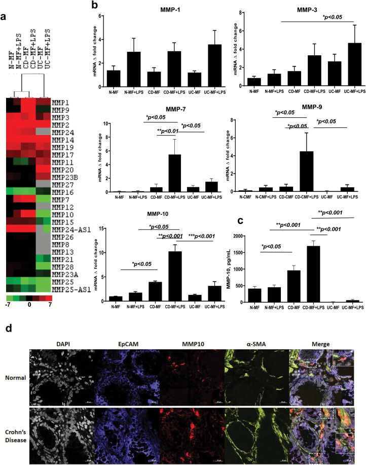
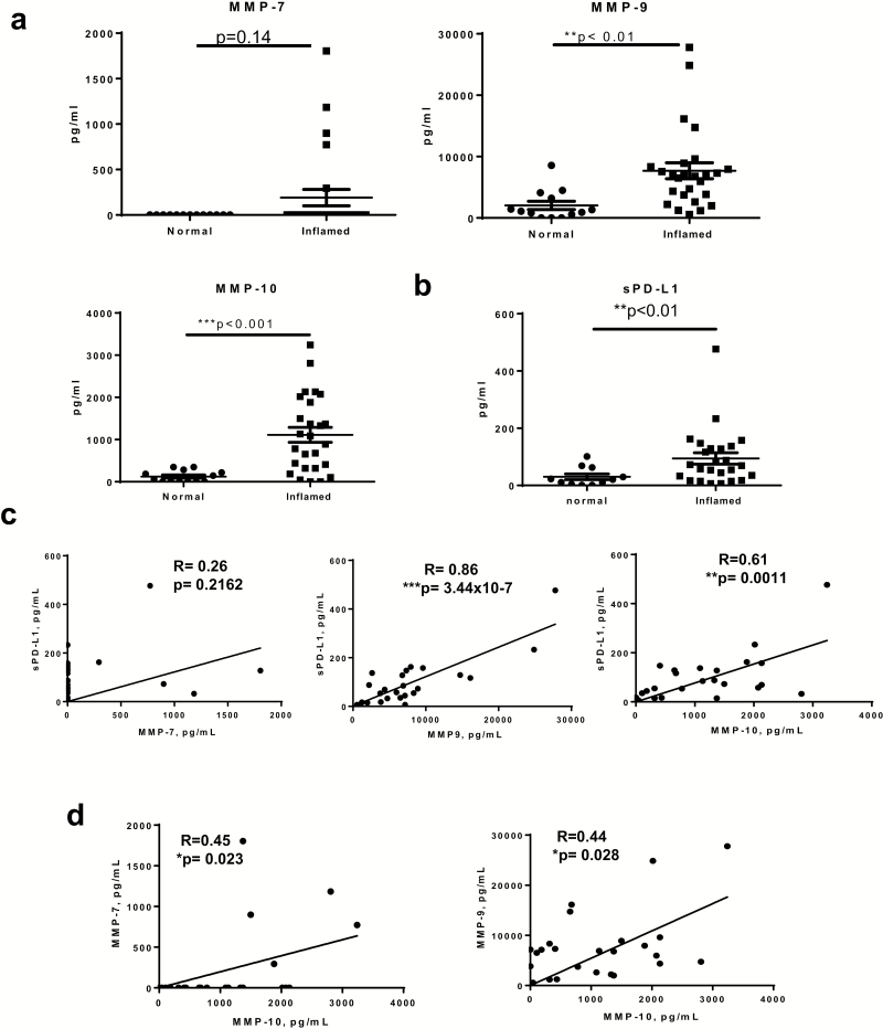
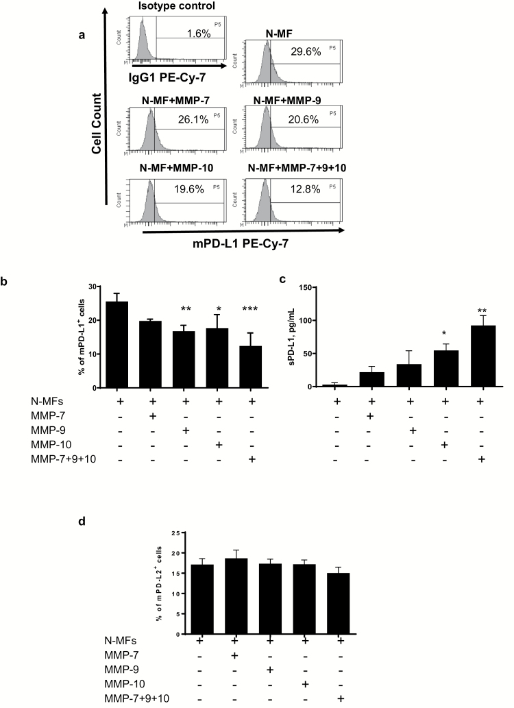
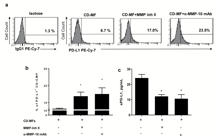
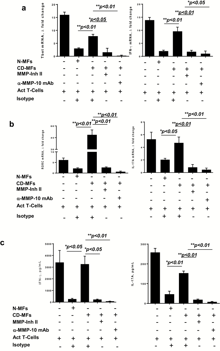
Increased T helper (Th)1/Th17 immune responses are a hallmark of Crohn's disease (CD) immunopathogenesis. CD90+ (myo-)fibroblasts (MFs) are abundant cells in the normal (N) intestinal mucosa contributing to mucosal tolerance via suppression of Th1 cell activity through cell surface membrane-bound PD-L1 (mPD-L1). CD-MFs have a decreased level of mPD-L1. Consequently, mPD-L1-mediated suppression of Th1 cells by CD-MFs is decreased, yet the mechanism responsible for the reduction in mPDL-1 is unknown. Increased expression of matrix metalloproteinases (MMPs) has been reported in CD. Herein we observed that when compared to N- and ulcerative colitis (UC)-MFs, CD-MFs increase in LPS-inducible levels of MMP-7 and -9 with a significant increase in both basal and inducible MMP-10. A similar pattern of MMP expression was observed in the CD-inflamed mucosa. Treatment of N-MFs with a combination of recombinant human MMP-7, -9 and -10 significantly decreased mPD-L1. In contrast, inhibition of MMP activity with MMP inhibitors or anti-MMP-10 neutralizing antibodies restores mPD-L1 on CD-MFs. CD-MFs demonstrated reduced capacity to suppress Th1 and Th17 responses from activated CD4+ T cells. By contrast, supplementation of the CD-MF:T-cell co-cultures with MMP inhibitors or anti-MMP neutralizing antibodies restored the CD-MF-mediated suppression. Our data suggest that (i) increased MMP-10 expression by CD-MFs and concomitant cleavage of PD-L1 from the surface of CD-MFs are likely to be one of the factors contributing to the decrease of mPD-L1-mediated suppression of Th1/Th17 cells in CD; and (ii) MMPs are likely to have a significant role in the intestinal mucosal immune responses.

Keywords: Crohn’s disease; matrix metalloproteinases; membrane-bound and soluble PD-L1; myo-/fibroblasts.


Figures

Fig. 1.
Fig. 1.
Expression of basal and LPS-inducible patterns of MMPs differs in CD-MFs when compared to N- and UC-MFs. (a) mRNA microarray analysis shows differential MMP expression in CD-MFs compared to N-MFs and UC-MFs at basal and TLR4-stimulated conditions. Sample-based hierarchical clustering was carried out as described in Methods. The color chart at the bottom of the figure shows levels of fold decrease and increase, log2 fold changes are shown. (b) Basal and LPS-inducible differential expression of MMP-7, -9 and -10 by MFs isolated from normal, CD and UC mucosa was analyzed on mRNA level using real-time PCR. (c) Basal and LPS-inducible secretion of MMP-10 by MFs isolated from CD mucosa is increased when compared to normal, and UC mucosa-derived MFs, multiplex MMP array analysis. LPS was used at concentration 1 μg ml−1, 72-h exposure. Data are shown as means ± SEM, n = 5, *P < 0.05; **P < 0.01; ***P < 0.001. (d) MMP-10 expression is increased within α-SMA+ MFs in CD intestinal mucosa when compared to the healthy controls. Confocal microscopy images of representative cross-sections from CD and normal human intestinal mucosa (n = 4 per group) are shown. MFs were detected by anti-α-SMA mAb (in green), and epithelial cells were identified with anti-EpCAM mAb (in blue). Co-localization of MMP-10 (in red) with α-SMA+ MFs results in formation of yellow-orange color on merged images. High resolution areas in the merged images are outlined in the boxes.
Fig. 2.
Fig. 2.
Secreted MMP-9 and -10 are significantly increased within inflamed CD mucosa and associated with an increase in tissue secretion of soluble (s) PD-L1. Mucosa of surgical specimens from CD patients were used to compare inflamed versus non-inflamed normal (normal) adjacent tissue. (a) Multiplexed MMP array assay showed significant increases in the secretion of MMP-9 and -10, but not -7 in the inflamed CD mucosa when compared to adjacent non-inflamed controls. Data are shown as mean ± SEM, n = 11–25 per group. (b) ProcartaPlex™ soluble PD-L1 immunoassay analysis showed increased sPD-L1 in the inflamed CD mucosa when compared to adjacent non-inflamed mucosa. Data are shown as mean ± SEM, n = 11–25 per group. (c) Linear regression and Pearson correlation analysis show that increased sPD-L1 is significantly correlated with secreted MMP-9 and -10 in inflamed CD mucosa, n = 25 per group. (d) Linear regression and Pearson correlation show that increases in MMP-7 and -9 correlate with the increase of MMP-10 in the inflamed CD mucosa, n = 25 per group, **P < 0.01; ***P < 0.001.
Fig. 3.
Fig. 3.
Recombinant human-activated MMP-9 and -10 reduce levels of membrane-bound (m) PD-L1 on N-MFs. Primary human isolates of N-MFs were treated with MMP-7, -9, -10 or combinations of these MMPs for 3 h as described in Methods, then immunostained and used for multi-color flow cytometry. Condition media from these cultures were used to determine sPD-L1 by PD-L1 singleplex assay. Live events were gated based on forward and side scatters, as well as negativity for the incorporation of the viability dye (conjugated to eFluor™ 780) and were analyzed for the surface expression of mPD-L1 and mPD-L2. (a) Representative histograms of mPD-L1 expression on the surface of N-MFs following treatment with activated MMPs are shown. Summary of the (b) mPD-L1 and (c) sPD-L1 expressed/produced by N-MFs following treatment with activated MMPs. Summary of the (d) mPD-L2 protein expression on surface of N-MFs following treatment with activated MMPs. Data are shown as mean ± SEM, n = 5, *P < 0.05; **P < 0.01; ***P < 0.001.
Fig. 4.
Fig. 4.
The proteolytic activity of MMPs contributes to the reduction of the mPD-L1 on cell surface of CD-MFs. Primary human isolates of CD-MFs were treated or not with the 30 nM of MMP Inhibitor II (CAS 203915-59-7) or anti-human MMP-10 mAbs (clone LA-12) for 72 h, then immunostained and used for multi-color flow cytometry. Live events were gated based on forward and side scatters, as well negativity for the incorporation of the viability dye (conjugated to eFluor™ 780) and were analyzed for the surface expression of mPD-L1. (a) Representative histograms and (b) summary of the flow cytometry analysis show that expression of mPD-L1 is reduced on cell surface of CD-MFs. (c) Summary of sPD-L1 shows an increase in sPD-L1 in the conditioned media from CD-MFs. Addition of broad spectrum MMP Inhibitor-II, which targets MMP-1, -3, -7, -9 or anti-MMP-10 mAb restores mPD-L1 level on the surface of CD-MFs and decreases sPD-L1 concentration in the condition media derived from these cells. Data are shown as mean ± SEM, n = 4, *P < 0.05.
Fig. 5.
Fig. 5.
MMP inhibition restores the capacity of CD-MFs to suppress activated Th1/Th17 cell responses. MFs were co-cultured with CD3/CD28 pre-activated allogeneic CD4+ T cells for 5 days in the presence or absence of the MMP inhibitor II (30 nM) or anti-MMP-10 (1 μg ml−1) blocking mAbs. (a) Tbet and IFN-γ mRNA expression by the T cells was analyzed using real-time RT–PCR analysis. (b) RORC and IL-17A mRNA expression by the T cells was analyzed using real-time RT–PCR analysis. (c) IFN-γ and IL-17A production was analyzed using multiplex cytokine analysis. The means ± SEM are shown, n = 4 allogeneic donor pairs per group, two experimental replicates each, *P < 0.05; **P < 0.01.

References

    1. Fuss, I. J., Neurath, M., Boirivant, M.et al. 1996. Disparate CD4+ lamina propria (LP) lymphokine secretion profiles in inflammatory bowel disease. Crohn’s disease LP cells manifest increased secretion of IFN-gamma, whereas ulcerative colitis LP cells manifest increased secretion of IL-5. J. Immunol. 157:1261. - PubMed
    1. Strober, W. and Fuss, I. J. 2011. Proinflammatory cytokines in the pathogenesis of inflammatory bowel diseases. Gastroenterology 140:1756. - PMC - PubMed
    1. Heller, F., Florian, P., Bojarski, C.et al. 2005. Interleukin-13 is the key effector Th2 cytokine in ulcerative colitis that affects epithelial tight junctions, apoptosis, and cell restitution. Gastroenterology 129:550. - PubMed
    1. Pinchuk, I. V., Saada, J. I., Beswick, E. J.et al. 2008. PD-1 ligand expression by human colonic myofibroblasts/fibroblasts regulates CD4+ T-cell activity. Gastroenterology 135:1228. - PMC - PubMed
    1. Owens, B. M., Steevels, T. A., Dudek, M.et al. 2013. CD90(+) stromal cells are non-professional innate immune effectors of the human colonic mucosa. Front. Immunol. 4:307. - PMC - PubMed

Publication types

MeSH terms