. 2020 Feb 15;458(2):141-152.
doi: 10.1016/j.ydbio.2019.10.013. Epub 2019 Oct 18.

Cytoglobin deficiency potentiates Crb1-mediated retinal degeneration in rd8 mice

Affiliations

Cytoglobin deficiency potentiates Crb1-mediated retinal degeneration in rd8 mice

Young Sam Kwon et al. Dev Biol. .

Abstract

Purpose: The purpose of this study is to determine the effect of Cytoglobin (Cygb) deficiency on Crb1-related retinopathy. The Crb1 cell polarity complex is required for photoreceptor function and survival. Crb1-related retinopathies encompass a broad range of phenotypes which are not completely explained by the variability of Crb1 mutations. Genes thought to modify Crb1 function are therefore important targets of research. The biological function of Cygb involves oxygen delivery, scavenging of reactive oxygen species, and nitric oxide metabolism. However, the relationship of Cygb to diseases involving the Crb1 cell polarity complex is unknown.

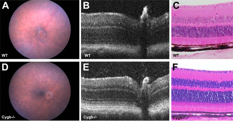
Methods: Cygb knockout mice homozygous for the rd8 mutation (Cygb-/-rd8/rd8) were screened for ocular abnormalities and imaged using optical coherence tomography and fundus photography. Electroretinography was performed, as was histology and immunohistochemistry. Quantitative PCR was used to determine the effect of Cygb deficiency on transcription of Crb1 related cell polarity genes.

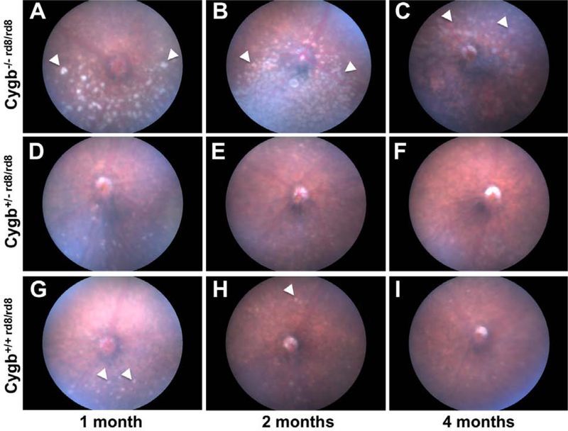
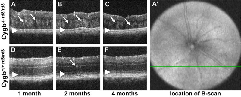
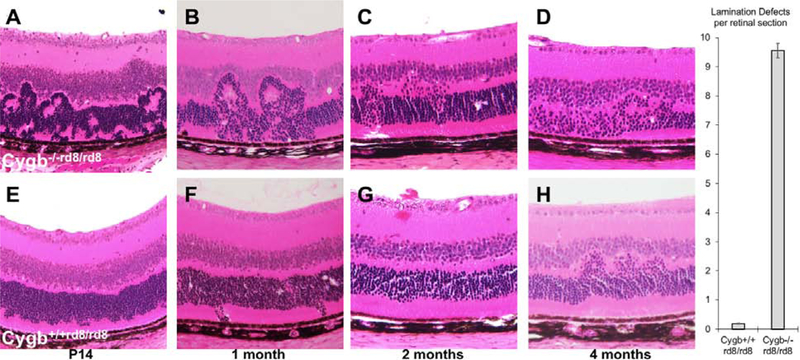
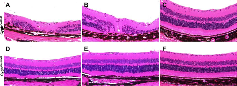
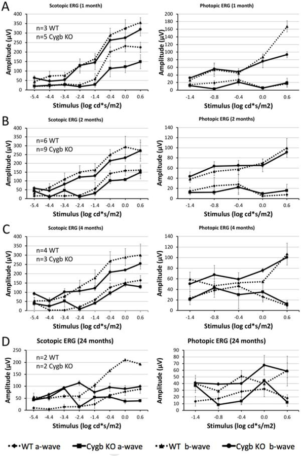
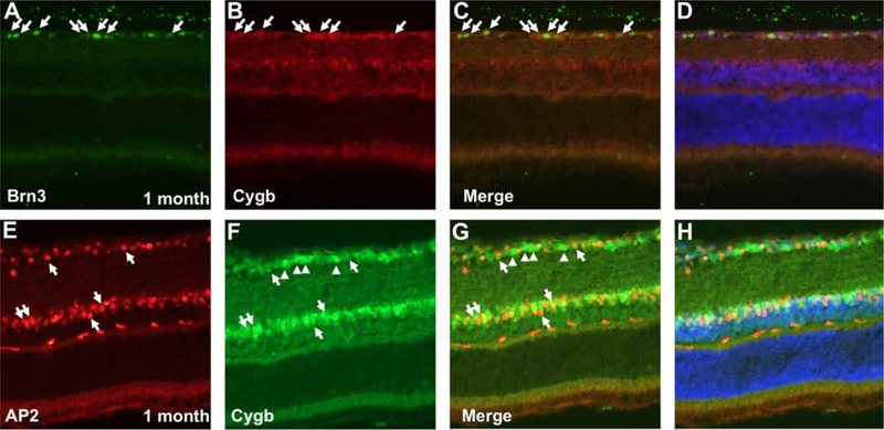
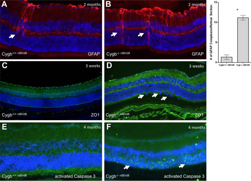
Results: Cygb-/-rd8/rd8 mice develop an abnormal retina with severe lamination abnormalities. The retina undergoes progressive degeneration with the ventral retina more severely affected than the dorsal retina. Cygb expression is in neurons of the retinal ganglion cell layer and inner nuclear layer. Immunohistochemical studies suggest that cell death predominates in the photoreceptors. Electroretinography amplitudes show reduced a- and b-waves, consistent with photoreceptor disease. Cygb deficient retinas had only modest transcriptional perturbations of Crb1-related cell polarity genes. Cygb-/- mice without the rd8 mutation did not exhibit obvious retinal abnormalities.

Conclusions: Cygb is necessary for retinal lamination, maintenance of cell polarity, and photoreceptor survival in rd8 mice. These results are consistent with Cygb as a disease modifying gene in Crb1-related retinopathy. Further studies are necessary to investigate the role of Cygb in the human retina.


Figures

Figure 1:
Figure 1:
Cytoglobin deficiency potentiates the severity of the rd8 fundus phenotype. Cygb−/−rd8/rd8 mice (A, B, C) accumulate an increased number and size of fundus flecks (arrowheads) when compared to Cygb+/+rd8/rd8 mice (G, H, I) at all ages examined. By 2 months of age, the inferior fundus of Cygb−/−rd8/rd8 animals develops confluent flecks (B), which undergo pigmentary atrophy (C) by 4 months. The number and size of flecks in Cygb+/−rd8/rd8 mice (D, E, F) is indistinguishable from Cygb+/+rd8/rd8 littermates (G, H, I) at all ages examined. See quantification of white spots in Supplemental Figure 1. For each genotype n > 15.
Figure 2:
Figure 2:
Optical Coherence Tomography of Cygb−/−rd8/rd8 mice shows outer retinal hyperreflective foci and loss of outer retinal bands. Areas of hyperreflectivity are seen as early as post-natal month 1 (A, arrows), and persist at postnatal month 2 (B, arrows) and 4 (C, arrows). Similar lesions are occasionally see in Cygb+/+rd8/rd8 animals (D,E,F) at these ages (arrow in E), but to a lesser degree. The outer retinal bands (D,E,F, arrowheads) representing the external limiting membrane, the inner segment/outer segment junction (aka ellipsoid zone), and the retinal pigmented epithelium are not discernable in Cygb−/−rd8/rd8 mice (A,B,C arrowheads). Also, the total thickness of the retina decreases with age in Cygb−/−rd8/rd8 mice (A,B,C). The b-scans shown here are taken from horizontal scans in the inferior retina. For example, the green line in A’ corresponds to the cross section in panel A.
Figure 3:
Figure 3:
Cygb deficiency increases the severity of photoreceptor lamination defects in rd8 mice. At several postnatal ages, hematoxylin and eosin histological analysis of the outer nuclear layer in Cygb−/−rd8/rd8 retina on the rd8 background (A-C) has profound outer retinal lamination defects with photoreceptor rosettes that eventually lead to photoreceptor loss (D). Cygb+/+rd8/rd8 mice (E-H) have less severe lamination defects that progress less rapidly and without obvious cell loss at these time points. (n = 5 each group).
Figure 4:
Figure 4:
Cygb deficiency leads to severe retinal degeneration in aged rd8 mice. At 24 months postnatal age, Cygb−/−rd8/rd8 mice on the rd8 background (A-C) have areas of mild, moderate, and severe retinal degeneration within the same eye. Cygb+/+rd8/rd8 mice (D-F) have areas of normal to mild retinal degeneration at this stage. (n = 6 each group).
Figure 5:
Figure 5:
Rod retinal function declines with age in Cygb deficient rd8 mice. Scotopic (dark-adapted) and photopic (light-adapted) electroretinography was performed in Cygb−/−rd8/rd8 (designated Cygb KO) mice and Cygb+/+rd8/rd8 (designated WT) controls at one (A), 2 (B), 4 (C), and 24 (D) months postnatal age. Stimulus-response curves are shown with absolute values of the a- and b-waves for Cygb−/−rd8/rd8 (solid line) and Cygb+/+rd8/rd8 (dashed line) on the same graphs. At 1- and 2-months postnatal age, rod and cone mediated retinal pathways appear similar in both groups. By 4 months of age (C) the scotopic b-wave declines in Cygb−/−rd8/rd8 mice when compared to Cygb+/+rd8/rd8 mice at this age. By 24 months postnatal age, the scotopic b-wave is markedly reduced in comparison to Cygb+/+rd8/rd8 mice. Photopic responses appear similar at all stages in both groups, suggesting less involvement of the cone-mediated responses. (Exact n is shown in figure for each time point. For A-C, n≥3 in each group. n=2 in each group for panel D).
Figure 6:
Figure 6:
Cygb is expressed in the inner retina. Immunohistochemistry at postnatal age 1 month using anti-ß-galactosidase antibodies as a surrogate for Cygb expression (A) shows expression in the retinal ganglion cell layer and the inner aspect of the inner nuclear layer. Anti-Cygb staining (B) is seen in a similar pattern as anti-ß-galactosidase at the same age. LacZ histochemistry corroborates this expression pattern in postnatal day 14 heterozygous mice (C, left), and in neonatal heterozygous mice at postnatal day 3 (C, right).
Figure 7:
Figure 7:
Cygb is expressed in retinal ganglion cells and amacrine cells. Double-labeling at postnatal age 1 month using (A-D) anti-Cygb and anti-Brn3, or (E-H) anti-Cygb and anti-AP2 show co-labeling in both retinal ganglion cells and amacrine neurons, respectively. Cytoglobin is localized both to the cytoplasmic and nuclear compartments in these neurons. Arrows point to cells expressing both markers. Arrowheads in E-H point to cells in the ganglion cell layer that are Cygb-positive but AP2-negative, and likely represent RGCs.
Figure 8:
Figure 8:
Cygb deficiency results in disruption of the outer limiting membrane, reactive Muller glia, and cell death. Typical anti-GFAP staining is show in (A) Cygb+/+rd8/rd8 mice and (B) Cygb−/−rd8/rd8 mice at postnatal age 2 months. Quantification of GFAP-positive activated Muller glial complexes per retinal section is shown (*P<0.05 was considered significant, student’s t-test). The outer limiting membrane as stained by anti-ZO1 immunohistochemistry show relative preservation of this structure in (A) Cygb+/+rd8/rd8 mice, but with frequent areas of discontinuity seen in (B) Cygb−/−rd8/rd8 mice at postnatal age 3 weeks. Virtually no anti-activated Caspase 3 staining is seen at postnatal age 4 months in (E) the Cygb+/+rd8/rd8 retina, while robust staining is seen in the outer nuclear layer at this age in (F) Cygb−/−rd8/rd8 mice.
Figure 9:
Figure 9:
Cygb knockout mice free from the rd8 mutation have no obvious retinal abnormalities. Color fundus photos of (A) WT and (B) Cygb−/− mice on the C57BL/6J background confirmed to be free of the rd8 mutation are indistinguishable at 4 months postnatal age. Spectral domain optical coherence tomography (SD-OCT) of (C) WT and (D) Cygb−/− mice show similar retinal lamination, which is consistent with (E,F) the hematoxylin and eosin histological analysis at 4 months postnatal.

References

    1. Gosens I, den Hollander AI, Cremers FP, Roepman R. Composition and function of the Crumbs protein complex in the mammalian retina. Exp Eye Res 2008. May; 86(5):713–26. - PubMed
    1. Tepass U, Theres C, Knust E. crumbs encodes an EGF-like protein expressed on apical membranes of Drosophila epithelial cells and required for organization of epithelia. Cell 1990. June 1; 61(5):787–99. - PubMed
    1. Grawe F, Wodarz A, Lee B, Knust E, Skaer H. The Drosophila genes crumbs and stardust are involved in the biogenesis of adherens junctions.Development 1996. March; 122(3):951–9. - PubMed
    1. den Hollander AI, Heckenlively JR, van den Born LI, de Kok YJ, van der Velde-Visser SD, Kellner U, Jurklies B, van Schooneveld MJ, Blankenagel A, Rohrschneider K, Wissinger B, Cruysberg JR, Deutman AF, Brunner HG, Apfelstedt-Sylla E, Hoyng CB, Cremers FP. Leber congenital amaurosis and retinitis pigmentosa with Coats-like exudative vasculopathy are associated with mutations in the crumbs homologue 1 (CRB1) gene. Am J Hum Genet 2001. July; 69(1):198–203. - PMC - PubMed
    1. van den Hurk JA, Rashbass P, Roepman R, Davis J, Voesenek KE, Arends ML, Zonneveld MN, van Roekel MH, Cameron K, Rohrschneider K, Heckenlively JR, Koenekoop RK, Hoyng CB, Cremers FP, den Hollander AI. Characterization of the Crumbs homolog 2 (CRB2) gene and analysis of its role in retinitis pigmentosa and Leber congenital amaurosis. Mol Vis 2005. April 15; 11():263–73. - PubMed

Publication types

MeSH terms