. 2019 Oct 21;10(1):4731.
doi: 10.1038/s41467-019-12888-x.

Identification and characterization of Cardiac Glycosides as senolytic compounds

Affiliations

Identification and characterization of Cardiac Glycosides as senolytic compounds

Francisco Triana-Martínez et al. Nat Commun. .

Erratum in

  • Author Correction: Identification and characterization of Cardiac Glycosides as senolytic compounds.
    Triana-Martínez F, Picallos-Rabina P, Da Silva-Álvarez S, Pietrocola F, Llanos S, Rodilla V, Soprano E, Pedrosa P, Ferreirós A, Barradas M, Hernández-González F, Lalinde M, Prats N, Bernadó C, González P, Gómez M, Ikonomopoulou MP, Fernández-Marcos PJ, García-Caballero T, Del Pino P, Arribas J, Vidal A, González-Barcia M, Serrano M, Loza MI, Domínguez E, Collado M. Triana-Martínez F, et al. Nat Commun. 2020 Sep 16;11(1):4771. doi: 10.1038/s41467-020-18714-z. Nat Commun. 2020. PMID: 32938939 Free PMC article.

Abstract

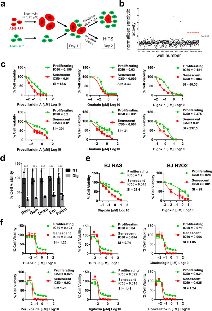
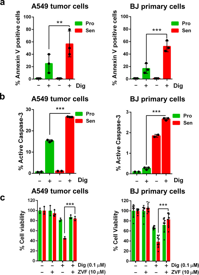
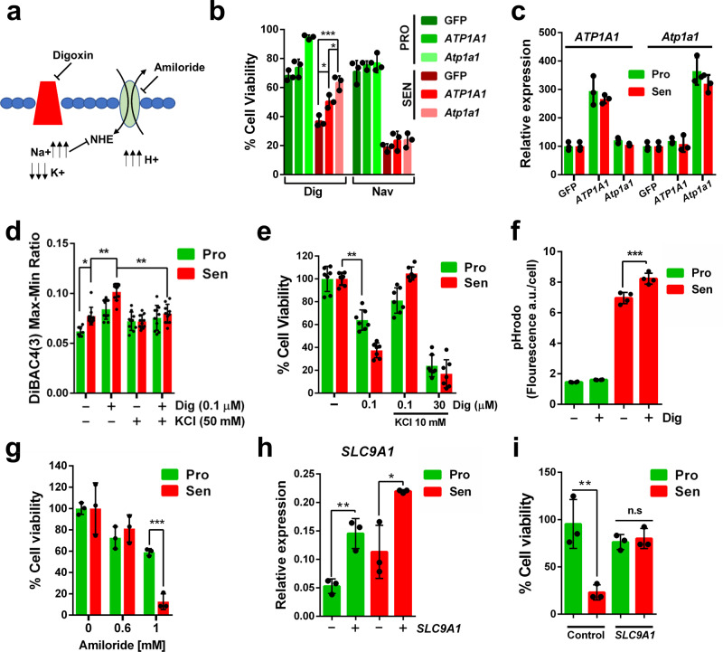
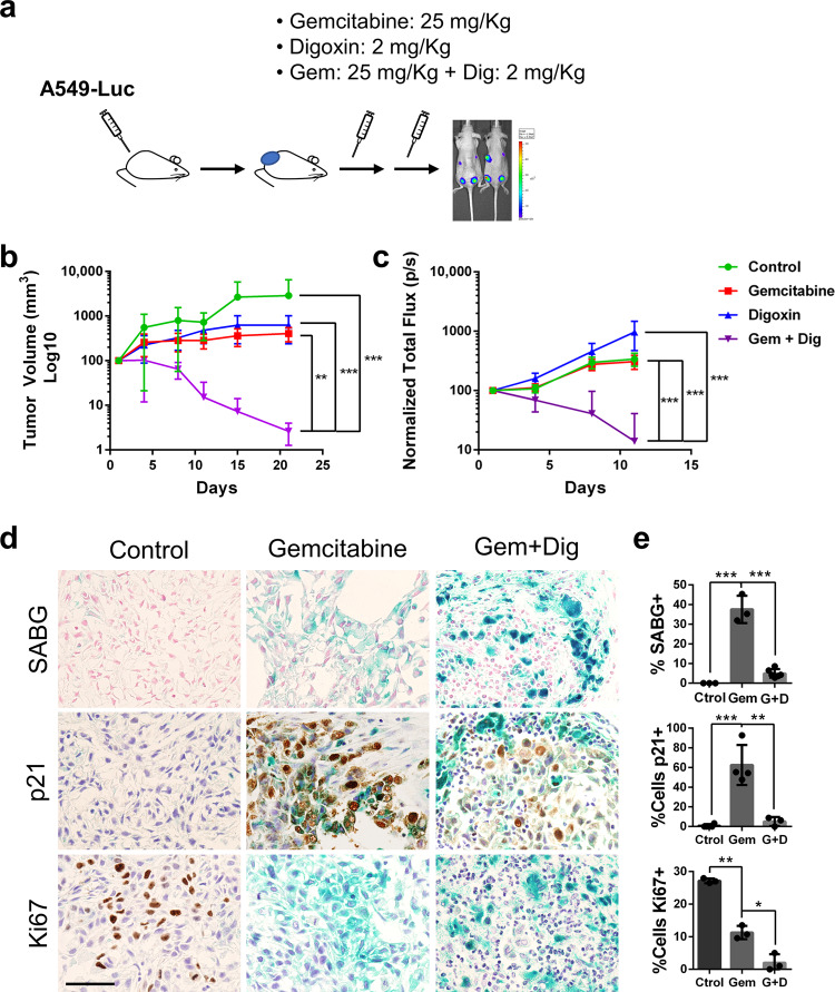
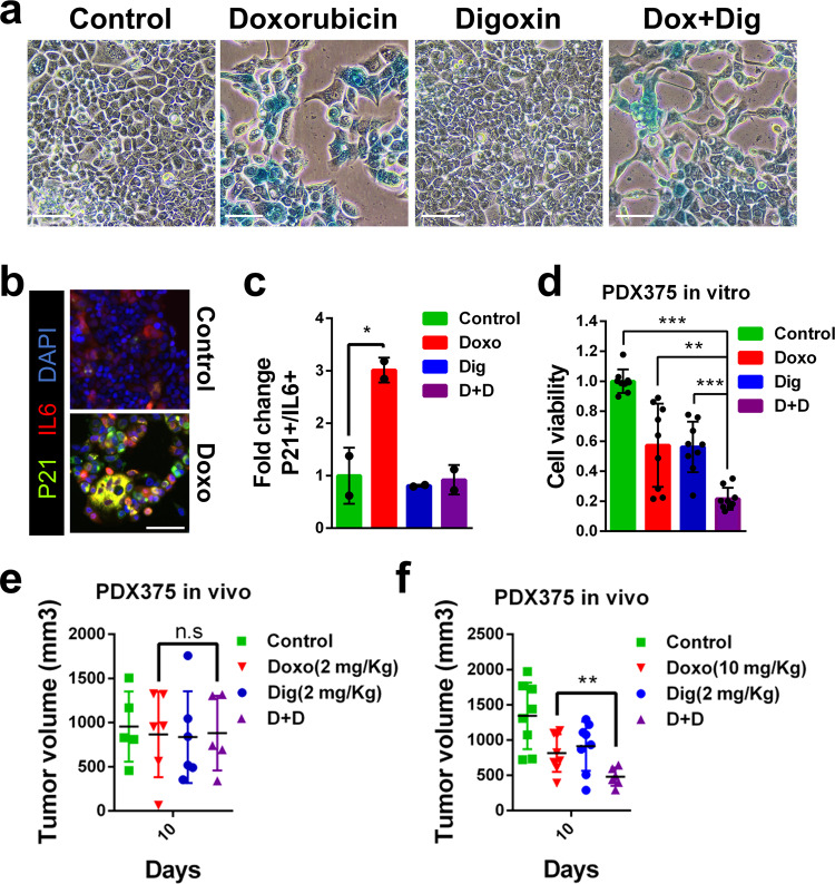
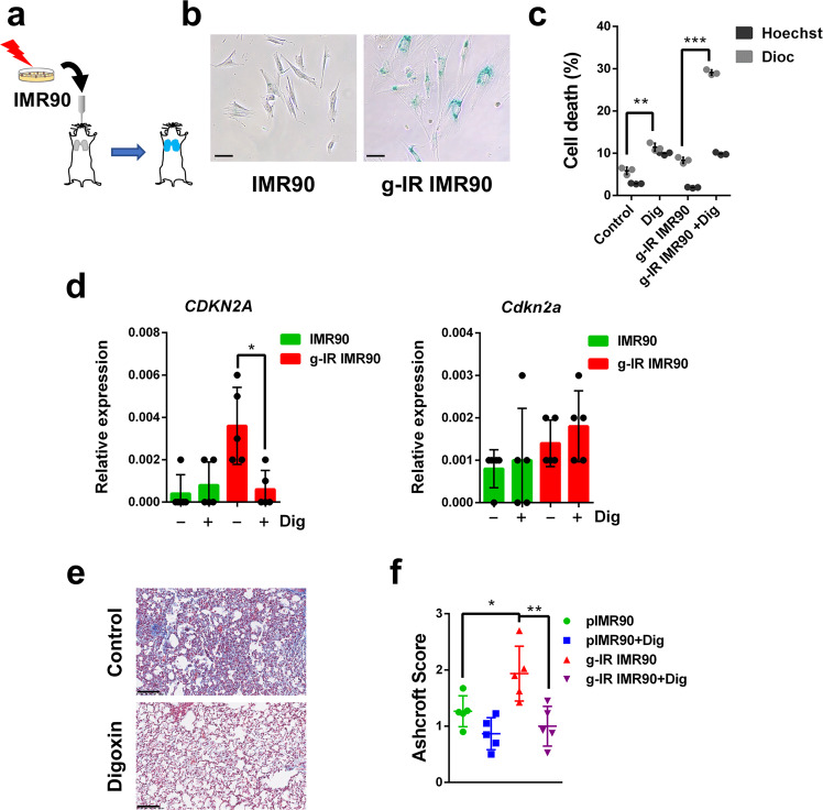
Compounds with specific cytotoxic activity in senescent cells, or senolytics, support the causal involvement of senescence in aging and offer therapeutic interventions. Here we report the identification of Cardiac Glycosides (CGs) as a family of compounds with senolytic activity. CGs, by targeting the Na+/K+ATPase pump, cause a disbalanced electrochemical gradient within the cell causing depolarization and acidification. Senescent cells present a slightly depolarized plasma membrane and higher concentrations of H+, making them more susceptible to the action of CGs. These vulnerabilities can be exploited for therapeutic purposes as evidenced by the in vivo eradication of tumors xenografted in mice after treatment with the combination of a senogenic and a senolytic drug. The senolytic effect of CGs is also effective in the elimination of senescence-induced lung fibrosis. This experimental approach allows the identification of compounds with senolytic activity that could potentially be used to develop effective treatments against age-related diseases.


Conflict of interest statement

M.S. is founder and advisor of Senolytic Therapeutics, Inc. The remaining authors declare no competing interests.

Figures

Fig. 1
Fig. 1
High-throughput screenings of senolytic compounds. a Schematic diagram of the experimental system used to screen compounds for potential senolytic activity. b Scatter plot representing the normalized senolytic activity obtained with each compound present in the Prestwick Chemical Library. Proscillaridin A value highlighted in red. c IC50 curves obtained with increasing concentrations of Proscillaridin A, Ouabain and Digoxin (from left to right) in A549 tumor cells (upper panels) and BJ primary fibroblasts (bottom panels). Actual IC50 values and Senolytic Indexes (SI) are also shown for each condition. d Relative cell viability (%) after Digoxin or vehicle treatment of A549 cells rendered senescent by different chemotherapeutic reagents (Bleo: Bleomycin; Gem: Gemcitabine; Doxo: Doxorubicin; Eto: Etoposide; and Palbo: Palbociclib). n = 3 independent experiments. Statistical significance was assessed by the two-tailed Student's t-test: ***p < 0.001. e IC50 curves of BJ primary fibroblasts induced to senescence by oncogenic RAS overexpression (left panel) or H2O2 treatment (right panel) treated with increasing concentrations of Digoxin. f IC50 curves of SK-MEL-103 melanoma cells induced to senescence by Palbociclib and treated with increasing concentrations of the indicated CGs identified on a screening for senolytic activities present in compounds obtained from plant extracts. Data correspond to the average ± s.d. Source data for these experiments are provided as a Source Data file
Fig. 2
Fig. 2
CGs kill senescent cells by inducing apoptosis. a Annexin V positive cells (%) in proliferating (Pro, in green) and senescence (Sen, in red) A549 cells (left panel) or primary BJ fibroblasts (right panel) after Digoxin treatment. b Active Caspase-3 positive cells (%) in proliferating (Pro, in green) and senescence (Sen, in red) A549 cells (left panel) or primary BJ fibroblasts (right panel) after Digoxin treatment. c Relative cell viability (%) of proliferative (Pro) or senescence (Sen) A549 cells treated with pan caspase inhibitor Z-VAD-FMK (ZVF), Digoxin (Dig) or the combination, as indicated. n = 3 independent experiments. All data correspond to the average ± s.d. Statistical significance was assessed by the two-tailed Student's t-test: ***p < 0.001; **p < 0.01. Source data for these experiments are provided as a Source Data file
Fig. 3
Fig. 3
Mechanism of action of Digoxin-induced senolysis. a Diagram showing the activity of Digoxin. b Relative cell viability (%) of proliferative (green) or senescence (red) A549 cells overexpressing human or mouse ATP1A1 or Atp1a1, respectively, and treated with Digoxin or Navitoclax. Statistical analysis with Anova with Tuckey test: ***p < 0.001; *p < 0.05; c and mRNA expression (relative to GAPDH) of ATP1A1 (left) and Atp1a1 (right). n = 3 independent experiments for both d Membrane potential determination of proliferative (green) or senescence (red) A549 cells treated with Digoxin and KCl, using fluorescent probe DiBAC4(3). n = 10 biologically independent samples. Statistical significance by two-tailed Student's t-test: **p < 0.01; *p < 0.05. e Relative cell viability (%) of proliferative (green) or senescence (red) A549 cells treated with Digoxin and KCl. n = 7 biologically independent samples. Statistical analysis with Anova with Tuckey test: **p < 0.01. f Determination of the relative intracellular H+concentration in proliferative (green) or senescence (red) A549 cells treated or not with Digoxin. n = 4 biologically independent samples. Statistical significance by two-tailed Student's t-test: ***p < 0.001. g Relative cell viability (%) of proliferative (green) or senescence (red) A549 cells treated or not with Amiloride. n = 3 biologically independent samples. Statistical significance by two-tailed Student's t-test: ***p < 0.001. h Relative mRNA expression of SLC9A1 in proliferative (green) or senescence (red) A549 cells overexpressing or not SLC9A1 as indicated. n = 3 biologically independent samples. Statistical significance by two-tailed Student's t-test: **p < 0.01; *p< 0.05. i Relative cell viability (%) of proliferative (green) or senescence (red) A549 cells treated with Digoxin and exogenously expressing SLC9A1 or GFP. n = 3 independent experiments. Statistical significance by two-tailed Student's t-test: **p < 0.01; n.s. not significant. All data correspond to the average ± s.d. Source data for these experiments are provided as a Source Data file
Fig. 4
Fig. 4
Senolytic activity of Digoxin on xenografts. a Diagram showing the experimental plan to test the effect of administrating Gemcitabine, Digoxin or the combination of both (Gem+Dig) in the growth of A549 cells expressing luciferase (A549-Luc) as subcutaneous tumors in nude mice. b Tumor volume (mm3) progression with time after intraperitoneal injection of Gemcitabine (n = 12), Digoxin (n = 8) or the combination of both (Gem+Dig) (n = 12), or vehicle (n = 4) as negative control as indicated. Statistical analysis was performed with Anova with Tuckey test: ***p < 0.001; **p < 0.01. c Tumor growth determined by measuring normalized total flux of luminescence as detected by IVIS with the same groups of animals. Statistical analysis was performed with Anova with Tuckey test: ***p < 0.001. d Representative images of SABG staining (upper panels) and immunohistochemical analysis of p21 (middle panels) and Ki67 (bottom panels) in tumors obtained after injection of A549 cells in nude mice and treated with Gemcitabine, Gemcitabine plus Digoxin (Gem+Dig), or control group (scale bar = 50 μm). e Quantifications of stainings shown in (d). n = 3 biologically independent samples. Statistical significance was assessed by the two-tailed Student's t-test: ***p < 0.001; **p < 0.01; *p< 0.05. All data correspond to the average ± s.d. Source data for these experiments are provided as a Source Data file
Fig. 5
Fig. 5
Senolytic activity of Digoxin on human PDX. a In vitro SABG staining of PDX375 in control, Doxorubicin, Digoxin and Doxorubicin plus Digoxin (Dox+Dig) treated cells (scale bar = 100 μm). b Representative immunofluorescent images of PDX375 cells transduced with lentiviral reporter constructs for the expression of p21 (in green) and IL6 (in red) in control condition or after Doxorubicin treatment. DAPI (in blue) was used to stained nuclei (scale bar = 100 μm). c Quantification (fold change) of double positive p21/IL6 cells in control, or Doxorubicin (Doxo), Digoxin (Dig), or Doxorubicin plus Digoxin (D+D) treatment (n = 3 biologically independent samples). d In vitro cell viability (relative to control) of PDX375 cells treated with Doxorubicin (Doxo), Digoxin (Dig), or the combination (D+D) (n = 3 biologically independent samples). e In vivo growth (tumor volume) of PDX375 subcutaneously injected into nude mice and treated with Doxorubicin (Doxo: 2 mg/kg; n = 6), Digoxin (Dig: 2 mg/kg; n = 6), the combination (D+D; n = 5), or vehicle (Control; n = 5). f Same in vivo growth analysis in mice treated with Doxorubicin (Doxo: 10 mg/kg; n = 8), Digoxin (Dig: 2 mg/kg; n = 8), the combination (D+D; n = 6), or vehicle (Control; n = 8). Statistical significance was assessed by the two-tailed Student's t-test: ***p < 0.001; **p < 0.01; *p< 0.05. All data correspond to the average ± s.d. Source data for these experiments are provided as a Source Data file
Fig. 6
Fig. 6
Senolytic activity of Digoxin in lung fibrosis. a Schematic diagram of the experimental system to induce lung fibrosis in mice by intratracheal instillation of proliferative or senescent gamma-irradiated IMR90 cells. b In vitro SABG staining of control or gamma-irradiated (g-IR) IMR90 cells (scale bar = 100 μm). c In vitro analysis of cell death in control or (g-IR) IMR90 cells treated or not with Digoxin (Dig). Black bars represent the % of DiOC6(3) low and grey bars represent Hoechst 33342 positive cells. n = 3 independent experiments. d Relative expression of the mRNA coding for CDKN2A (left panel) or Cdkn2a (right panel) in lung cell extracts from mice injected with control proliferative (green) or gamma-irradiated (g-IR, red) IMR90 cells, and treated or not with Digoxin, as indicated. n = 5 independent experiments. e Representative images of lung sections stained with Masson Trichrome from mice injected with gamma-irradiated IMR90 cells and treated (bottom panel) or not (upper panel) with Digoxin (scale bar = 100 μm). f Ashcroft score of Masson Trichrome staining in sections from mice injected with control proliferative or gamma-irradiated (g-IR) IMR90 cells, treated or not with Digoxin (+Dig). n = 5 independent experiments. Statistical significance was assessed by the two-tailed Student's t-test: ***p < 0.001; **p < 0.01; *p < 0.05. Data correspond to the average ± s.d. Source data for these experiments are provided as a Source Data file

Comment in

  • Adding to the senolytic arsenal.
    Paiva SL. Paiva SL. Nat Rev Drug Discov. 2019 Nov;18(12):901. doi: 10.1038/d41573-019-00181-x. Nat Rev Drug Discov. 2019. PMID: 31780845 No abstract available.

References

    1. Muñoz-Espín D, Serrano M. Cellular senescence: from physiology to pathology. Nat. Rev. Mol. Cell Biol. 2014;15:482–496. - PubMed
    1. Hernandez-Segura A, Nehme J, Demaria M. Hallmarks of cellular senescence. Trends Cell Biol. 2018;28:436–453. - PubMed
    1. Demaria M, et al. An essential role for senescent cells in optimal wound healing through secretion of PDGF-AA. Dev. Cell. 2014;31:722–733. - PMC - PubMed
    1. Sun Y, Coppé J-P, Lam EW-F. Cellular senescence: the sought or the unwanted? Trends Mol. Med. 2018;24:871–885. - PubMed
    1. Lujambio A. To clear, or not to clear (senescent cells)? That is the question. BioEssays. 2016;38:S56–S64. - PubMed

Publication types

MeSH terms