. 2019 Oct 21;9(1):15047.
doi: 10.1038/s41598-019-51517-x.

Mitochondrial dysfunction increases pro-inflammatory cytokine production and impairs repair and corticosteroid responsiveness in lung epithelium

Affiliations

Mitochondrial dysfunction increases pro-inflammatory cytokine production and impairs repair and corticosteroid responsiveness in lung epithelium

R F Hoffmann et al. Sci Rep. .

Abstract

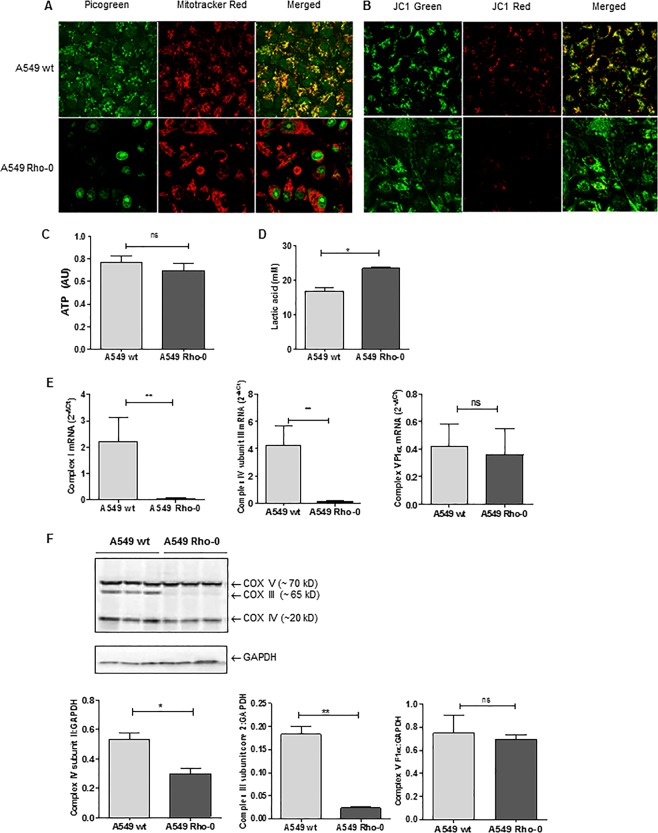
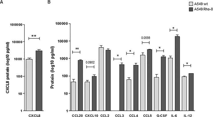
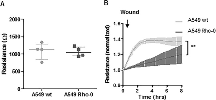
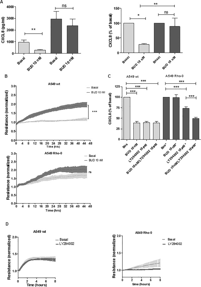
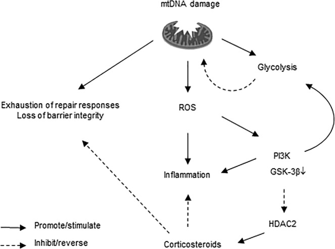
COPD is characterized by chronic lung inflammation and irreversible lung tissue damage. Inhaled noxious gases, including cigarette smoke, are the major risk factor for COPD. Inhaled smoke first encounters the epithelial lining of the lungs, causing oxidative stress and mitochondrial dysfunction. We investigated whether a mitochondrial defect may contribute to increased lung epithelial pro-inflammatory responses, impaired epithelial repair and reduced corticosteroid sensitivity as observed in COPD. We used wild-type alveolar epithelial cells A549 and mitochondrial DNA-depleted A549 cells (A549 Rho-0) and studied pro-inflammatory responses using (multiplex) ELISA as well as epithelial barrier function and repair (real-time impedance measurements), in the presence and absence of the inhaled corticosteroid budesonide. We observed that A549 Rho-0 cells secrete higher levels of pro-inflammatory cytokines than wild-type A549 cells and display impaired repair upon wounding. Budesonide strongly suppressed the production of neutrophil attractant CXCL8, and promoted epithelial integrity in A549 wild-type cells, while A549 Rho-0 cells displayed reduced corticosteroid sensitivity compared to wild-type cells. The reduced corticosteroid responsiveness may be mediated by glycolytic reprogramming, specifically glycolysis-associated PI3K signaling, as PI3K inhibitor LY294002 restored the sensitivity of CXCL8 secretion to corticosteroids in A549 Rho-0 cells. In conclusion, mitochondrial defects may lead to increased lung epithelial pro-inflammatory responses, reduced epithelial repair and reduced corticosteroid responsiveness in lung epithelium, thus potentially contributing to the pathogenesis of COPD.


Conflict of interest statement

This study was funded by and performed within the framework of the Top Institute Pharma project T1-201 “COPD, transition of systemic inflammation into multi-organ pathology”, with partners of the University of Groningen, University Medical Center Groningen, University Medical Center Utrecht, University Medical Center Maastricht, Nycomed BV, GlaxoSmithKline, Danone, AstraZeneca and Foundation TI Pharma. We have no non-financial competing interests.

Figures

Figure 1
Figure 1
Mitochondrial (mt)DNA depletion and mitochondrial dysfunction in A549 Rho-0 cells. A549 wild-type (wt) and Rho-0 cells were grown to confluence and serum deprived for 24 hrs. (A) Cells were stained with Picogreen for detection of the DNA and Mitotracker DeepRed. Co-localization of DNA and mitochondrial bodies indicates mtDNA. Representatives of 3 independent experiments are shown. (B) Cells were stained with JC1 staining mitochondrial membrane potential is lowered by depletion of mtDNA in the A549 Rho-0 cells when compared to control cells. (C) ATP (mean ± SEM, n = 6–7) and (D) lactate (mean ± SEM, n = 3) were measured in cell-free culture supernatants. (E) RNA was isolated, cDNA synthesized, mRNA expression of NADH dehydrogenase (complex I), cytochrome c oxidase (complex IV) subunit III and ATPase subunit F1α (complex V) analyzed by qPCR and related to the expression of housekeeping genes β2µG and PPIA. (F) Cell lysates were prepared and complex III, Complex IV and the ATPase subunit components were detected by western blotting, analysed by densitometry and related to GAPDH (mean ± SEM, n = 3). *p < 0.05 and **p < 0.01 between the indicated values as measured by the one-tailed Mann-Whitney test.
Figure 2
Figure 2
Increased pro-inflammatory activity in A549 Rho-0 compared to wild-type A549 cells. A549 wild-type (wt) and Rho-0 cells were grown to confluence, serum deprived overnight and supernatants were collected 24 hours later. (A) CXCL8 (mean ± SEM, n = 5) and (B) CCL20, CXCL10, CCL2, CCL3, CCL4, CCL5, G-CSF, IL-6 and IL-12 (mean ± SEM, n = 5) levels (pg/ml) were measured in cell-free supernatants using ELISA and multiplex ELISA respectively. *p < 0.05 and **p < 0.01 between the indicated values as analysed by the two-tailed Mann-Whitney test.
Figure 3
Figure 3
A549 Rho-0 cells display reduced wound repair compared to wild-type A549 cells. (A) A549 wild-type and Rho-0 cells were seeded in a density of 100.000 cells/well in duplicates in ECIS arrays and grown to confluence for 24 hours. Resistance was measured at a frequency of 400 Hz (mean ± SEM, n = 4). (B) Confluent monolayers were wounded by electroporation. Resistance was monitored for 8 hours at 400 Hz and levels were normalized to the values immediately after wounding (mean ± SEM, n = 4). **p < 0.01 between the indicated values as analyzed by 2-way ANOVA.
Figure 4
Figure 4
A549 Rho-0 cells are less sensitive to budesonide than wild-type A549 cells, which is restored by blocking of PI3K/Akt signaling. (A) A549 wild-type and Rho-0 cells were grown to confluence, serum deprived overnight and incubated with/without budesonide (BUD, 10 nM) for 24 hours. CXCL8 was measured in cell-free supernatant and expressed as absolute values and as percentage of the levels without BUD (mean ± SEM, n = 56). (B) A549 wild-type and Rho-0 cells were grown to confluence in ECIS arrays for 24 hours. Subsequently, 10 nM budesonide (BUD) or vehicle was added and cells were cultured for another 48 hours. Resistance was monitored at 400 Hz and normalized to the levels immediately after the addition of BUD (mean ± SEM, n = 4). (C) A549 wild-type and Rho-0 cells were grown to confluence, serum deprived overnight and incubated with/without budesonide (BUD, 10 nM) for 24 hours. LY294002 (10 µM) was added 30 min before the exposure to BUD. CXCL8 was measured in cell-free supernatant. CXCL8 levels (mean ± SEM, n = 5) are expressed as percentage of the levels without BUD. (D) A549 wild-type and Rho-0 cells were grown in ECIS arrays. Confluent monolayers were wounded by electroporation. LY294002 (10 µM) was added 60 min prior to wounding. Resistance was monitored for 8 hours at 400 Hz and levels were normalized to the values immediately after wounding (mean ± SEM, n = 4). *p < 0.05, **p < 0.01 ***p < 0.001 between the indicated values as assessed by the Wilcoxon signed rank test within cell lines, the Mann Witney test between cell lines, 1-way ANOVA with Bonferroni’s multiple comparison test for panel c, 2-way ANOVA for ECIS data.
Figure 5
Figure 5
Schematic of proposed processes upon mitochondrial DNA (mtDNA) depletion in lung epithelial cells. During homeostasis, ATP is produced by oxidative phosphorylation and there is a balance between active and inactive PI3K/GSK-3βsignaling, ensuring limited glycolysis. Upon mitochondrial damage/dysfunction induced by mtDNA depletion, increased release of reactive oxygen species (ROS) production results in increased activity of redox sensitive kinases such as PI3K and a subsequent increase in production of pro-inflammatory mediators. Cells can no longer perform normal electron transport for ATP synthesis and rely on ATP derived from glycolysis, which is accompanied by impairment/exhaustion of repair responses. This glycolytic switch is promoted by the activation of PI3K signaling and subsequent inactivation of GSK-3β and HDAC2, resulting in reduced corticosteroid sensitivity of pro-inflammatory responses as well as reduced potential of corticosteroids to improve barrier integrity, the latter through mechanisms that need further elucidation.

References

    1. Barnes PJ, Ito K, Adcock IM. Corticosteroid resistance in chronic obstructive pulmonary disease: inactivation of histone deacetylase. Lancet. 2004;363:731–3. doi: 10.1016/S0140-6736(04)15650-X. - DOI - PubMed
    1. Heijink, I. et al. Oxidant-induced corticosteroid unresponsiveness in human bronchial epithelial cells. Thorax, 10.1136/thoraxjnl-2013-203520 (2013). - PubMed
    1. Ito K, et al. Decreased histone deacetylase activity in chronic obstructive pulmonary disease. N. Engl. J. Med. 2005;352:1967–76. doi: 10.1056/NEJMoa041892. - DOI - PubMed
    1. Barnes PJ, Adcock IM. Glucocorticoid resistance in inflammatory diseases. Lancet. 2009;373:1905–17. doi: 10.1016/S0140-6736(09)60326-3. - DOI - PubMed
    1. Schaaf MJ, Cidlowski JA. Molecular mechanisms of glucocorticoid action and resistance. J. Steroid Biochem. Mol. Biol. 2002;83:37–48. doi: 10.1016/S0960-0760(02)00263-7. - DOI - PubMed

Publication types