. 2020 Jan;13(1):110-117.
doi: 10.1038/s41385-019-0214-9. Epub 2019 Oct 21.

TGFβ1 single-nucleotide polymorphism C-509T alters mucosal cell function in pediatric eosinophilic esophagitis

Affiliations

TGFβ1 single-nucleotide polymorphism C-509T alters mucosal cell function in pediatric eosinophilic esophagitis

L D Duong et al. Mucosal Immunol. 2020 Jan.

Abstract

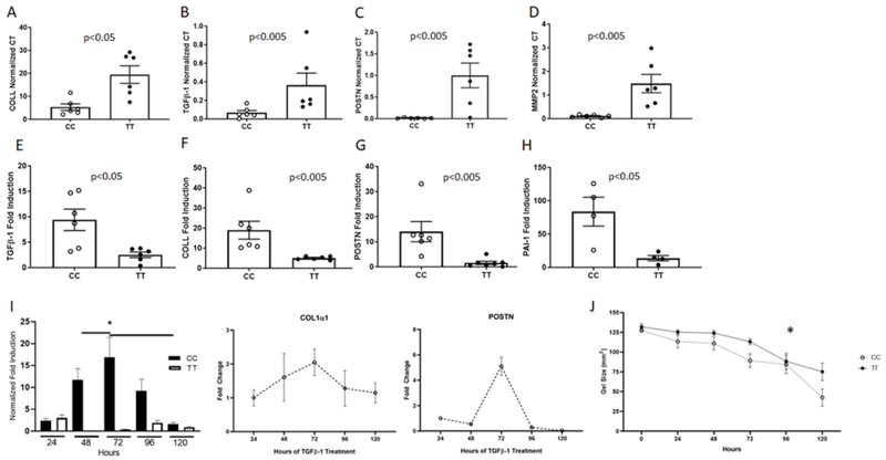
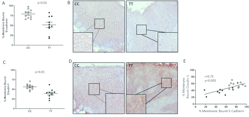
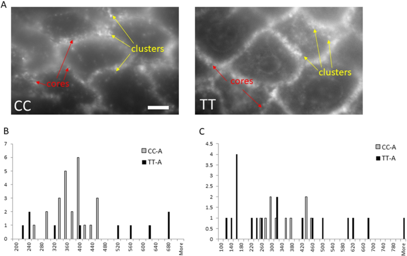
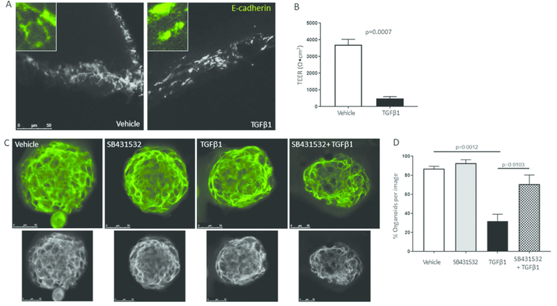
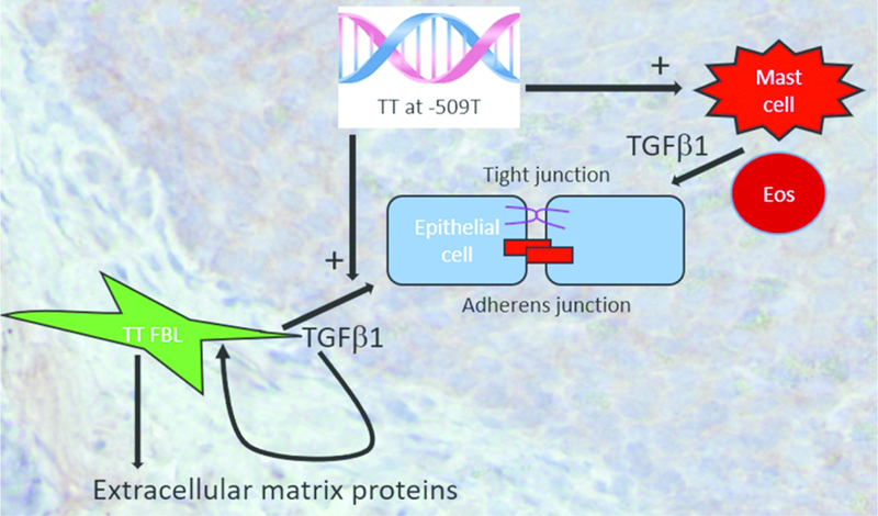
Eosinophilic esophagitis (EoE) is a chronic Th2 antigen-driven disorder associated with tissue remodeling. Inflammation and remodeling lead to esophageal rigidity, strictures, and dysphagia. TGFβ1 drives esophageal remodeling including epithelial barrier dysfunction and subepithelial fibrosis. A functional SNP in the TGFβ1 gene that increases its transcription (C-509T) is associated with elevated numbers of esophageal TGFβ1-expressing cells. We utilized esophageal biopsies and fibroblasts from TT-genotype EoE children to understand if TGFβ1 influenced fibroblast and epithelial cell function in vivo. Genotype TT EoE esophageal fibroblasts had higher baseline TGFβ1, collagen1α1, periostin, and MMP2 (p < 0.05) gene expression and distinct contractile properties compared with CC genotype (n = 6 subjects per genotype). In vitro TGFβ1 exposure caused greater induction of target gene expression in genotype CC fibroblasts (p < 0.05). Esophageal biopsies from TT-genotype subjects had significantly less epithelial membrane-bound E-cadherin (p < 0.01) and wider cluster distribution at nanometer resolution. TGFβ1 treatment of stratified primary human esophageal epithelial cells and spheroids disrupted transepithelial resistance (p < 0.001) and E-cadherin localization (p < 0.0001). A TGFβ1-receptor-I inhibitor improved TGFβ1-mediated E-cadherin mislocalization. These data suggest that EoE severity can depend on genotypic differences that increase in vivo exposure to TGFβ1. TGFβ1 inhibition may be a useful therapy in subsets of EoE patients.


Conflict of interest statement

COI: None of the authors has a relevant conflict of interest

Figures

Figure 1.
Figure 1.. Genotype CC and TT fibroblasts have different transcription profiles.
Baseline transcript levels for TGFβ1 (A), collagen1α1 (COLL) (B), periostin (C), and matrix metalloproteinase 2 (MMP-2) (D) in CC and TT primary esophageal fibroblasts matched for passage number, culture conditions, and EoE disease activity. TGFβ1-induced gene expression of TGFβ1 (E), collagen1α1 (COLL) (F), periostin (POSTN) (G), and plasminogen activator inhibitor-1 (PAI-1) (H). All gene expression was normalized to the housekeeping gene ribosomal protein L13a (RPL13a) and shown as normalized Ct (A–D) or as fold induction (E-H) for the matched vehicle treated well. Each data point represents a distinct patient. Each experiment was repeated in biological triplicates on different days and in qPCR technical replicates (–3). Chronic exposure to TGFβ1 in CC versus TT (representative assay, left panel I) and over time in CC fibroblasts (right panel, I; n=5 different individuals, bars represent SEM). *p<.01. Contraction of TT versus CC fibroblasts done in triplicate wells in esophageal fibroblasts from 10 different individuals (n=5 CC and n=5 TT) (J). *:p<.05
Figure 2.
Figure 2.. Genotype CC and TT esophageal biopsies have distinct patterns of cell adhesion proteins.
The percentage of cells per total epithelial height that expressed membrane bound E-cadherin-1 (A) or claudin-1 (C) was quantified using immunohistochemistry (red stain) and ImageJ analysis. Representative images of CC and TT subject epithelium are shown (B, D). Each point on the graphs represents the average of 5 different epithelial regions in 10 different subject biopsies with stains repeated on 3 different days. The degree of membrane bound E-cadherin and claudin-1 correlate positively regardless of genotype (closed circles=TT, open circles=CC) (E).
Figure 3.
Figure 3.. Total internal reflection fluorescence (TIRF) imaging shows differences in CC and TT E-cadherin.
TIRF high resolution imaging of CC and TT genotype biopsies (A) demonstrates more variability in the central core (B) and cluster (C) sizes in active genotype TT biopsies. Graphs represent quantification using TIRF imaging of core and cluster sizes and distances in at least 20 images (5 to 6 areas per tissue section that had the sharpest focus) from 6 different subjects. Representative image is shown. Scale bar = 5 microns.
Figure 4.
Figure 4.. TGFβ1 alters epithelial resistance and E-cadherin.
Representative images of vehicle and TGFβ1 treated epithelial cells on transwells (A) and the effect on transepithelial electrical resistance (TEER) (B). Quantification of the percent of spheroids with properly localized E-cadherin during treatment with vehicle, the ALKV inhibitor SB431532, TGFβ1 or TGFβ1+SB431532 (C). Representative image of spheroids treated with vehicle, SB431532, TGFβ1 or both. Images converted to gray scale below. Each image is a single slice of a 3-dimensional confocal image (D). Spheroids were treated in 2–3 replicate wells using 2 different donors on 2 different days. TEER experiments were repeated 3 times on different days in triplicate wells using epithelial cells from 2 different donors. Bars represent means and standard error from a representative experiment.
Figure 5.
Figure 5.. Model of the effects of TT genotype at TGFβ1 SNP −509 on EoE remodeling.
Genotype TT at −509 in the TGFβ1 promoter causes increased TGFβ1 expression from resident esophageal fibroblasts and associates with increased numbers of mast cells which, along with eosinophils, are reservoirs of TGFβ1. TGFβ1 functions in an auto- or paracrine manner on resident fibroblasts to augment production of pro-fibrotic factors such as collagen1α1, especially in genotype TT EoE patients. TGFβ1 perpetuates EoE by causing barrier breakdown of both adherens and tight junctions.

References

    1. Dellon ES, Liacouras CA, Molina-Infante J, Furuta GT, Spergel JM, Zevit N, et al. Updated international consensus diagnostic criteria for eosinophilic esophagitis: Proceedings of the AGREE conference. Gastroenterology. 2018. - PMC - PubMed
    1. Liacouras CA, Furuta GT, Hirano I, Atkins D, Attwood SE, Bonis PA, et al. Eosinophilic esophagitis: updated consensus recommendations for children and adults. The Journal of allergy and clinical immunology. 2011;128(1):3–20 e6; quiz 1–2. - PubMed
    1. Dellon ES. Epidemiology of Eosinophilic Esophagitis. Gastroenterology clinics of North America. 2014;43(2):201–18. - PMC - PubMed
    1. Spergel JM, Dellon ES, Liacouras CA, Hirano I, Molina-Infante J, Bredenoord AJ, et al. Summary of the updated international consensus diagnostic criteria for eosinophilic esophagitis: AGREE conference. Annals of allergy, asthma & immunology : official publication of the American College of Allergy, Asthma, & Immunology. 2018;121(3):281–4. - PMC - PubMed
    1. Schoepfer AM, Safroneeva E, Bussmann C, Kuchen T, Portmann S, Simon HU, et al. Delay in diagnosis of eosinophilic esophagitis increases risk for stricture formation in a time-dependent manner. Gastroenterology. 2013;145(6):1230–6 e1–2. - PubMed

Publication types

Substances