. 2019 Oct 4:11:271.
doi: 10.3389/fnagi.2019.00271. eCollection 2019.

Role of Microglial Cells in Alzheimer's Disease Tau Propagation

Affiliations

Role of Microglial Cells in Alzheimer's Disease Tau Propagation

Ena Španić et al. Front Aging Neurosci. .

Abstract

Uncontrolled immune response in the brain contributes to the progression of all neurodegenerative disease, including Alzheimer's disease (AD). Recent investigations have documented the prion-like features of tau protein and the involvement of microglial changes with tau pathology. While it is still unclear what sequence of events is causal, it is likely that tau seeding potential and microglial contribution to tau propagation act together, and are essential for the development and progression of degenerative changes. Based on available evidence, targeting tau seeds and controlling some signaling pathways in a complex inflammation process could represent a possible new therapeutic approach for treating neurodegenerative diseases. Recent findings propose novel diagnostic assays and markers that may be used together with standard methods to complete and improve the diagnosis and classification of these diseases. In conclusion, a novel perspective on microglia-tau relations reveals new issues to investigate and imposes different approaches for developing therapeutic strategies for AD.

Keywords: Alzheimer’s disease; blood-brain barrier; inflammation; microglia; neurodegeneration; tau protein propagation.


Figures

Figure 1
Figure 1
Immunocytochemical expression of the Iba1 marker visualizes the morphology of microglia. Illustrative examples of Iba1-expressing microglia in various stages of activation in the adult Wistar rat brain 3 days after inoculation of 4 μg of preformed synthetic tau fibrils (gift of Dr. Rakez Kayed, Galveston, TX, USA) into the entorhinal cortex. (A) Hippocampus (CA1), (B) entorhinal cortex, (C) activated microglia, (D) resting microglia. Scale bars (A,B) 100 μm, (C,D) 50 μm.
Figure 2
Figure 2
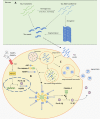
Schematic representation of Panel (A). Tau seeds formation (based on Nizynski et al., 2017). (B) NLRP3 inflammasome activation (based on data by Stancu et al., 2019), and (C) microglia releasing tau seeds in exosomes (based on Hopp et al., 2018). Aβ, amyloid β; ASC, apoptosis-associated speck-like protein containing a C-terminal caspase recruitment domain (CARD); DAMPs, damage-associated molecular patterns; mtROS, mitochondrial reactive oxygen species (ROS); NLRP3, NACHT, LRR and PYD domains-containing protein 3; PAMPs, pathogen-associated molecular patterns; TLR, toll-like receptor. See text for details.

References

    1. Aguzzi A., Rajendran L. (2009). The transcellular spread of cytosolic amyloids, prions and prionoids. Neuron 64, 783–790. 10.1016/j.neuron.2009.12.016 - DOI - PubMed
    1. Appel S., Beers D., Zhao W. (2014). Role of inflammation in neurodegenerative diseases. Neurobiol. Brain Dis. 380–395. 10.1016/B978-0-12-398270-4.00025-2 - DOI
    1. Arriagada P. V., Growdon J. H., Hedley-Whyte E. T., Hyman B. T. (1992). Neurofibrillary tangles but not senile plaques parallel duration and severity of Alzheimer’s disease. Neurology 42, 631–639. 10.1212/wnl.42.3.631 - DOI - PubMed
    1. Asai H., Ikezu S., Tsunoda S., Medalla M., Luebke J., Haydar T., et al. . (2015). Depletion of microglia and inhibition of exosome synthesis halt tau propagation. Nat. Neurosci. 18, 1584–1593. 10.1038/nn.4132 - DOI - PMC - PubMed
    1. Babić M., ŠvobŠtrac D., Mück-Šeler D., Pivac N., Stanić G., Hof P. R., et al. . (2014). Update on the core and developing cerebrospinal fluid biomarkers for Alzheimer’s disease. Croat. Med. J. 55, 347–365. 10.3325/cmj.2014.55.347 - DOI - PMC - PubMed

LinkOut - more resources