

DEEP LEARNING-BASED ASSESSMENT OF TUMOR-ASSOCIATED STROMA FOR DIAGNOSING BREAST CANCER IN HISTOPATHOLOGY IMAGES
- PMID: 31636811
- PMCID: PMC6802272
- DOI: 10.1109/ISBI.2017.7950668
DEEP LEARNING-BASED ASSESSMENT OF TUMOR-ASSOCIATED STROMA FOR DIAGNOSING BREAST CANCER IN HISTOPATHOLOGY IMAGES
Abstract
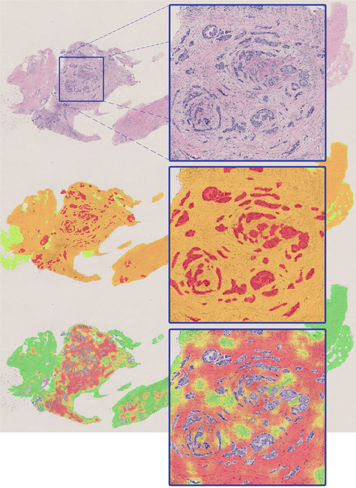
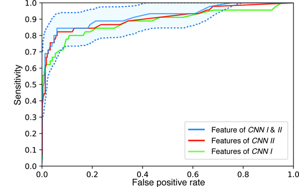
Diagnosis of breast carcinomas has so far been limited to the morphological interpretation of epithelial cells and the assessment of epithelial tissue architecture. Consequently, most of the automated systems have focused on characterizing the epithelial regions of the breast to detect cancer. In this paper, we propose a system for classification of hematoxylin and eosin (H&E) stained breast specimens based on convolutional neural networks that primarily targets the assessment of tumor-associated stroma to diagnose breast cancer patients. We evaluate the performance of our proposed system using a large cohort containing 646 breast tissue biopsies. Our evaluations show that the proposed system achieves an area under ROC of 0.92, demonstrating the discriminative power of previously neglected tumor associated stroma as a diagnostic biomarker.
Keywords: Breast Cancer; Convolutional Neural Networks; Digital pathology; Tumor Associated Stroma.
Figures
References
-
- Patey DH and Scarff RW, “The position of histology in the prognosis of carcinoma of the breast.,” The Lancet, vol. 211, no. 5460, pp. 801–804, 1928.
-
- Beck AH, Sangoi AR, Leung S, Marinelli RJ, Nielsen TO, van de Vijver MJ, West RB, van de Rijn M, and Koller D, “Systematic analysis of breast cancer morphology uncovers stromal features associated with survival,” Science translational medicine, vol. 3, no. 108, pp. 108ra113–108ra113, 2011. - PubMed
-
- Kalluri R and Zeisberg M, “Fibroblasts in cancer,” Nature Reviews Cancer, vol. 6, no. 5, pp. 392–401, 2006. - PubMed
Grants and funding
LinkOut - more resources
Full Text Sources
Other Literature Sources