Review
. 2019 Aug 23:4:29.
doi: 10.1038/s41392-019-0063-8. eCollection 2019.

History and progress of hypotheses and clinical trials for Alzheimer's disease

Affiliations
Review

History and progress of hypotheses and clinical trials for Alzheimer's disease

Pei-Pei Liu et al. Signal Transduct Target Ther. .

Erratum in

Abstract

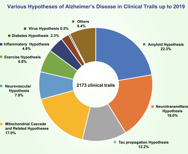
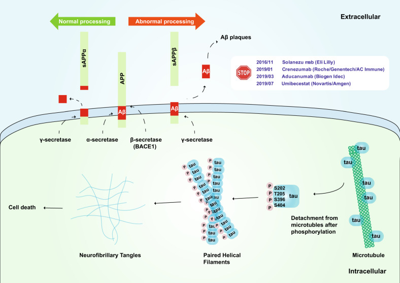
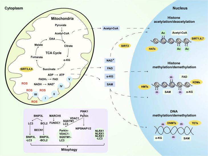
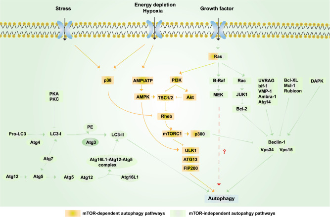
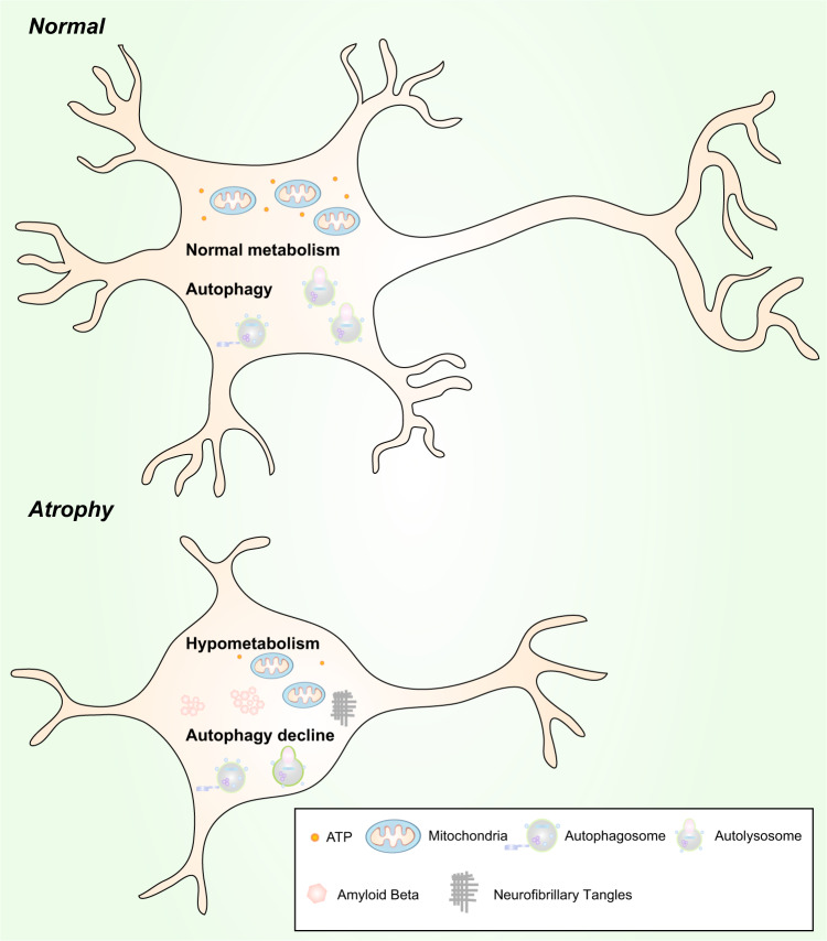
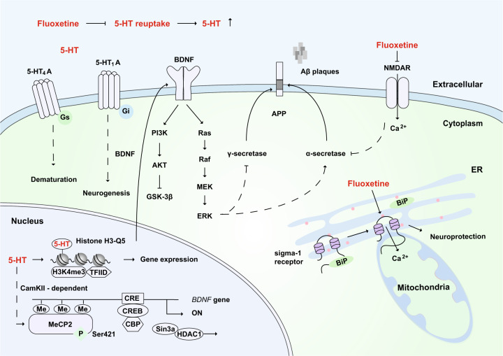
Alzheimer's disease (AD) is a neurodegenerative disease characterized by progressive memory loss along with neuropsychiatric symptoms and a decline in activities of daily life. Its main pathological features are cerebral atrophy, amyloid plaques, and neurofibrillary tangles in the brains of patients. There are various descriptive hypotheses regarding the causes of AD, including the cholinergic hypothesis, amyloid hypothesis, tau propagation hypothesis, mitochondrial cascade hypothesis, calcium homeostasis hypothesis, neurovascular hypothesis, inflammatory hypothesis, metal ion hypothesis, and lymphatic system hypothesis. However, the ultimate etiology of AD remains obscure. In this review, we discuss the main hypotheses of AD and related clinical trials. Wealthy puzzles and lessons have made it possible to develop explanatory theories and identify potential strategies for therapeutic interventions for AD. The combination of hypometabolism and autophagy deficiency is likely to be a causative factor for AD. We further propose that fluoxetine, a selective serotonin reuptake inhibitor, has the potential to treat AD.

Keywords: Neurological disorders; Neuroscience.


Conflict of interest statement

Competing interestsThe authors declare no competing interests.

Figures

Fig. 1
Fig. 1
Percentage of clinical trials in which each of the various hypotheses for AD were tested up to 2019. The amyloid hypothesis was the most heavily tested (22.3% of trials); the neurotransmitter hypothesis was the second most tested (19.0% of trials); the percentage of trials that tested the tau propagation hypothesis was ~12.7%; 17.0% of trials tested the mitochondrial cascade hypothesis and related hypotheses; 7.9% of trials tested the neurovascular hypothesis; 6.6% of trials tested the exercise hypothesis; 4.6% of trials tested the inflammatory hypothesis; 0.5% of trials tested the virus hypothesis; and the other uncatalogued trials made up approximately 8.4% of all trials
Fig. 2
Fig. 2
Schematic of the amyloid hypothesis and tau hypothesis. Upper: The transmembrane APP protein can be cleaved by two pathways. Under normal processing, APP is hydrolyzed by α-secretase and then by γ-secretase, which does not produce insoluble Aβ; under abnormal processing, APP is hydrolyzed by β secretase (BACE1) and then by γ secretase, which produces insoluble Aβ. Phase III clinical trials of solanezumab (Eli Lilly), crenezumab (Roche/Genentech/AC Immune), aducanumab (Biogen Idec), and umibecestat (Novartis/Amgen), which target the amyloid hypothesis, have all been terminated thus far. Lower: The tau protein can be hyperphosphorylated at amino residues Ser202, Thr205, Ser396, and Ser404 (which are responsible for tubulin binding), thereby leading to the release of tau from microtubules and the destabilization of microtubules. Hyperphosphorylated tau monomers aggregate to form complex oligomers and eventually neurofibrillary tangles, which may cause cell death
Fig. 3
Fig. 3
Mitochondrial cascade and related hypotheses. Mitochondria are the main contributors to ROS production, which is significantly increased in AD. The metabolites of mitochondrial TCA, such as pyruvate, fumarate, malate, OAA, and α-KG, not only directly regulate energy production but also play an important role in the epigenetic regulation of neurons and longevity.,,– For example, SAM provides methyl groups for histone and DNA methyltransferases (HMTs and DNMTs)., α-KG is a necessary cofactor for TET DNA methylases, histone demethylases (HDMs), and lysine demethylases KDMs/JMJDs., Mitochondria also regulate the levels and redox state of FAD, a cofactor of the histone demethylase LSD1. Dysfunctional mitochondria can be removed by mitophagy, which is also very important in the progression of AD. BNIP3L interacts with LC3 or GABARAP and regulates the recruitment of damaged mitochondria to phagophores. In addition, Beclin 1 is released from its interaction with Bcl-2 to activate autophagy after BNIP3L competes with it. PINK1 promotes autophagy by recruiting the E3 ligase PARK2. Then, VDAC1 is ubiquitinated and then binds to SQSTM1. SQSTM1 can interact with LC3 and target this complex to the autophagosome. L. monocytogenes can promote the aggregation of NLRX1 and the binding of LC3, thus activating mitophagy. The MARCH5-FUNDC1 axis mediates hypoxia-induced mitophagy. The mitochondrial proteins NIPSNAP1 and NIPSNAP2 can recruit autophagy receptors and bind to autophagy-related proteins. ROS: reactive oxygen species; TCA: tricarboxylic acid cycle; OAA: oxaloacetate; α-KG: α-ketoglutarate; SAM: S-adenosyl methionine; TET: ten–eleven translocation methylcytosine dioxygenase; FAD: flavin adenine dinucleotide
Fig. 4
Fig. 4
Schematic representation of autophagy. Yellow box: mTOR-dependent autophagy pathways. Growth factors can inhibit autophagy via activating the PI3K/Akt/mTORC1 pathway; under nutrient-rich conditions, mTORC1 is activated, whereas under starvation and oxidative stress, mTORC1 is inhibited. AMPK-dependent autophagy activation can be induced by starvation and hypoxia. Ras can also activate autophagy via activating PI3K, while p300 can inhibit autophagy. p38 promotes autophagy by phosphorylating and inactivating Rheb and then inhibiting mTOR under stress. Green boxes: mTOR-independent autophagy pathways. The PI3KCIII complex (also called the Beclin 1–Vps34–Vps15 complex) is essential for the induction of autophagy and is regulated by interacting proteins, such as the negative regulators Rubicon, Mcl-1, and Bcl-XL/Bcl-2, while proteins including UVRAG, Atg14, Bif-1, VMP-1, and Ambra-1 induce autophagy by binding Beclin 1 and Vps34 and promoting the activity of the PI3KCIII complex. In addition, various kinases also regulate autophagy. ERK and JNK-1 can phosphorylate Bcl-2, release its inhibition, and consequently induce autophagy; the phosphorylation of Beclin 1 by Akt inhibits autophagy, whereas the phosphorylation of Beclin 1 by DAPK promotes autophagy. Autophagy can be inhibited by the action of PKA and PKC on LC3. Finally, Atg4, Atg3, Atg7, and Atg10 are autophagy-related proteins that mediate the formation of the Atg12–Atg5–Atg16L1 complex and LC3-II. RAS and p300 can also regulate autophagy via the mTOR-independent pathway
Fig. 5
Fig. 5
In addition to mitochondrial dysfunction, hypometabolism may underlie the cholinergic hypothesis, metal ion hypothesis, and neurovascular hypothesis. a Glucose is enzymatically catalyzed to produce pyruvate. Pyruvate is converted to acetyl-CoA and then enters the TCA cycle or is used in the cytoplasm to synthesize acetylcholine. However, in AD patients, because of hypometabolism, the production of acetyl-CoA and ATP is insufficient, which leads to a reduction in acetylcholine synthesis. b Mitochondrial complexes I–III require Fe/S clusters, and complexes II–IV need hemoproteins for electron transfer and the oxidative phosphorylation of the respiratory chain. When iron deficiency occurs, the production of Fe/S and hemoproteins decreases, thereby affecting mitochondrial function and resulting in hypometabolism. In addition, copper is essential for the function of complex IV. Clearly, Cu–Zn superoxide dismutase (SOD1) requires copper and zinc., c Hypoperfusion and hypoxia in vascular diseases leads to insufficient oxygen supply, which in turn leads to insufficient ATP synthesis, resulting in hypometabolism in AD patients. TCA: tricarboxylic acid cycle; SOD1: superoxide dismutase 1
Fig. 6
Fig. 6
Hypometabolism and autophagy decline are likely to be causative factors of neuronal atrophy. Normal neurons vs. atrophic neurons. Upper: Normal levels of autophagy and metabolism exist in neurons to maintain their morphology and function. Lower: Hypometabolism and a reduction in autophagy are found in atrophic neurons
Fig. 7
Fig. 7
The potential mechanisms of fluoxetine in the remission of AD. As a selective 5-HT reuptake inhibitor, fluoxetine can increase the extraneuronal concentration of 5-HT. 5-HT binds to the 5-HT4A receptor to promote neuronal dematuration through a Gs-mediated pathway. 5-HT binds to the 5-HT1A receptor, which is involved in BDNF-dependent neurogenesis through the Gi-mediated signaling pathway. After 5-HT stimulation, MeCP2 is phosphorylated at Ser421 through CaMKII-dependent signaling, and this promotes the dissociation of CREB from HDAC and then increases the expression of BDNF. BDNF activates downstream signaling pathways, including the MEK-ERK pathway, which might promote the activity of α-secretase, inhibit γ-secretase, and reduce the production of toxic amyloid Aβ. Moreover, the serotonylation of histone H3 at glutamine 5 (Q5) enhances the binding of H3K4me3 and TFIID and allows gene expression. Fluoxetine has been reported to bind and inhibit NMDA receptors directly, which can reduce the inhibition of α-secretase and thus prevent the production of Aβ. In addition, fluoxetine can bind to the endoplasmic reticulum protein sigma-1 receptor, which induces the dissociation of Bip from the sigma-1 receptor and promotes neuroprotection. 5-HT: serotonin; ER: endoplasmic reticulum

References

    1. Blennow K, de Leon MJ, Zetterberg H. Alzheimer’s disease. Lancet. 2006;368:387–403. - PubMed
    1. Nelson PT, Braak H, Markesbery WR. Neuropathology and cognitive impairment in Alzheimer disease: a complex but coherent relationship. J. Neuropathol. Exp. Neurol. 2009;68:1–14. - PMC - PubMed
    1. Brookmeyer R, Gray S, Kawas C. Projections of Alzheimer’s disease in the United States and the public health impact of delaying disease onset. Am. J. Public Health. 1998;88:1337–1342. - PMC - PubMed
    1. Breteler MM, van den Ouweland FA, Grobbee DE, Hofman A. A community-based study of dementia: the Rotterdam elderly study. Neuroepidemiology. 1992;11(Suppl 1):23–28. - PubMed
    1. Ferri CP, et al. Global prevalence of dementia: a Delphi consensus study. Lancet. 2005;366:2112–2117. - PMC - PubMed

Publication types