. 2020 Feb;177(3):642-655.
doi: 10.1111/bph.14880. Epub 2020 Jan 17.

Activation of oestrogen receptor α induces a novel form of LTP at hippocampal temporoammonic-CA1 synapses

Affiliations

Activation of oestrogen receptor α induces a novel form of LTP at hippocampal temporoammonic-CA1 synapses

Leigh Clements et al. Br J Pharmacol. 2020 Feb.

Abstract

Background and purpose: 17β estradiol (E2) rapidly regulates excitatory synaptic transmission at the classical Schaffer collateral (SC) input to hippocampal CA1 neurons. However, the impact of E2 on excitatory synaptic transmission at the distinct temporoammonic (TA) input to CA1 neurons and the oestrogen receptors involved is less clear.

Experimental approach: Extracellular recordings were used to monitor excitatory synaptic transmission in hippocampal slices from juvenile male (P11-24) Sprague Dawley rats. Immunocytochemistry combined with confocal microscopy was used to monitor the surface expression of the AMPA receptor (AMPAR) subunit, GluA1 in hippocampal neurons cultured from neonatal (P0-3) rats.

Key results: Here, we show that E2 induces a novel form of LTP at TA-CA1 synapses, an effect mirrored by the ERα agonist, PPT, and blocked by an ERα antagonist. ERα-induced LTP is NMDA receptor (NMDAR)-dependent and involves a postsynaptic expression mechanism that requires PI 3-kinase signalling and synaptic insertion of GluA2-lacking AMPARs. ERα-induced LTP has overlapping expression mechanisms with classical Hebbian LTP, as HFS-induced LTP occluded PPT-induced LTP and vice versa. In addition, activity-dependent LTP was blocked by the ERα antagonist, suggesting that ERα activation is involved in NMDA-LTP at TA-CA1 synapses.

Conclusion and implications: ERα induces a novel form of LTP at juvenile male hippocampal TA-CA1 synapses. As TA-CA1 synapses are implicated in episodic memory processes and are an early target for neurodegeneration, these findings have important implications for the role of oestrogens in CNS health and neurodegenerative disease.


Conflict of interest statement

The authors declare no conflicts of interest.

Figures
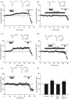
Figure 1
Figure 1
ERα activation induces LTP at juvenile TA‐CA1 synapses. (a–e) Pooled data showing the effects of E2 (1 μM; 15 min; a,b), PPT (25–50 nM; c,d) and DPN (10 nM; e) on excitatory synaptic transmission at TA‐CA1 synapses in juvenile hippocampal slices. At the end of experiments, application of dopamine (100 μM; 5 min) inhibits synaptic transmission confirming TA stimulation. In this and subsequent figures, each point is the average of four successive responses, and representative fEPSPs are shown above each plot and for the time indicated. Application of E2 caused a persistent increase in synaptic transmission (a) that was blocked by selective inhibition of ERα with MPP (b). (c) Addition of the ERα agonist PPT induced a persistent increase in synaptic efficacy, an effect that was blocked by the ERα antagonist MPP (d). (e) In contrast, the ERβ agonist DPN has no effect on excitatory synaptic transmission. (f) Bar chart of pooled data illustrating the effects of PPT alone and in the presence of MPP or the ERβ antagonist PHTPP on synaptic transmission. ERα activation induced a novel form of LTP at TA‐CA1 synapses
Figure 2
Figure 2
PPT‐induced LTP is NMDAR‐dependent and involves a postsynaptic expression mechanism. (a) Plot of pooled data illustrating the effects of 25‐nM PPT (15 min) on synaptic transmission at TA‐CA1 synapses. (b) Corresponding plot of the pooled paired‐pulse ratio (PPR) against time for experiments shown in (a). The effects of PPT were not accompanied by any change in PPR, indicating a postsynaptic mechanism. (c–e) Plots of pooled data illustrating the effects of PPT (25 nM; 15 min) on synaptic transmission in hippocampal slices. NMDAR activation was involved as PPT failed to induce LTP in the presence of the competitive NMDAR antagonist D‐AP5 (50 μM; c). (d,e) The ability of PPT to induce LTP was blocked following inhibition of GluN2B subunits with ifenprodil (d) but was unaffected by the GluN2A inhibitor, NVP‐AAM0077 (e). (f) Bar chart of pooled data illustrating the relative effects of PPT on synaptic transmission in control conditions and after treatment with D‐AP5, ifenprodil, or NVP‐AAM0077. PPT‐induced LTP involves activation of GluN2B subunits
Figure 3
Figure 3
Synaptic insertion of GluA2‐lacking AMPARs underlies ERα‐induced LTP. (a–d) Plots of pooled data illustrating effects on synaptic transmission in hippocampal slices. (a) LTP was evoked by addition of 25‐nM PPT, but prior exposure to Phtx (1 μM; b) prevented PPT‐induced LTP. (c) Application of Phtx immediately after addition of PPT resulted in reversal of PPT‐induced. (d) Addition of Phtx, 45 min after PPT failed to reverse PPT‐induced LTP. (e) Representative confocal images of surface GluA1 labelling in control hippocampal neurons and after PPT, MPP or PPT plus MPP (DIV7‐13). (f) Pooled data illustrating the relative effects on GluA1 surface labelling in control conditions and after addition of PPT, MPP, or PPT plus MPP. ERα activation increases GluA1 surface expression (DIV7–13). (g) Pooled data showing the effects on % GluA1/PSD‐95 co‐localisation. The ERα agonist, PPT, increased GluA1 expression at hippocampal synapses (DIV7‐13)
Figure 4
Figure 4
ERα‐induced LTP involves PI 3‐kinase signalling. (a–c) Pooled data illustrating the effects on excitatory synaptic transmission in slices. The ability of PPT to induce LTP (a) was blocked after PI3K inhibition with LY294002 (b) but was unaffected in the presence of the ERK inhibitor, PD98059 (c). (d) Pooled data illustrating the effects of PPT on synaptic transmission in control conditions and after incubation with inhibitors of PI3‐kinase (LY294002; wortmannin) or ERK (PD98059; U0126). PPT‐induced LTP involves a PI 3‐kinase‐driven process. (e) Representative confocal images of surface GluA1 labelling in control neurons and after PPT, PPT plus LY294002 or PPT plus wortmannin (DIV8‐15). (f) Pooled data illustrating the relative effects on GluA1 surface labelling in control conditions, after addition of PPT and in the combined presence of PPT plus LY294002 or PPT plus wortmannin (DIV8‐15). The ability of ERα to increase GluA1 surface expression involves PI 3‐kinase signalling
Figure 5
Figure 5
ERα activation is involved in activity‐dependent LTP at TA‐CA1 synapses. (a–e) Plots of pooled data illustrating effects on synaptic transmission at TA‐CA1 synapses. (a) Application of PPT induces LTP, however subsequent delivery of HFS (shown by arrow) failed to increase synaptic strength further. (b) LTP was evoked by HFS, however subsequent addition of PPT failed to alter synaptic transmission. ERα‐induced LTP and activity‐dependent LTP at TA‐CA1 synapses share similar expression mechanisms. (c) In control slices, delivery of HFS induced LTP. (d) In interleaved slices treated with MPP, delivery of HFS failed to induce LTP. (e) HFS fails to induce LTP in slices treated with letrozole. (f) Bar chart of pooled data illustrating the effects of HFS on synaptic transmission in control conditions (black bar) and following treatment with either MPP, PHTPP, or letrozole (grey bars)
Figure 6
Figure 6
Activation of ERα induces a novel form of NMDA‐dependent LTP at juvenile TA‐CA1 synapses. Schematic representation of the key cellular mechanisms involved in the novel form of LTP induced by ERα at juvenile TA‐CA1 synapses. Activation of ERα by selective agonists or endogenous E2 results in stimulation of PI 3‐kinase signalling. This in turn increases the synaptic activation of GluN2B‐containing NMDA receptors which ultimately promote insertion of GluA2‐lacking AMPA receptors into hippocampal TA‐CA1 synapses

References

    1. Aksoy‐Aksel, A. , & Manahan‐Vaughan, D. (2015). Synaptic strength at the temporoammonic input to the hippocampal CA1 region in vivo is regulated by NMDA receptors, metabotropic glutamate receptors and voltage‐gated calcium channels. Neuroscience, 309, 191–199. 10.1016/j.neuroscience.2015.03.014 - DOI - PubMed
    1. Alexander, S. P. H. , Cidlowski, J. A. , Kelly, E. , Mathie, A. , Peters, J. A. , Vealerc, E. L. , … CGTP Collaborators (2019). THE CONCISE GUIDE TO PHARMACOLOGY 2019/20: Nuclear hormone receptors. British Journal of Pharmacology, 176, S229–S246. 10.1111/bph.14750 - DOI - PMC - PubMed
    1. Alexander, S. P. H. , Roberts, R. E. , Broughton, B. R. S. , Sobey, C. G. , George, C. H. , Stanford, S. C. , … Ahluwalia, A. (2018). Goals and practicalities of immunoblotting and immunohistochemistry: A guide for submission to the British Journal of Pharmacology. British Journal of Pharmacology, 175, 407–411. 10.1111/bph.14112 - DOI - PMC - PubMed
    1. Anderson, W. W. , & Collingridge, G. L. (2007). Capabilities of the WinLTP data acquisition program extending beyond basic LTP experimental functions. Journal of Neuroscience Methods, 162, 346–356. 10.1016/j.jneumeth.2006.12.018 - DOI - PubMed
    1. Barnes, L. L. , Wilson, R. S. , Bienias, J. L. , Schneider, J. A. , Evans, D. A. , & Bennet, D. A. (2005). Sex differences in the clinical manifestations of Alzheimer disease pathology. Archives of General Psychiatry, 62, 685–691. 10.1001/archpsyc.62.6.685 - DOI - PubMed

Publication types

Substances