Review
. 2019 Dec;235(6):1036-1044.
doi: 10.1111/joa.13069. Epub 2019 Oct 21.

The masks of Lorenzo Tenchini: their anatomy and surgical/bioengineering clues

Affiliations
Review

The masks of Lorenzo Tenchini: their anatomy and surgical/bioengineering clues

Fulvio Barbaro et al. J Anat. 2019 Dec.

Abstract

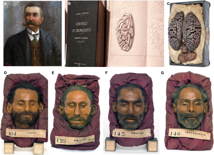
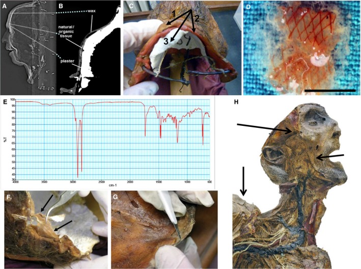
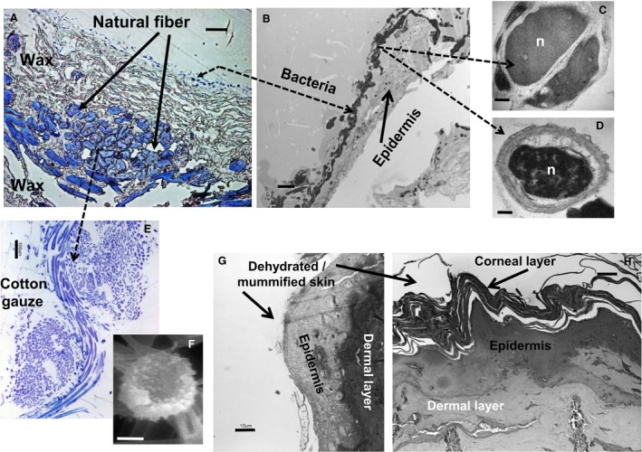
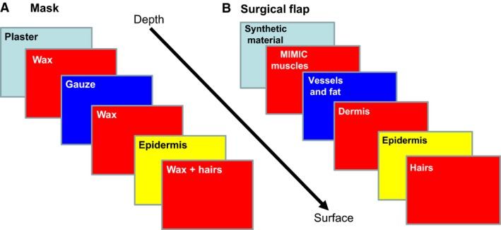
An academic, anatomist, and Lombrosian psychiatrist active at the University of Parma in Italy at the end of the 19th century, Lorenzo Tenchini produced ceroplastic-like masks that are unique in the anatomical Western context. These were prepared from 1885 to 1893 with the aim of 'cataloguing' the behaviour of prison inmates and psychiatric patients based on their facial surface anatomy. Due to the lack of any reference to the procedure used to prepare the masks, studies were undertaken by our group using X-ray scans, infrared spectroscopy, bioptic sampling, and microscopy analysis of the mask constituents. Results showed that the masks were stratified structures including plaster, cotton gauze/human epidermis, and wax, leading to a fabrication procedure reminiscent of 'additive layer manufacturing'. Differences in the depths of these layers were observed in relation to the facial contours, suggesting an attempt to reproduce, at least partially, the three-dimensional features of the facial soft tissues. We conclude the Tenchini masks are the first historical antecedent of the experimental method for face reconstruction used in the early 2000s to test the feasibility of transferring a complete strip of face and scalp from a deceased donor to a living recipient, in preparation for a complete face transplant. In addition, the layering procedure adopted conceptually mimics that developed only in the late 20th century for computer-aided rapid prototyping, and recently applied to bioengineering with biomaterials for a number of human structures including parts of the skull and face. Finally, the masks are a relevant example of mixed ceroplastic-cutaneous preparations in the history of anatomical research for clinical purposes.

Keywords: additive layer manufacturing; bioengineering; ceroplastics; face transplant; facial reconstruction; surface anatomy.


Conflict of interest statement

All authors confirm that they have no conflicts of interest to declare.

Figures

Figure 1
Figure 1
(A) Portrait of Lorenzo Tenchini, Oil on canvas, 1911, BIOMED, Parma, Italy. To our knowledge, this is the only image currently available of Tenchini, although dated 5 years after his sudden death at age 54. (B) Frontispiece of an original copy of the book by Tenchini on Criminal's Brains (kept in the Historical Library at BIOMED), and the brain of subject no. 72 (catalogued as ‘murderer’, whose mask is lacking) prepared using Giacomini's method, as displayed at the International Exposition: ‘The Fabric of the Bodies: from Anatomy to Robotics’, Palace of the Governor, 14 October–17 December 2017, Parma, Italy (link at http://lafabricadeicorpi.unipr.it/index.php). (C) Mask of subject no. 104 (catalogued as ‘thief’). (D) Mask of subject no. 125 (catalogued as ‘injurer’). (E) Mask of subject no. 143 (catalogued as ‘wilful murderer’). (F) Mask of subject no. 146 (catalogued as ‘arsonist). Note the striking empathic tone of the physiognomy. Judicial and autopsy histories of most of the portrayed subjects are recorded in handwritten entries prepared by Tenchini himself and kept in an archive at BIOMED in Parma, Italy. To ensure ethical confidentiality, none of the masks shown here contain biographically sensitive data nor does their exhibition lead to violation of current privacy issues (from Toni et al. 2016a, with permission).
Figure 2
Figure 2
(A) Computed tomography sagittal section of the mask from subject no. 143 (T1‐weighted image, CS = 0.5, Somatom Emotion 6, Siemens, courtesy of Prof. Cristina Rossi, Radiology Center, Parma University Hospital, Parma, Italy). Note three layers with different levels of radiopacity. (B) Grey densitometry revealed wax on the surface, cotton/hemp or human skin at the intermediate level, and plaster at the bottom (partly modified from Toni et al. 2016a, with permission). (C) Macroscopically, the stratification corresponded to (1,2) superficial and intermediate layers rich in wax and (3) a deep layer of plaster (mask of subject no. 69). (D) Temperature‐dependent melting (60 °C) of wax fragments from layers (1,2) of the mask from subject no. 153. The fragments contained different pigments, and a fiber network likely of vegetal origin (bar = 12 mm). (E) FTIR spectrum of layers (1,2) from the mask of subject no. 69. Peaks are compatible with pigmented beeswax. In other masks (e.g. subjects nos 149 and 155) linseed oil and sealing wax were recognized. (F) Bioptic sample of the intermediate layer (arrows) from the mask of subject no. 143. The material is similar to parchment. (G) Bioptic sample of the intermediate layer (arrows) from the mask of subject no. 122, at the level of the neck. Note that it is covered by reddish‐pigmented wax. (H) Dehydrated anatomical specimen from the Tenchini collection at BIOMED, in Parma, Italy, whose facial and shoulder skin (arrows point to the areas of sampling) was used as a control for comparison with the material of the intermediate layer found in the masks.
Figure 3
Figure 3
(A) LM image of an Epon 812‐embedded, 1‐μm section of a fragment of the intermediate layer (transverse cut through the thickness) from the mask of subject no. 149 (toluidine blue staining). Note the presence of the stained fibrous material, covered on both sides by unstained wax. On top of the superficial layer of wax, a thin line of blue nuclei and cell fragments from bacteria and microorganismic spores is visible. (B) LM section analogous to that in (A), depicting the same line of bacterial cells covering a strip of compact, sclerotic, and partly degenerated epithelial tissue containing scattered cell nuclei compatible with epidermis without stratum corneum. (C) TEM image of an Epon 812‐embedded, 70–90 nm section (high contrast preparation as described in Toni et al. 1990), of the same fragment shown in (A) and (B), depicting Staphylococcus aureus, a common saprofyte of the human skin. (D) TEM image of the same section as in (C), showing a microorganismic spore typical of the human skin. (E) LM control image of standard cotton gauze, prepared as the material fragment shown in (A). Note that the structure of natural fibers recapitulates that of the stained fibrous material in (A), indicating that both are cotton or hemp. (F) SEM image of a cell with a typical biconcave morphology compatible with a human erythrocyte (sample prepared and analysed as described in Zamparelli et al. 2014). This suggests that dermal fragments including blood cells were accidentally transferred to the masks during the ‘transplant’ of the cadaveric epidermis. (G,H) LM images of Epon 812‐embedded, 1‐μm sections of dehydrated skin (face and shoulder, respectively) from an anatomical specimen of Tenchini's collection used as internal control (toluidine blue staining). Note the striking similarity of the facial epidermis between the samples (G) and (B). Scale bars: (A,B,G,H) 10 μm; (E) 100 μm; (F) 1 μm; (C,D) 0.1 μm.
Figure 4
Figure 4
(A) Schematic of the layered organization of the facial wall in the masks compared with (B), the layered anatomical organization of the surgical flap (epidermis + dermis + hypodermal structures) used for experimental transfer onto a synthetic 3D resin model of the recipient's face in preparation for a real face transplant (see Siemionow & Agaoglu, 2006; Siemionow, 2015). Note the similarity in the 3D organization of this layering between that of the masks and human face and frontal scalp, in a sort of ‘recapitulation’ for reconstructive purposes.

References

    1. Burman J (2016) The aesthetics of Tenchini's maschere: some comparisons In: Lorenzo Tenchini and his Masks: An Anatomical Clinical Collection of the Late 19th Century at the Universities of Parma and Turin (eds Toni R, Bassi E, Montaldo S, Porro A.), pp. 38–42. Milan: Skyrà.
    1. Chang BS, Uhm CS, Park CH, et al. (2006) Preserved skin structure of a recently found fifteenth‐century mummy in Daejeon, Korea. J Anat 209, 671–680. - PMC - PubMed
    1. Churchland PS (2011) Braintrust: What Neuroscience Tells Us about Morality. Princeton: Princeton University Press.
    1. Claes P, Vandermeulen D, De Greef S, et al. (2006) Craniofacial reconstruction using a combined statistical model of face shape and soft tissue depths: methodology and validation. Forensic Sci Int 159(suppl 1), S147–S158. - PubMed
    1. Claes P, Vandermeulen D, De Greef S, et al. (2010) Computerized craniofacial reconstruction: conceptual framework and review. Forensic Sci Int 201, 138–145. - PubMed