

Extracellular matrix and biomimetic engineering microenvironment for neuronal differentiation
- PMID: 31638079
- PMCID: PMC6975142
- DOI: 10.4103/1673-5374.266907
Extracellular matrix and biomimetic engineering microenvironment for neuronal differentiation
Abstract
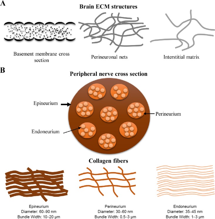
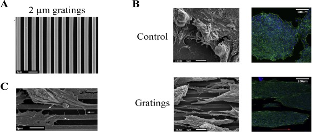
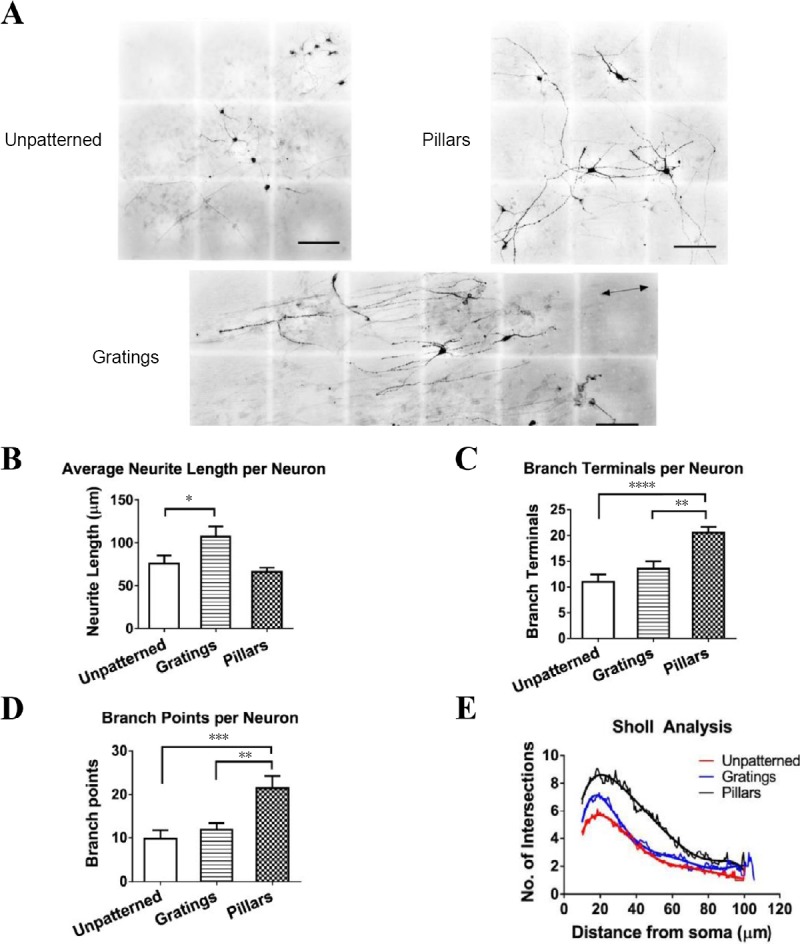
Extracellular matrix (ECM) influences cell differentiation through its structural and biochemical properties. In nervous system, neuronal behavior is influenced by these ECMs structures which are present in a meshwork, fibrous, or tubular forms encompassing specific molecular compositions. In addition to contact guidance, ECM composition and structures also exert its effect on neuronal differentiation. This short report reviewed the native ECM structure and composition in central nervous system and peripheral nervous system, and their impact on neural regeneration and neuronal differentiation. Using topographies, stem cells have been differentiated to neurons. Further, focussing on engineered biomimicking topographies, we highlighted the role of anisotropic topographies in stem cell differentiation to neurons and its recent temporal application for efficient neuronal differentiation.
Keywords: biomimetic platforms; biophysical cues; contact guidance; extracellular matrix; neural regeneration; neural stem cell niche; neuronal development; neuronal differentiation; neuronal maturation; stem cell; topography.
Conflict of interest statement
None
Figures
References
-
- Abbasi N, Hashemi SM, Salehi M, Jahani H, Mowla SJ, Soleimani M, Hosseinkhani H. Influence of oriented nanofibrous PCL scaffolds on quantitative gene expression during neural differentiation of mouse embryonic stem cells. J Biomed Mater Res A. 2016;104:155–164. - PubMed
-
- Adarsh KG. Evaluation of acellular and cellular nerve grafts in repair of rat peripheral nerve. J Neurosurg. 1988;68:117–123. - PubMed
-
- Anh Tuan N, Sharvari RS, Evelyn KFY. From nano to micro: topographical scale and its impact on cell adhesion, morphology and contact guidance. J Phys Condens Matter. 2016;28:183001. - PubMed
-
- Ankam S, Lim CK, Yim EK. Actomyosin contractility plays a role in MAP2 expression during nanotopography-directed neuronal differentiation of human embryonic stem cells. Biomaterials. 2015;47:20–28. - PubMed
Publication types
LinkOut - more resources
Full Text Sources
