. 2020 Apr;93(1108):20190523.
doi: 10.1259/bjr.20190523. Epub 2019 Oct 31.

Coronary artery fistulas detected with coronary CT angiography: a pictorial review of 73 cases

Affiliations

Coronary artery fistulas detected with coronary CT angiography: a pictorial review of 73 cases

Ning Li et al. Br J Radiol. 2020 Apr.

Abstract

Coronary artery fistulas (CAFs) are abnormal connections of the coronary arteries that bypass the myocardial capillary bed and terminate into chambers of the heart or major blood vessels. CAFs are rare, and most of them are congenital. Because CAFs can be asymptomatic and detected incidentally, the true incidence is difficult to evaluate. CAFs usually have various and complicated image features, and the clinical symptoms mainly depend on the size, origin and drainage site of the fistulas. Thus, accurate imaging assessment of these characteristics is crucial for therapeutic planning and post-operative evaluation. Due to the high temporal and spatial resolution, coronary CT angiography has recently become more widely used in cardiovascular disease diagnosis, and more asymptomatic CAFs are accidentally found. Furthermore, with multiplanar reconstruction images, some complicated and subtle structures can be displayed more accurately. In this article, we reviewed the imaging features of CAFs on coronary CT angiography, mainly focusing on the pre- and post-operative anatomy displaying of these abnormalities.


Figures

Figure 1.
Figure 1.
A 61-year-old female with right coronary artery to pulmonary artery fistula detected during evaluation of atypical precordial discomfort. Maximumintensity projection (MIP) image (a) shows an right coronary artery (RCA) to pulmonary artery (PA) fistulous vessel (arrow) taking an anterolateral course of the pulmonary trunk with mild dilatation before entering the main pulmonary artery (MPA); persistent high-density jet at the drainage site was displayed (curved arrow). Virtual reality (VR) image (b) shows the whole body of the fistulous connection (arrows) from RCA to MPA with a tortuous form.
Figure 2.
Figure 2.
A complex CPAF detected in a 46-year-old male who had no history of cardiac disease but experienced chest discomfort recently. VR images (a) show the fistula track between the right coronary artery (arrow, 1), proximal left anterior descending artery (arrow, 2), bronchial artery (arrow, 3) and MPA. An aneurysmal dilatation formed before the fistula tracks entering the MPA; a high-density flow jet inserted into the MPA was well demonstrated. MRP image (b) shows jet-like blood flow into the MPA at the drainage site (curved arrow). CPAF, coronary pulmonary artery fistula; MPA, main pulmonary artery; VR,virtual reality.
Figure 3.
Figure 3.
RCA-RA fistula in a 1-year-old child with continuous murmur shortly after birth. VR image (a) demonstrates the dilated RCA draining into the RA with two fistulas at the termination. CPR image (b) shows the whole shape of the RCA and the irregularly dilated distal segment finally leading to the RA with two fistulas. MRP image (c) shows high-density flow jet at the two adjacent drainage sites (curved arrow), which were confirmed in the following surgery with areas of 3 and 2 mm2. CPR, curved multiplanar reformation; RCA-RA, right coronary artery to right atrium; VR, virtual reality.
Figure 4.
Figure 4.
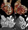
RCA-LV fistula in a 5-month-old child, which was detected at his foetal period. Axial images demonstrate that the RCA drained into the LV through a 7 mm fistula (a). 1 week after the operation, the RCA and occluder were clearly shown (b); then, after 1 year, thrombosis was seen filling almost the entire vessel (c). VR images (d, e, f) demonstrated the changes of the right canal before operation and 1 week and 1 year after the operation. RCA-LV, right coronary artery to left ventricle; VR, virtual reality.
Figure 5.
Figure 5.
LAD-RV fistula in a 21-year-old male who had a history of continuous cardiac murmur since his childhood. VR and MIP images show the right and left main coronary artery all originate from the dilated right sinus of Valsalva. The dilated LAD finally drained into the RV, and the fistula was clearly shown on the MIP image. LAD-RV, left coronary artery to right ventricle; MIP, maximum intensity projection; VR, virtual reality.
Figure 6.
Figure 6.
Complex CCF between RCA/LAD and the right ventricle detected in a 5-year-old girl with Glenn procedure history because of pulmonary atresia. LM and RCA (a, b) both originate from the left coronary sinus with a tortuous and dilated from, especially the LAD branch and the diagonal branch (D). They were all finally drained into the right ventricle (curved arrow). CCF, coronary-cameral fistula; LAD, left anterior descending artery; LM, left main; RCA, right coronary artery.
Figure 7.
Figure 7.
LCX-RV fistula in an 11-year-old girl with persistent cardiac murmurs. MIP image (a) shows dilated and tortuous LCX coronary artery draining into the RV (curved black arrow). Then, she underwent interventional treatment with a symmetrical umbrella blocker through the right ventricle. Thrombosis was seen at the distal end of the circumflex branch on the follow-up MIP image (b) 1 month later, and the patient had no symptoms of myocardial ischaemia. VR images (c, d) clearly displayed the LCX-RV fistula with an obviously dilated and tortuous form and the change of LCX canal (e) after operation. As shown in the VR image, we can see normal blunt branch visualisation at the proximal end of thrombosis. LCX-RV, left circumflex coronary artery to right ventricular; MIP, maximum intensityprojection; VR, virtual reality.
Figure 8.
Figure 8.
RCA-LV fistula in a 5-month-old boy with cardiac murmur. MIP images before operation (a), 1 month (b) and 6 months (c) after operation demonstrate an RCA-LV fistula, and the expansion degree of the RCA gradually decreased during the follow-up time. RCA-LV, right coronary artery to left ventricle.

References

    1. Yun G, Nam TH, Chun EJ. Coronary artery fistulas: pathophysiology, imaging findings, and Management-Erratum. Radiographics 2018; 38: 2214. doi: 10.1148/rg.2018184013 - DOI - PubMed
    1. Krause W. Uber den ursprung einer accessorischen A. coronaria AUS Der A. pulmonalis. Z Ratl Med 1865; 24: 225–7.
    1. Loukas M, Germain AS, Gabriel A, John A, Tubbs RS, Spicer D. Coronary artery fistula: a review. Cardiovasc Pathol 2015; 24: 141–8. doi: 10.1016/j.carpath.2014.01.010 - DOI - PubMed
    1. Mavroudis C, Backer CL, Rocchini AP, Muster AJ, Gevitz M. Coronary artery fistulas in infants and children: a surgical review and discussion of coil embolization. Ann Thorac Surg 1997; 63: 1235–42. doi: 10.1016/S0003-4975(97)00251-8 - DOI - PubMed
    1. Ghandour A, Rajiah P. Unusual fistulas and connections in the cardiovascular system: a pictorial review. World J Radiol 2014; 6: 169–76. doi: 10.4329/wjr.v6.i5.169 - DOI - PMC - PubMed

MeSH terms