. 2019 Nov 21;123(46):9773-9785.
doi: 10.1021/acs.jpcb.9b07502. Epub 2019 Nov 11.

Salt Dependence of A-Form RNA Duplexes: Structures and Implications

Affiliations

Salt Dependence of A-Form RNA Duplexes: Structures and Implications

Yen-Lin Chen et al. J Phys Chem B. .

Abstract

The biological functions of RNA range from gene regulation through catalysis and depend critically on its structure and flexibility. Conformational variations of flexible, non-base-paired components, including RNA hinges, bulges, or single-stranded tails, are well documented. Recent work has also identified variations in the structure of ubiquitous, base-paired duplexes found in almost all functional RNAs. Duplexes anchor the structures of folded RNAs, and their surface features are recognized by partner molecules. To date, no consistent picture has been obtained that describes the range of conformations assumed by RNA duplexes. Here, we apply wide angle, solution X-ray scattering (WAXS) to quantify these variations, by sampling length scales characteristic of helical geometries under different solution conditions. To identify the radius, helical rise, twist, and length of dsRNA helices, we exploit molecular dynamics generated structures, explicit solvent models, and ensemble optimization methods. Our results quantify the substantial and salt-dependent deviations of double-stranded (ds) RNA duplexes from the assumed canonical A-form conformation. Recent experiments underscore the need to properly describe the structures of RNA duplexes when interpreting the salt dependence of RNA conformations.


Figures

Figure 1.
Figure 1.
Explicit solvent and ion models. The left panel shows one RNA12 conformation in 500 mM KCl and 5.0 mM MgCl2. Solvent and ion models in the absence of solute are shown in the middle and right columns, at the quoted salt concentrations. The color schemes are defined as follows. Green: K+ from 3D-RISM. Dark green: bulk K+. Yellow: Mg2+ ions from 3D-RISM. Dark yellow: bulk Mg2+ ions. Red: bulk Cl ions. The number of ions directly corresponds to the experimental bulk concentrations. Notice that for 0.25 mM MgCl2, the bulk solvent contains no ions due to the low concentration.
Figure 2.
Figure 2.
The flowchart for the extended ensemble optimization method (eEOM) and the 5-fold cross validation (CV) strategy, including hyperparameters. The l’s are the lth fold of the truncated SWAXS curve and C(l) and El are the chromosome and error from the lth CV. See main text for a detailed explanation.
Figure 3.
Figure 3.
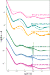
The fitted results from eEOM and the corresponding RNA12 conformations shown as phosphate backbones. (a) The experimental SWAXS curves and fits for duplexes in solutions containing different concentrations of KCl. The χ2 value for each condition (from 30 (cyan) to 500 (black) mM) is 1.169, 0.932, 0.946, 1.152 and 1.037. (b) The representative RNA12 conformations in the KCl refinement. (c) The experimental SWAXS curves and fits for duplexes in solutions containing different concentrations of MgCl2. The χ2 value for each condition (from 0.25 (pink) to 5.0 (dark red) mM) is 1.434, 1.124 and 1.400. (d) The representative RNA12 conformations in the MgCl2 refinement. Note that the SWAXS curves are offset for visualization. The green duplex represents the canonical A-form RNA12 helix. The backbone structures are rendered by PyMOL (Schrodinger Inc., New York, NY). (e) This panel compares experimental SWAXS curves in solutions containing 100 mM KCl and 1 mM MgCl2. The differences measured between the salts is larger than the deviations within either salt series, over the range tested. All data have been deposited in the SASBDB with accession codes: SAS SASDHG2, SASDHH2, SASDHJ2, SASDHK2, SASDHGL2, SASDHM2, SASDHN2, SASDHP2, SASDHQ2, SASDHS2, SASDHR2, SASDHT2, SASDHU2, SASDHV2.
Figure 4.
Figure 4.
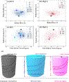
The mapping of a multidimensional conformational spaces into two, 2-dimensional parameter spaces using helical rise and radius for KCl (a) and MgCl2 (b) and total helical extension and twist for KC l(c) and MgCl2 (d) with the same color schemes as in Fig. 3. Parameters for all structures in the MD pools are shown as small transparent points in the background. (e) The schematic summary of the RNA12 duplex in canonical A-form, KCl and MgCl2 as suggested by the helical parameters in (a)-(d). Note that the cartoon illustration is not drawn to scale. Solution conformations of RNA duplexes differ from the A-form structure. Results of our refinements suggest that higher-valent cations unwind and further compress the duplex.
Figure 5.
Figure 5.
The results from 5-fold cross validation and from varying tests of experimental errors. (a) The refined conformations of the full SWAXS data (bars) and every fold of validation test (dots) from solutions containing 30 (top) to 500 (bottom) mM KCl. Figs. S10–S14 provide additional information about the robustness of the 5-fold cross validation analysis. (b) The same plot as (a) for solutions containing MgCl2 at concentrations of 0.25 (top) to 5.0 (bottom) mM. (c) The traces of E¯ and one standard deviation from 100 CV runs for all the salt concentrations explored, using previously defined color scheme. Note that the range of the y-axis differs for KCl and MgCl2 to show the non-convergent behavior and the poor fit obtained for the 0.5 mM MgCl2 condition, whose interpretation is dropped in the main text. With the exception of this point, the others have a good average error on the validation set, and display the consistent refined conformations shown in (a) and (b), which bolsters the robustness of our results. (d) The refined RNA12 conformations extracted from data synthesized with added experimental noise to achieve a S/N ratio of 23.2, 9.28, 3.09 and 1.21. The inset displays the fit of our eEOM procedure to this fabricated, noisy data set. The representative RNA conformations are well-recapitulated despite the different S/N levels, even when the signal is comparable in size to the noise. As expected, larger noise introduces a mix of conformations into the refined ensemble, rendering a larger confidence interval in the conformational spaces. The S/N level of our SWAXS experiments is about 3–5.
Figure 6.
Figure 6.
Direct comparison of experimental SWAXS measurements of 25-bp RNA and DNA duplexes to calculated SWAXS curves from a canonical A-form RNA25 and B-form DNA25 following the same procedures as the RNA12. These comparisons suggest that the dsDNA assumes a canonical B-form duplex under a broad range of solution conditions. In contrast, and consistent with our findings on the shorter duplex, the 25 bp RNA duplex has significant structural variations in different solutions and does not adopt a canonical A-form. The obvious deviations suggest that RNA duplex is highly dynamic, with ion-dependent conformations.
Figure 7.
Figure 7.
The A-form ratios of the refined ensembles using 11 dinucleotide steps for KCl (a) and MgCl2 (b) containing solutions at the quoted concentrations. The perfect A-form duplex has an A-form ratio of 1.0. The normalized fractions indicate the abundance of A-form structure in the derived ensembles. Note the broader distribution of structures in KCl, and the overall (average) lower A -ratio in MgCl2 which reflects the duplex compression in divalent salt.
Figure 8.
Figure 8.
Comparison of twist and radius of 23 dsRNA duplexes from the online protein data bank (pdb) with our results. These 23 dsRNA duplexes have different lengths and are found in both crystals (black) and in solution (yellow) with various agents, ions and even bound proteins. More information about these 23 duplex structures can be found in Table S1. Our refined RNA12 duplex data in KCl (blue) and MgCl2 (red) are also shown. The canonical A-form RNA duplex is shown as green diamond. Most (but not all) of PDB derived duplex structures have larger helical radii and are more unwound than the A-form.
Figure 9.
Figure 9.
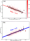
The empirical correlation between the features in the calculated SWAXS curves and geometrical parameters of the duplex. (a) A linear relationship is found between the position and contrast (defined in the text) of the first maximum in the scattering profile, and the helical radius. Since helical radius gives rise to the strongest periodicity in the RNA12 duplex, the smaller the radius, the higher q the maximum shifts to with less contrast due to more structural contributions. (b) A linear relationship is found between the position of the first minimum in the profile and the average helical rise. Although the length scale of the rise in the dinucleotide step is below our resolution, the periodicity still appears in multi-nucleotide step, which falls in the resolution of our q-range.

References

    1. Dock-Bregeon AC, Chevrier B, Podjarny A, Moras D, deBear JS, Gough GR, Gilham PT and Johnson JE High resolution structure of the RNA duplex [U(U-A)6A]2. Nature. 335(6188): 375–8 (1988). - PubMed
    1. Holbrook SR, Cheong C, Tinoco I and Kim SH Crystal structure of an RNA double helix incorporating a track of non-Watson-Crick base pairs. Nature. 353(6344): 579–81 (1991). - PubMed
    1. Tolbert BS, Miyazaki Y, Barton S, Kinde B, Starck P, Singh R, Bax A, Case DA and Summers MF Major groove width variations in RNA structures determined by NMR and impact of13C residual chemical shift anisotropy and 1H-13C residual dipolar coupling on refinement. J. Biomol. NMR 47(3):205–19 (2010). - PMC - PubMed
    1. Chu VB, Lipfert J, Bai Y, Pande VS, Doniach S and Herschlag D Do conformational biases of simple helical junctions influence RNA folding stability and specificity? RNA 15(12): 2195–205 (2009). - PMC - PubMed
    1. Daher M, Mustoe AM, Morriss-Andrews A, Brooks CL and Walter NG Tuning RNA folding and function through rational design of junction topology. Nucleic Acids Res. 45(16): 9706–9715 (2017). - PMC - PubMed

Publication types