Case Reports
. 2019 Oct 21;14(1):113.
doi: 10.1186/s13000-019-0896-z.

Plasmacytoid urothelial carcinoma: a rapid autopsy case report with unique clinicopathologic and genomic profile

Affiliations
Case Reports

Plasmacytoid urothelial carcinoma: a rapid autopsy case report with unique clinicopathologic and genomic profile

Caroline T Simon et al. Diagn Pathol. .

Abstract

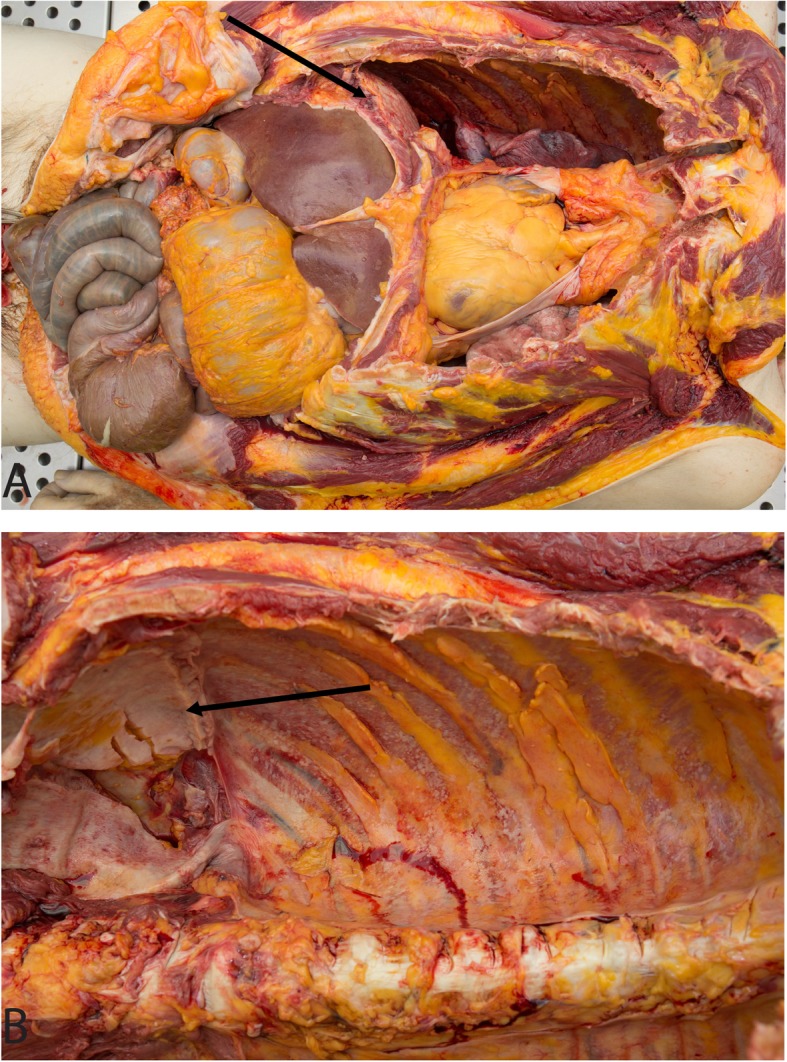
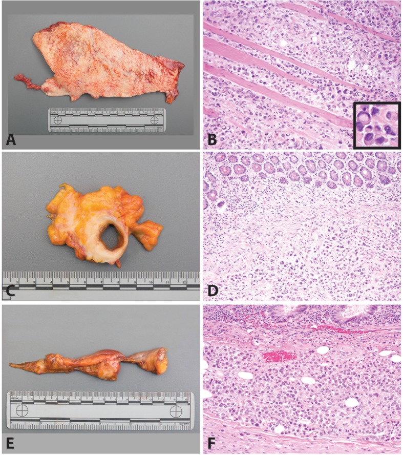
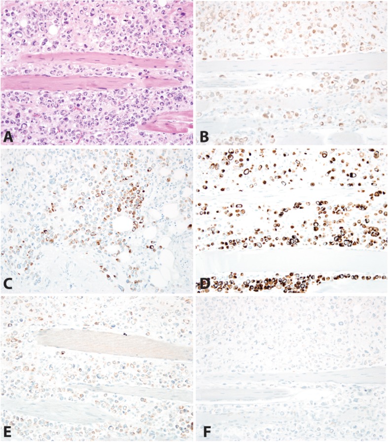
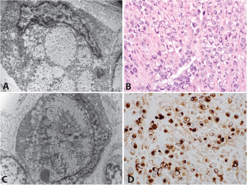
Background: Rapid ("warm") autopsies of patients with advanced metastatic cancer provide important insight into the natural history, pathobiology and histomorphology of disease in treatment-resistant tumors. Plasmacytoid urothelial carcinoma (PUC) is a rare variant of urothelial carcinoma characterized by neoplastic cells morphologically resembling plasma cells. PUC is typically aggressive, high-stage at presentation, and associated with poor outcomes. Recurrence is common in PUC, with the majority of recurrences occurring in the peritoneum.

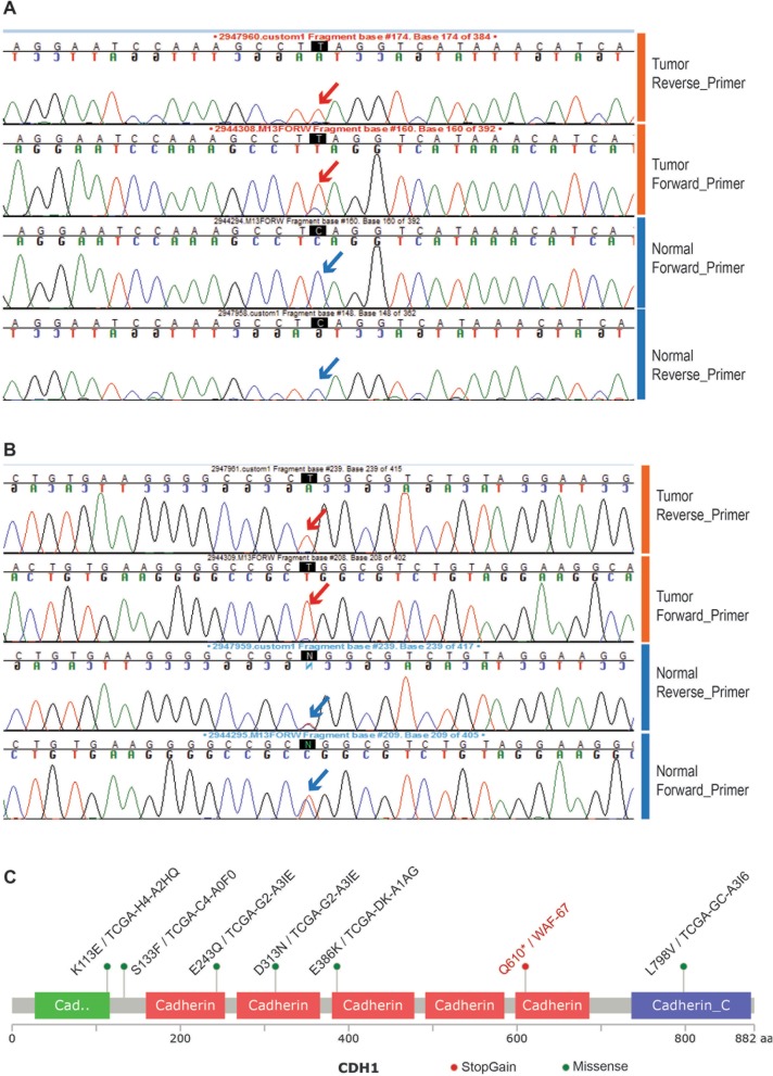
Case presentation: Here, we report rapid autopsy findings from a patient with recurrent PUC. The patient had persistent pain after cystoprostatectomy, although initial post-operative imaging showed no evidence of disease. Imaging obtained shortly before his death showed only subtle growth along vascular tissue planes; however, extensive disease was seen on autopsy. Plasmacytoid tumor cells formed sheets involving many serosal surfaces. Molecular interrogation confirmed a mutation in CDH1 exon 12 leading to early truncation of the CDH1 protein in the tumor cells.

Conclusions: The sheet-like growth pattern of PUC makes early phases of disease spread much more difficult to capture on cross-sectional imaging. Alternative forms of surveillance may be required for detection of recurrent PUC, and providers may need to treat based on symptoms and clinical suspicion.

Keywords: CDH1; Plasmacytoid; Rapid autopsy; Urothelial carcinoma.


Conflict of interest statement

The authors declare that they have no competing interests.

Figures

Fig. 1
Fig. 1
Plasmacytoid Urothelial Carcinoma in the Abdomen (in Situ). The diaphragm was diffusely thickened in a white plaque of tumor (a, shown before evisceration by arrow) and the peritoneal/serosal surface was diffusely thickened in a white plaque of tumor (b, shown after evisceration by arrow)
Fig. 2
Fig. 2
Plasmacytoid Urothelial Carcinoma in the Abdomen. The abdominal wall and peritoneum were grossly thickened with a white plaque (a). The skeletal muscle of the right abdominal wall was diffusely infiltrated by plasmacytoid tumor cells (b) [H&E 200x; inset 400x]. The lower gastrointestinal tract was diffusely involved with tumor encasing the ileum (c) with involvement of the submucosa (d) [H&E 200x] and the appendix (e) with tumor extending into the muscularis (f) [H&E 200x]. Tumor was present throughout the abdomen in the omentum, gallbladder, colon and rectum. Extra-abdominal metastases were present in the lungs, pleura, diaphragm and lymph nodes (not pictured)
Fig. 3
Fig. 3
Immunohistochemical Profile of Plasmacytoid Urothelial Carcinoma in the Right Abdominal Wall. Hematoxylin and Eosin demonstrates carcinoma in sheets splaying the skeletal muscle (a). The neoplastic cells are diffusely positive for GATA-3 (b), CK20 (c), and CK7 (d). A subset of neoplastic cells is positive for CD 138 (e). E-cadherin is negative (f). Additional immunohistochemical stains show the tumor cells are diffusely positive for pancytokeratin, patchy expression of CK903, and p53, and focal expression of PAX-8 and negative for CDX-2, p63, NKX3.1, PAX-2 and PSA
Fig. 4
Fig. 4
Deleterious mutation in CDH1 exon-12 in the index tumor sample. Chromatograms indicate the mutant allele present in the tumor sample (red arrows), both in the forward and reverse strand sequencing. The tumor specific mutation in exon-12; C1828T_Q610Stop gain, leads to early truncation of the CDH1 protein. The wild type allele (blue arrows) in the corresponding nucleotide position, detected in the adjacent normal sample from this index case, both in the forward and reverse strands are shown alongside for comparison (a). A nearby single nucleotide polymorphism (SNP) position in Exon 13 T/C2076T A692A_SNP_position (rs1801552) shows loss of heterozygosity of CDH1 loci in the tumor (b). Location of the deleterious stop gain mutation (red lollipop) observed in the index tumor (Sample name in red) and various missense mutations (green lollipops; of unknown significance) observed among the TCGA Bladder Urothelial Carcinomas (6/121 samples)- is represented in the CDH1 pfam protein domain structure schematic diagram using the Mutation Mapper tool (www.cbioportal.org) (c)
Fig. 5
Fig. 5
Electron microscopy analysis and CD10 immunostain of plasmacytoid urothelial carcinoma. Electron microscopy was performed on a representative section of tumor demonstrated nuclei pushed aside by intracellular vacuoles with microvilli lining the inner surface (a,c), Hematoxylin and eosin of the tumor (b) and a CD10 immunostain showing an apical cytoplasmic staining pattern, similar to those reported in microvillous inclusion disease (d)

References

    1. Rubin MA, Putzi M, Mucci N, et al. Rapid (“warm”) autopsy study for procurement of metastatic prostate cancer. Clin Cancer Res. 2000;6:138–1038-1045. - PubMed
    1. Pisapia DJ, Salvatore S, Pauli C, et al. Next-generation rapid autopsies enable tumor evolution tracking and generation of preclinical models. JCO Precis Oncol. 2017;2017. 10.1200/PO.16.00038. Epub 2017 Jun 14. - PMC - PubMed
    1. Shah RB, Mehra R, Chinnaiyan AM, et al. Androgen-independent prostate cancer is a heterogeneous group of diseases: lessons from a rapid autopsy program. Cancer Res. 2004;64:9209–9216. doi: 10.1158/0008-5472.CAN-04-2442. - DOI - PubMed
    1. Varambally S, Yu J, Laxman B, et al. Integrative genomic and proteomic analysis of prostate cancer reveals signatures of metastatic progression. Cancer Cell. 2005;8:393–406. doi: 10.1016/j.ccr.2005.10.001. - DOI - PubMed
    1. Tomlins SA, Laxman B, Dhanasekaran SM, et al. Distinct classes of chromosomal rearrangements create oncogenic ETS gene fusions in prostate cancer. Nature. 2007;448:595–599. doi: 10.1038/nature06024. - DOI - PubMed

Publication types

LinkOut - more resources