. 2019 Oct 22;14(10):e0223148.
doi: 10.1371/journal.pone.0223148. eCollection 2019.

Sealing agent reduces formation of single and dual-species biofilms of Candida albicans and Enterococcus faecalis on screw joints at the abutment/implant interface

Affiliations

Sealing agent reduces formation of single and dual-species biofilms of Candida albicans and Enterococcus faecalis on screw joints at the abutment/implant interface

Cecília Alves de Sousa et al. PLoS One. .

Erratum in

Abstract

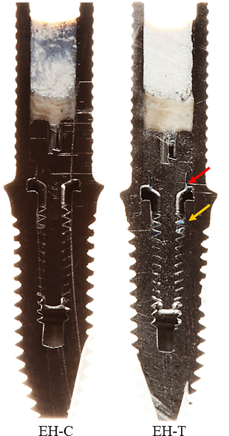
The aim of this research was to evaluate the efficacy of a commercial sealing agent at the abutment/implant interface against microleakage of single and dual-species biofilms of Candida albicans and Enterococcus faecalis into external hexagon (EH) and Morse taper (MT) prosthetic connections. A total of 216 samples of implants and their abutments were tested. Six groups (n = 36) were evaluated based on biofilm and period of incubation (7 and 14 days). The implant connections EH and MT (n = 18) were divided according to the use of the material (n = 9) (EH-T and MT-T: with the sealing agent; EH-C and MT-C: control). The biofilms were analyzed by microbial counting (CFU/mL) and SEM analysis and photographs of the material in the screw joints were also taken. Data were analyzed by Student t test, two-way ANOVA and Bonferroni test. For the single-species biofilms, there was a significant reduction in the growth of E. faecalis when compared MT-C and MT-T or EH-C and EH-T at 7 and 14 days. The same was observed for C. albicans biofilms. For dual-species biofilms of E. faecalis and C. albicans, the sealing agent was more effective in preventing microbial infiltration into the MT connection at 14 days, while microbial infiltration did not occur into EH connections even in absence of the sealing agent for both periods of evaluation. Overall, these data suggest that the presence of the sealing agent reduces or eliminates the microleakage of E. faecalis and C. albicans biofilms into the implants regardless of the period of incubation.


Conflict of interest statement

The authors have declared that no competing interests exist.

Figures

Fig 1
Fig 1. Organization chart illustrating the division of the groups.
Fig 2
Fig 2
A. Application of the material to the hexagonal portion of the implant HE; B. Application in the indexing portion of the prosthetic abutment CM; C. Application of the material to the threads of the retaining screw.
Fig 3
Fig 3. Single-species biofilms.
A. E. faecalis biofilms—Mean (standard deviation) of E. faecalis counts (in logarithmic scale) recovered from implants (MT and EH) of control (C) and test (T) groups. B. Candida albicans biofilms. Mean (standard deviation) of C. albicans counts (in logarithmic scale) recovered from implants (MC and EH) of control (C) and test (T) groups. Different lowercase letters show statistical difference between each group and the time of evaluation, according to ANOVA/Bonferroni tests, considering p< 0.05 as statistically significant.
Fig 4
Fig 4. Dual-species biofilms.
Mean (standard deviation) of E. faecalis and C. albicans counts (in logarithmic scale) recovered from implants (MT and EH) of control (C) and test (T) groups. Different lowercase letters show statistical difference between each group and time of evaluation, according to ANOVA/Bonferroni tests, considering p< 0.05 as statistically significant.
Fig 5
Fig 5. Representative micrographs of the MT specimens after 7 days of incubation with E. faecalis and C. albicans, in single or dual-species biofilms, obtained by SEM.
A. External region of the CM-C implant; B. CM-C with E. faecalis in single-species biofilm formed in the internal region of the implant; C. CM-C with C. albicans in single-species biofilm formed in the internal region of the implant; D. CM-C with dual-species biofilm and extracellular matrix of E. faecalis and C. albicans formed in the internal region of the implant; E. External region of CM-T implant with the presence of the sealing agent at the platform; F. CM-T implant with absence of E. faecalis in single-species biofilm and presence of the sealing agent in the internal region of the implant; G. CM-T with absence of C. albicans biofilm in single-species biofilm and presence of the sealing agent in the internal region of the implant; H. CM-T with the absence of E. faecalis and C. albicans in dual-species biofilm.
Fig 6
Fig 6. Representative micrographs of the EH specimens after 7 days of incubation with E. faecalis and C. albicans, in single or dual-species biofilms, obtained by SEM.
A. External region of the EH-C implant; B. HE-C with E. faecalis in single-species biofilm formed in the internal region of the implant; C. EH-C with C. albicans in single-species biofilm formed in the internal region of the implant; D. EH-C with dual-species biofilm of E. faecalis and C. albicans in the internal region of the implant; E. External region of the EH-T implant with presence of the sealing agent; F. EH-T with presence of colonies of E. faecalis dispersed on the surface and particles of the sealing agent in the internal region of the implant; G. EH-T with absence of C. albicans biofilm and presence of particles of the wax agent throughout the surface; H. EH-T with colonies dispersed on the surface in the dual-species biofilm with E. faecalis and C. albicans.
Fig 7
Fig 7. Representative micrographs of the MT specimens after 14 days of incubation with E. faecalis and C. albicans, in single or dual-species biofilms, obtained by SEM.
A. External region of the MT-C implant; B. MT-C with E. faecalis in single-species biofilm formed in the internal region of the implant; C. MT-C with C. albicans in single-species biofilm formed in the internal region of the implant; D. MT-C with dual-species biofilm of E. faecalis and C. albicans formed in the internal region of the implant; E. External region of the MT-T implant with presence of the agent in the platform; F. MT-T with presence of the sealing agent and some cells of E. faecalis in single-species biofilm in the internal region of the implant; G. MT-T with absence of C. albicans in single-species biofilm and presence of particles of the sealing agent near the extracellular matrix in the internal region of the implant; H. CM-T without colonies of E. faecalis or C. albicans on the surface of the internal region of the implant and the presence of the agent layer formation.
Fig 8
Fig 8. Representative micrographs of the EH specimens after 14 days of incubation with E. faecalis and C. albicans, in single or dual-species biofilms, obtained by SEM.
A. External region of the EH-C implant; B. EH-C with intense extracellular matrix and biofilm of E. faecalis formed in the internal region of the implant; C. EH-C with intense extracellular matrix and biofilm of C. albicans formed in the internal region of the implant; D. EH-C without cells of E. faecalis and C. albicans in the internal region of the implant, only particles of the sealing agent; E. External region of the EH-T implant with the presence of the sealing agent in the region of the hexagon and the connection platform; F. EH-T with presence of the sealing agent in the internal region of the implant; G. EH-T with absence of C. albicans biofilm and presence of the surface-wrapping agent; H. EH-T without colonies of E. faecalis and C. albicans, but the presence of the sealing agent.
Fig 9
Fig 9. Longitudinally sectioned images obtained from the specimens with MT connection.
The red arrow indicates the presence of the sealing agent at the abutment-implant interface, while the yellow arrow points to the sealant between the threads of the retaining screw and the internal thread of the implant at the uppermost portion of the screw joint.
Fig 10
Fig 10. Longitudinally sectioned images obtained from the specimens with EH connection.
The red arrow indicates the presence of the sealing agent at the abutment-implant interface, while the yellow arrow points to the sealant between the threads of the retaining screw and the internal thread of the implant at the uppermost portion of the screw joint.

References

    1. Berglundh T, Persson L, Klinge B. A systematic review of the incidence of biological and technical complications in implant dentistry reported in prospective longitudinal studies of at least 5 years. Journal of clinical periodontology. 2002;29 Suppl 3:197–212; discussion 32–3. Epub 2003/06/06. 10.1034/j.1600-051x.29.s3.12.x . - DOI - PubMed
    1. Swierkot K, Lottholz P, Flores-de-Jacoby L, Mengel R. Mucositis, peri-implantitis, implant success, and survival of implants in patients with treated generalized aggressive periodontitis: 3- to 16-year results of a prospective long-term cohort study. Journal of periodontology. 2012;83(10):1213–25. Epub 2012/01/24. 10.1902/jop.2012.110603 . - DOI - PubMed
    1. Schou S. Implant treatment in periodontitis-susceptible patients: a systematic review. Journal of oral rehabilitation. 2008;35 Suppl 1:9–22. Epub 2008/01/10. 10.1111/j.1365-2842.2007.01830.x . - DOI - PubMed
    1. Akcali A, Trullenque-Eriksson A, Sun C, Petrie A, Nibali L, Donos N. What is the effect of soft tissue thickness on crestal bone loss around dental implants? A systematic review. Clinical oral implants research. 2017;28(9):1046–53. Epub 2016/07/20. 10.1111/clr.12916 . - DOI - PubMed
    1. Costa FO, Ferreira SD, Cortelli JR, Lima RPE, Cortelli SC, Cota LOM. Microbiological profile associated with peri-implant diseases in individuals with and without preventive maintenance therapy: a 5-year follow-up. Clinical oral investigations. 2018. Epub 2018/11/06. 10.1007/s00784-018-2737-y . - DOI - PubMed

Publication types

MeSH terms