Review
. 2019 Oct 21;24(20):3781.
doi: 10.3390/molecules24203781.

G-Quadruplex-Forming Aptamers-Characteristics, Applications, and Perspectives

Affiliations
Review

G-Quadruplex-Forming Aptamers-Characteristics, Applications, and Perspectives

Carolina Roxo et al. Molecules. .

Abstract

G-quadruplexes constitute a unique class of nucleic acid structures formed by G-rich oligonucleotides of DNA- or RNA-type. Depending on their chemical nature, loops length, and localization in the sequence or structure molecularity, G-quadruplexes are highly polymorphic structures showing various folding topologies. They may be formed in the human genome where they are believed to play a pivotal role in the regulation of multiple biological processes such as replication, transcription, and translation. Thus, natural G-quadruplex structures became prospective targets for disease treatment. The fast development of systematic evolution of ligands by exponential enrichment (SELEX) technologies provided a number of G-rich aptamers revealing the potential of G-quadruplex structures as a promising molecular tool targeted toward various biologically important ligands. Because of their high stability, increased cellular uptake, ease of chemical modification, minor production costs, and convenient storage, G-rich aptamers became interesting therapeutic and diagnostic alternatives to antibodies. In this review, we describe the recent advances in the development of G-quadruplex based aptamers by focusing on the therapeutic and diagnostic potential of this exceptional class of nucleic acid structures.

Keywords: G-quadruplexes; anticoagulants; antiviral agents; aptamers; aptasensors; cancer; conjugates; diagnostics; therapeutics.


Conflict of interest statement

The authors declare no conflict of interest.

Figures
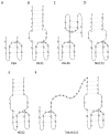
Figure 1
Figure 1
G-quadruplex models for some anticoagulant agents.
Figure 2
Figure 2
G-quadruplex models of some antiproliferative agents. (A) shows one of possible structures formed by AS1411; (B) APTA-12 aptamer, Z corresponds to gemcitabine; (C) AT11 aptamer; (D) AT11-L0 aptamer; (E) T40214 aptamer; (F) T40231 aptamer.
Figure 3
Figure 3
G-quadruplex models for some antiviral agents.
Figure 4
Figure 4
Schematic preparation of fluorescent aptasensor system for Cu2+ detection (top) and its application using SERS-imaging and photothermal therapy (bottom) [117]. pMBA: 4- mercaptobenzoic acid, TEOS: tetraethylorthosilicate, APTMS: (3-aminopropyl)trimethoxysilane.
Figure 5
Figure 5
Fluorometric approach to quantify the based on thioflavin T dye [118].
Figure 6
Figure 6
The Forster resonance energy transfer (FRET)-based ATP aptasensors. (a) ATP-binding oligonucleotide labeled with red quantum dots (RQDs) and gold nanorods (AuNRs), which in the presence of ATP and berberine, folds into a G4 structure. FRET process results in the quenching of RQD fluorescence and an increase of berberine fluorescence is simultaneously observed. (b) Ag- and CD-labeled ATP-binding oligonucleotide system forming the G4 structure in the presence of ATP and berberine [119].
Figure 7
Figure 7
Representation of the electrochemical sensor based on IGA3 aptamer (E-AB) for the detection of insulin levels [21].
Figure 8
Figure 8
Mechanism of thrombin aptamer beacon action: (a) Aptamer beacon in quenched stem-loop conformation, (b) unfolded conformation, (c) aptamer in G-quartet conformation, and (d) aptamer beacon with bound thrombin [125].
Figure 9
Figure 9
Fluorescent detection of thrombin based on BSA-Cds QDs [126].
Figure 10
Figure 10
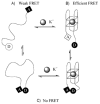
The model of action of aptasensor based on TBA conjugated with fluorophores: 6-carboxytetramethylrhodamine (TAMRA) (acceptor, A) and 6-carboxyfluorescein (FAM) (donor, D). FRET occurs when the distance between D and A is sufficient– (A) and (B). When the G4 is formed because of the presence of K+, the phenomenon is not observed (C) [136].
Figure 11
Figure 11
The chemical structure of potassium-sensing oligonucleotide (a) and the mechanism of K+ detection (b) [137].
Figure 12
Figure 12
G-quadruplex models of aptamers targeted toward myelin (A and B), codeine (C) and prion protein (D).

References

    1. Ellington A.D., Szostak J.W. In Vitro Selection of RNA Molecules That Bind Specific Ligands. Nature. 1990;346:818–822. doi: 10.1038/346818a0. - DOI - PubMed
    1. Gatto B., Palumbo M., Sissi C. Nucleic Acid Aptamers Based on the G-Quadruplex Structure: Therapeutic and Diagnostic Potential. Curr. Med. Chem. 2009;16:1248–1265. doi: 10.2174/092986709787846640. - DOI - PubMed
    1. Tuerk C., Gold L. Systematic Evolution of Ligands by Exponential Enrichement: RNA Ligands to Bacteriophage T4 DNA Polymerase. Science. 1990;249:505–510. doi: 10.1126/science.2200121. - DOI - PubMed
    1. Jenison R.D., Gill S.C., Pardi A., Polisky B. High-Resolution Molecular Discrimination by RNA. Science. 1994;263:1425–1429. doi: 10.1126/science.7510417. - DOI - PubMed
    1. Mendonsa S.D., Bowser M.T. In Vitro Evolution of Functional DNA Using Capillary Electrophoresis. J. Am. Chem. Soc. 2004;126:20–21. doi: 10.1021/ja037832s. - DOI - PubMed

Substances

LinkOut - more resources