. 2019 Oct 23;286(1913):20191649.
doi: 10.1098/rspb.2019.1649. Epub 2019 Oct 23.

Synchrotron X-ray absorption spectroscopy of melanosomes in vertebrates and cephalopods: implications for the affinity of Tullimonstrum

Affiliations

Synchrotron X-ray absorption spectroscopy of melanosomes in vertebrates and cephalopods: implications for the affinity of Tullimonstrum

Christopher S Rogers et al. Proc Biol Sci. .

Abstract

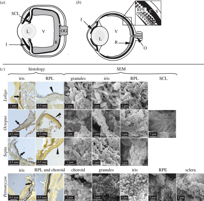
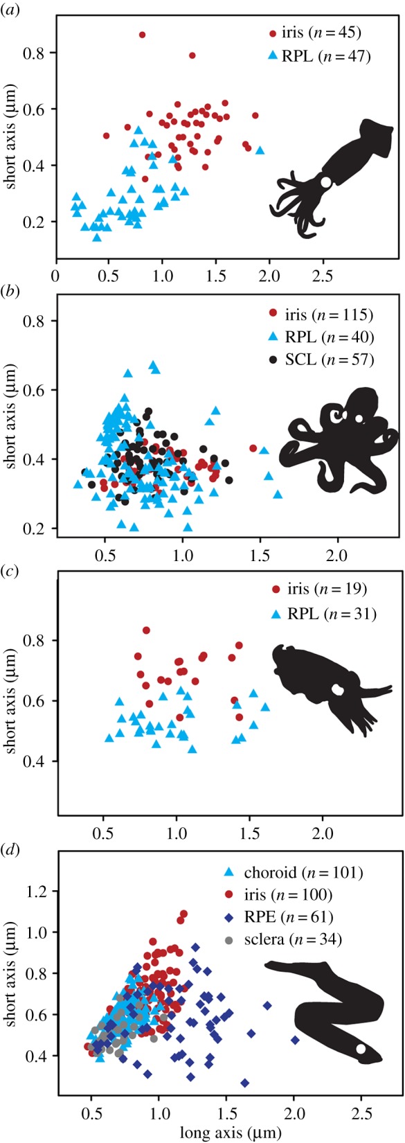
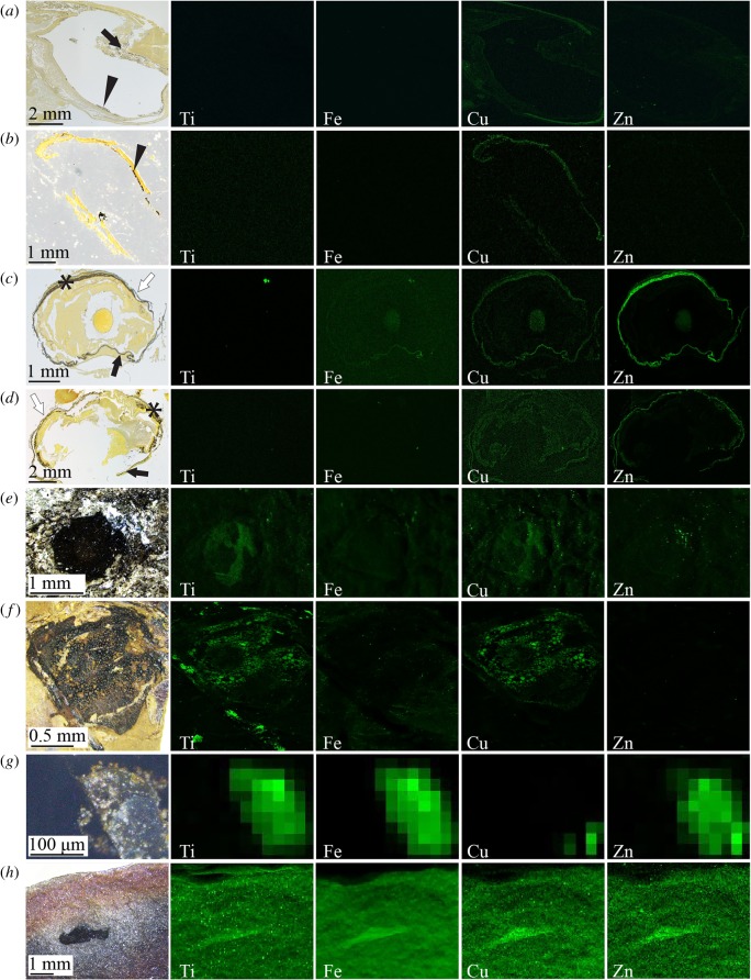
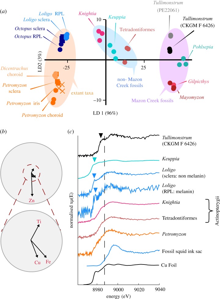
Screening pigments are essential for vision in animals. Vertebrates use melanins bound in melanosomes as screening pigments, whereas cephalopods are assumed to use ommochromes. Preserved eye melanosomes in the controversial fossil Tullimonstrum (Mazon Creek, IL, USA) are partitioned by size and/or shape into distinct layers. These layers resemble tissue-specific melanosome populations considered unique to the vertebrate eye. Here, we show that extant cephalopod eyes also show tissue-specific size- and/or shape-specific partitioning of melanosomes; these differ from vertebrate melanosomes in the relative abundance of trace metals and in the binding environment of copper. Chemical signatures of melanosomes in the eyes of Tullimonstrum more closely resemble those of modern cephalopods than those of vertebrates, suggesting that an invertebrate affinity for Tullimonstrum is plausible. Melanosome chemistry may thus provide insights into the phylogenetic affinities of enigmatic fossils where melanosome size and/or shape are equivocal.

Keywords: Konservat-Lagerstätten; fossil soft tissues; melanosomes; trace metals.


Conflict of interest statement

The authors declare no competing interests.

Figures

Figure 1.
Figure 1.
Anatomy of extant cephalopod and vertebrate eyes. Schematic illustrations of (a) cephalopod eye and (b) vertebrate eye with, inset, detail of tissue layers. (c) Histological sections and scanning electron micrographs (SEM) of eyes in Loligo, Octopus, Sepia and Petromyzon. Sections are stained with Warthin–Starry; melanin appears black. All tissues show melanosome-like microbodies. C, choroid; I/black arrows, iris; L, lens; O, optic nerve; OG, optic nerve ganglia; R, retina; RPE/*, retinal pigment epithelium; RPL/arrowheads, retinal pigmented layer; S/white arrow, sclera; SCL, subciliary layer; V, vitreous humour. (Online version in colour.)
Figure 2.
Figure 2.
Geometry of eye melanosomes. (a) Loligo, (b) Octopus, (c) Sepia and (d) Petromyzon.
Figure 3.
Figure 3.
SRS-XRF analysis of histological sections of Loligo (a), Octopus (b), Petromyzon (c) and Dicentrachus (d), and of fossil vertebrates (Teleostei) Tetradontiformes indet. (e) and Knightia (f), fossil cephalopod Keuppia (g) and Tullimonstrum (CKGM F 6426) (h). Histological sections are stained with Warthin–Starry; melanin appears black. SRS-XRF maps in (ah) are of regions shown in histological sections and photographs. See the electronic supplementary material, figure S2 for locations of regions in photographs of histological sections and for locations of regions in photographs of fossils shown. Black arrows, iris; arrowheads, RPL; asterisk, RPE; white arrows, sclera. Maximum concentration values for each SRS-XRF map are provided in the electronic supplementary material, table S5. (Online version in colour.)
Figure 4.
Figure 4.
Trace element chemistry of extant and fossil cephalopods and vertebrates and Tullimonstrum. (a) LDA based on measured concentrations of Ti, Fe, Cu and Zn. (b) Biplot of key elements contributing to variation in (a). (c) XANES spectra of selected specimens and standards at the Cu K edge (dashed line at 8987 eV). The greater variation in chemistry between Tullimonstrum and other Mazon Creek fossils present in (a) relative to the variation between other fossil biotas is present even when Fe is removed from the dataset (electronic supplementary material, figure S4c,d). Arrowheads in (c) indicate the position of pre-edge features in these spectra. Data for fossil squid ink sac are from [16].

References

    1. Oakley TH, Speiser DI. 2015. How complexity originates: the evolution of animal eyes. Annu. Rev. Ecol. Evol. Syst. 46, 237–260. (10.1146/annurev-ecolsys-110512-135907) - DOI
    1. Clements T, Dolocan A, Martin P, Purnell MA, Vinther J, Gabbott SE. 2016. The eyes of Tullimonstrum gregarium (Mazon Creek, Carboniferous) reveal a vertebrate affinity. Nature 532, 500–503. (10.1038/nature17647) - DOI - PubMed
    1. Liu Y, Hong L, Wakamatsu K, Ito S, Adhyaru BB, Cheng C-Y, Bowers CR, Simon JD. 2005. Comparisons of the structural and chemical properties of melanosomes isolated from retinal pigment epithelium, iris and choroid of newborn and mature bovine eyes. Photochem. Photobiol. 81, 510–516. (10.1562/2004-10-19-RA-345.1) - DOI - PubMed
    1. Durairaj C, Chastain JE, Kompella UB. 2012. Intraocular distribution of melanin in human, monkey, rabbit, minipig and dog eyes. Exp. Eye Res. 98, 23–27. (10.1016/j.exer.2012.03.004) - DOI - PMC - PubMed
    1. Zhang QX, Lu RW, Messinger JD, Curcio CA, Guarcello V, Yao XC. 2013. In vivo optical coherence tomography of light-driven melanosome translocation in retinal pigment epithelium. Sci. Rep. 3, 2644 (10.1038/srep02644) - DOI - PMC - PubMed

Publication types

LinkOut - more resources