. 2019 Oct 22;14(1):38.
doi: 10.1186/s13024-019-0340-6.

Targeting tauopathy with engineered tau-degrading intrabodies

Affiliations

Targeting tauopathy with engineered tau-degrading intrabodies

Gilbert Gallardo et al. Mol Neurodegener. .

Abstract

Background: The accumulation of pathological tau is the main component of neurofibrillary tangles and other tau aggregates in several neurodegenerative diseases, referred to as tauopathies. Recently, immunotherapeutic approaches targeting tau have been demonstrated to be beneficial in decreasing tauopathy in animal models. We previously found that passive immunotherapy with anti-tau antibody to human tau or expression of an anti-tau secreted single-chain variable fragment (scFv) in the central nervous system of a mouse model of tauopathy decreased but did not remove all tau-associated pathology. Although these and other studies demonstrate that conventional immunotherapeutic approaches targeting tau can influence tau pathogenesis, the majority of pathological tau remains in the cytosol of cells, not typically accessible to an extracellular antibody. Therefore, we reasoned targeting intracellular tau might be more efficacious in preventing or decreasing tauopathy.

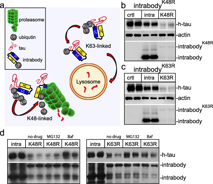
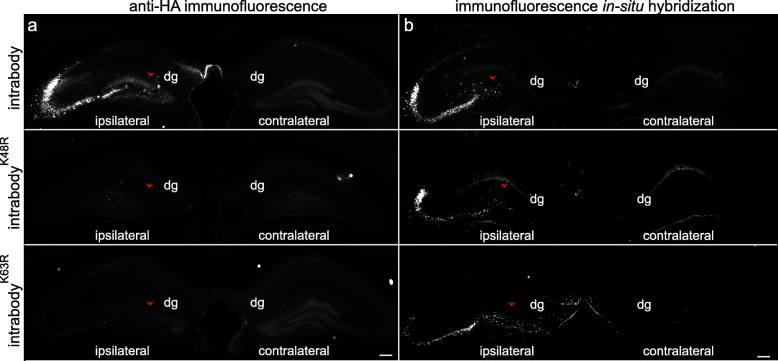
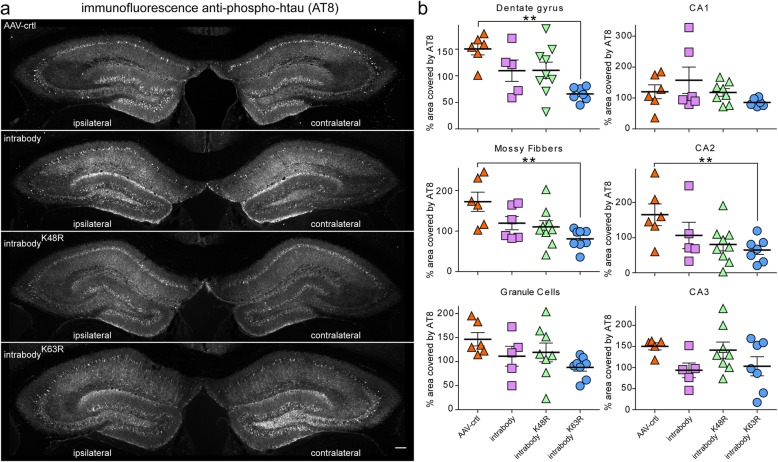
Methods: By utilizing our anti-tau scFv, we generated anti-tau intrabodies for the expression in the cytosol of neurons. To enhance the degradation capacity of conventional intrabodies, we engineered chimeric anti-tau intrabodies fused to ubiquitin harboring distinct mutations that shuttle intracellular tau for either the proteasome or lysosomal mediated degradation. To evaluate the efficacy in delaying or eliminating tauopathy, we expressed our tau degrading intrabodies or controls in human tau transgenic mice by adeno-associated virus prior to overt tau pathology and after tau deposition.

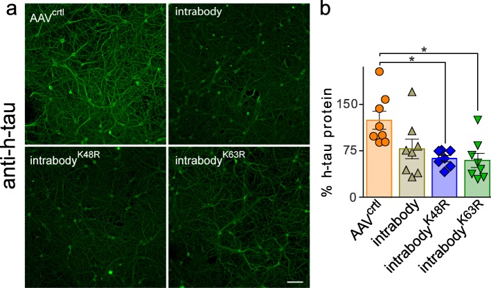
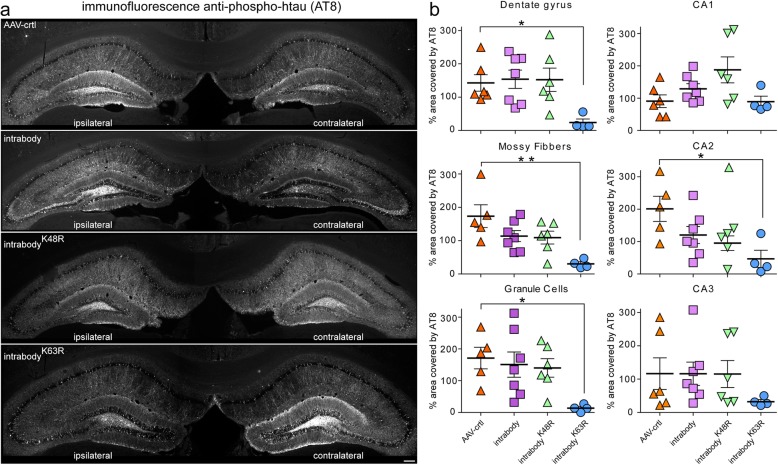
Results: Our results demonstrate, the expression of chimeric anti-tau intrabodies significantly reduce tau protein levels in primary neuronal cultures expression human tau relative to a non-modified anti-tau intrabody. We found the expression of engineered tau-degrading intrabodies destined for proteasomal-mediated degradation are more effective in delaying or eliminating tauopathy than a conventional intrabody in aged human tau transgenic mice.

Conclusion: This study, harnesses the strength of intrabodies that are amendable for targeting specific domains or modifications with the cell-intrinsic mechanisms that regulate protein degradation providing a new immunotherapeutic approach with potentially improved efficacy.

Keywords: Alzheimer’s disease; Immunotherapy; Intrabodies; Tau degradation; Tauopathy.


Conflict of interest statement

GG, HJ, CL, and DMH are listed as inventors on a patent licensed by Washington University to C2N Diagnostics on the therapeutic use of anti-tau antibodies. DMH co-founded and is on the scientific advisory board of C2N Diagnostics, LLC. C2N Diagnostics, LLC has licensed certain anti-tau antibodies to AbbVie for therapeutic development. DMH is on the scientific advisory board of Denali and Genentech and consults for Idorsia.

Figures

Fig. 1
Fig. 1
Engineering chimeric tau-degrading intrabodies fused to ubiquitin for proteasome or lysosomal-mediated degradation. a Diagram depicts that K48-linked polyubiquitin tau-intrabodies shuttle tau for proteasome degradation, whereas, K63-linked polyubiquitin tau-intrabodies shuttle tau for lysosomal degradation. b Immunoblotting analysis of HEK293t cell lysates co-expressing h-tau together with either a conventional anti-tau intrabody or the anti-tau intrabodyK48R revealed the chimeric anti-tau intrabody fused to ubiquitin harboring a K48R mutation decreased tau protein levels relative to the conventional intrabody or control. Protein levels are relative to actin. The expression of the anti-tau intrabodyK48R also revealed a lower band that potentially corresponds to a cleaved form of the chimeric intrabody. c Immunoblotting analysis of HEK293t cell lysates co-expressing h-tau together with either a conventional anti-tau intrabody or the anti-tau intrabodyK63R revealed the chimeric anti-tau intrabody fused to ubiquitin harboring a K63R mutation decreased tau protein levels relative to the conventional intrabody and control. Similarly, expression of the anti-tau intrabodyK63R also revealed a lower band that potentially corresponds to a cleaved form of the chimeric intrabody. d Immunoblotting analyses of HEK293t cell lysates co-expressing h-tau together with either a conventional anti-tau intrabody, anti-tau intrabodyK63R or anti-tau intrabodyK48R following proteasomal (MG132) or lysosomal (Baf) inhibition
Fig. 2
Fig. 2
Expression of tau-degrading intrabodies decreases human tau in neurons. a Representative images of primary neurons co-expressing P301S-htau and the various anti-tau intrabodies. Expression of the conventional anti-tau intrabody, intrabody-K48R or the intrabody-K63R displayed a marked decrease in h-tau protein levels relative to the AAV-control; scale bar 50 μm. b P301S-htau protein quantification by tau ELISA in primary neurons co-expressing the various intrabodies revealed a significant decrease in h-tau protein levels upon expression of each tau-degrading intrabody relative to the AAV-controls. Expression of the conventional anti-tau intrabody displayed an non-significant decrease in h-tau protein relative to the AAV-control. Results are representative of three independent experiments, all data are expressed as mean ± s.e.m. and one-way ANOVA was performed with Tukey’s Multiple Comparison. *p < 0.05
Fig. 3
Fig. 3
Immunofluorescence and fluorescence in situ analysis of anti-tau intrabody expression in aged P301S-tg mice. a Representative images of anti-HA for protein expression of the various anti-tau intrabodies in 9.5-month-old P301S-tg mice. Immunofluorescent detection of HA reveals clear-cut evidence of the non-targeting anti-tau intrabody within the ipsilateral hippocampus, whereas there was little to no immunofluorescence signal with the modified anti-tau intrabodies. Arrowhead indicating the injection site within the dentate gyrus (dg). b Representative images of in situ hybridization in 9.5-month-old P301S-tg mice validated the expression of the mRNA of the various anti-tau intrabodies within the hippocampus ipsilateral to the AAV injection. Scale bar 200 μM
Fig. 4
Fig. 4
Tau-degrading intrabodies prevent tauopathy at early-disease in P301S-tg mice. a Representative images of pathological tau accumulation as seen with AT8 staining in 9.5 month-old P301S-tg mice following the expression of anti-tau intrabodies prior to overt tau pathology (early-disease). b AAV-control displayed an increase in pathological tau accumulation in the ipsilateral hippocampal side. Expression of the conventional anti-tau intrabody displayed a non-significant increase in pathological tau accumulation in the dentate gyrus, granule cells, mossy fibers and CA2 region of the ipsilateral relative to the contralateral hippocampus. Expression of the chimeric tau-degrading intrabody fused to ubiquitin harboring a K48R mutation prone for lysosomal-meditated degradation displayed a non-significant increase in pathological tau accumulation in the granule cells, CA1 and CA3 region of the ipsilateral relative to the contralateral hippocampus. Expression of the chimeric tau-degrading intrabody fused to ubiquitin harboring a K63R mutation prone for proteasome-mediated degradation displayed a significant decrease in pathological tau accumulation in the ipsilateral hippocampal dentate gyrus, granule cells and mossy fibers ipsilateral relative to the contralateral hippocampus. Scale bar 200 μM. Quantification (n = 4–7) of pathological tau from the ipsilateral relative to the contralateral dentate gyrus. All data are mean ± s.e.m. one-way ANOVA with Tukey’s Multiple Comparison. *p < 0.05, **p < 0.01
Fig. 5
Fig. 5
Tau-degrading intrabodies eliminate tauopathy at mid-disease in P301S-tg mice. a. Representative images of pathological tau accumulation as seen with AT8 staining in 9.5 month-old P301S-tg mice following the expression of anti-tau intrabodies after tauopathy deposition (mid-disease). b AAV-control displayed an increase in pathological tau accumulation in the ipsilateral hippocampal side. Expression of the conventional anti-tau intrabody displayed a non-significant increase in pathological tau accumulation in the dentate gyrus, granule cells, mossy fibers and CA2 ipsilateral relative to the contralateral hippocampus. Expression of the chimeric tau-degrading intrabody fused to ubiquitin harboring a K48R mutation prone for lysosomal-meditated degradation displayed a non-significant increase in pathological tau accumulation in the granule cells, CA1 and CA3 regions of the ipsilateral relative to the contralateral hippocapus. Expression of the chimeric tau-degrading intrabody fused to ubiquitin harboring a K63R mutation prone for proteasome-mediated degradation displayed a significant decrease in pathological tau accumulation in the hippocampal dentate gyrus, granule cells and mossy fibers in the ipsilateral relative to the contralateral hippocampus. Scale bar 200 μM. Quantification (n = 6–9) of pathological tau from the contralateral to the ipsilateral dentate gyrus. All data are mean ± s.e.m. one-way ANOVA with Tukey’s Multiple Comparison. *p < 0.05, **p < 0.01

References

    1. Joachim CL, et al. Tau antisera recognize neurofibrillary tangles in a range of neurodegenerative disorders. Ann Neurol. 1987;22(4):514–520. doi: 10.1002/ana.410220411. - DOI - PubMed
    1. Lee VM, Goedert M, Trojanowski JQ. Neurodegenerative tauopathies. Annu Rev Neurosci. 2001;24:1121–1159. doi: 10.1146/annurev.neuro.24.1.1121. - DOI - PubMed
    1. Weingarten MD, et al. A protein factor essential for microtubule assembly. Proc Natl Acad Sci U S A. 1975;72(5):1858–1862. doi: 10.1073/pnas.72.5.1858. - DOI - PMC - PubMed
    1. Witman GB, et al. Tubulin requires tau for growth onto microtubule initiating sites. Proc Natl Acad Sci U S A. 1976;73(11):4070–4074. doi: 10.1073/pnas.73.11.4070. - DOI - PMC - PubMed
    1. Trojanowski JQ, et al. Distribution of tau proteins in the normal human central and peripheral nervous system. J Histochem Cytochem. 1989;37(2):209–215. doi: 10.1177/37.2.2492045. - DOI - PubMed

LinkOut - more resources