. 2019 Nov 29;294(48):18207-18219.
doi: 10.1074/jbc.RA119.010827. Epub 2019 Oct 22.

The R882H substitution in the human de novo DNA methyltransferase DNMT3A disrupts allosteric regulation by the tumor supressor p53

Affiliations

The R882H substitution in the human de novo DNA methyltransferase DNMT3A disrupts allosteric regulation by the tumor supressor p53

Jonathan E Sandoval et al. J Biol Chem. .

Abstract

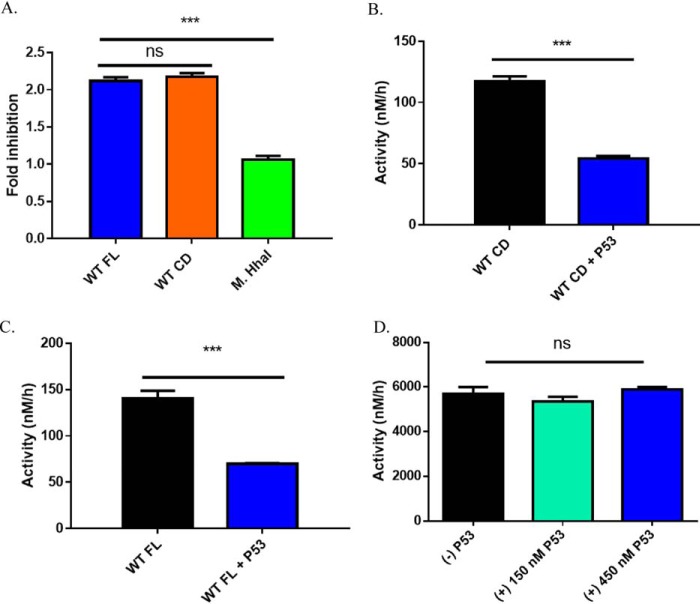
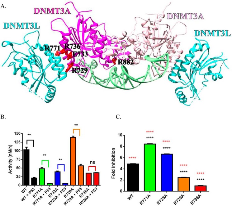
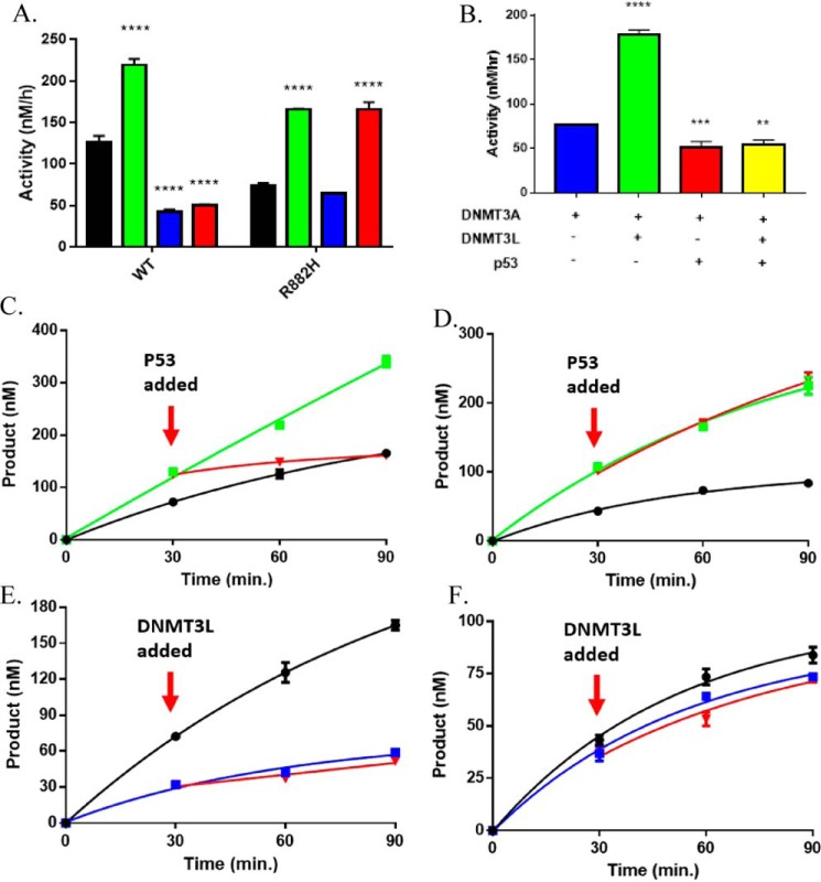
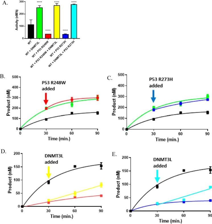
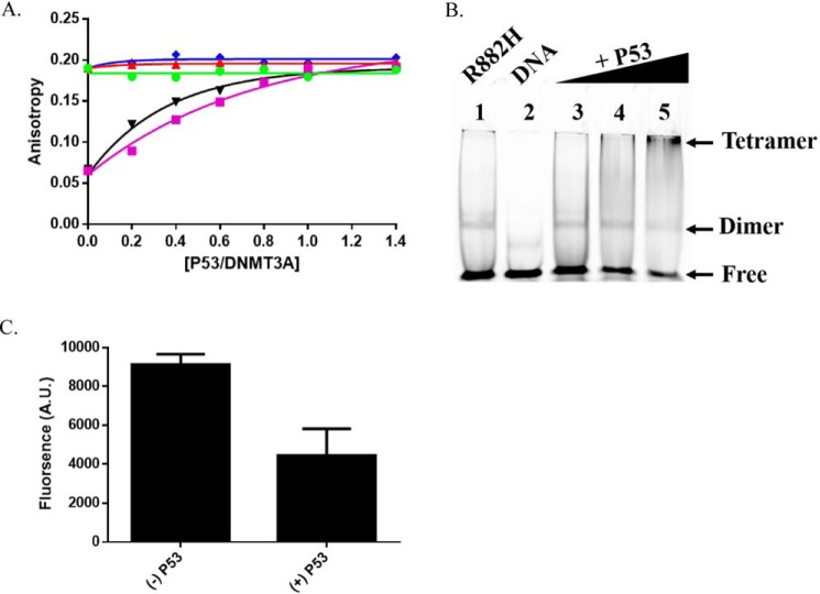
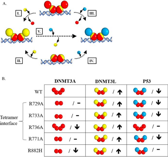
A myriad of protein partners modulate the activity of the human DNA methyltransferase 3A (DNMT3A), whose interactions with these other proteins are frequently altered during oncogenesis. We show here that the tumor suppressor p53 decreases DNMT3A activity by forming a heterotetramer complex with DNMT3A. Mutational and modeling experiments suggested that p53 interacts with the same region in DNMT3A as does the structurally characterized DNMT3L. We observed that the p53-mediated repression of DNMT3A activity is blocked by amino acid substitutions within this interface, but surprisingly, also by a distal DNMT3A residue, R882H. DNMT3A R882H occurs frequently in various cancers, including acute myeloid leukemia, and our results suggest that the effects of R882H and other DNMT3A mutations may go beyond changes in DNMT3A methylation activity. To further understand the dynamics of how protein-protein interactions modulate DNMT3A activity, we determined that p53 has a greater affinity for DNMT3A than for DNMT3L and that p53 readily displaces DNMT3L from the DNMT3A:DNMT3L heterotetramer. Interestingly, this occurred even when the preformed DNMT3A:DNMT3L complex was actively methylating DNA. The frequently identified p53 substitutions (R248W and R273H), whereas able to regulate DNMT3A function when forming the DNMT3A:p53 heterotetramer, no longer displaced DNMT3L from the DNMT3A:DNMT3L heterotetramer. The results of our work highlight the complex interplay between DNMT3A, p53, and DNMT3L and how these interactions are further modulated by clinically derived mutations in each of the interacting partners.

Keywords: DNA demethylation; DNMT3A; allosteric regulation; epigenetics; nucleic acid enzymology; p53; protein-protein interaction.


Conflict of interest statement

The authors declare that they have no conflicts of interest with the contents of this article

Figures

Figure 1.
Figure 1.
p53WT-dependent inhibition of DNA methylation is specific to DNMT3A. A, fold-inhibition calculated by product formed by WT DNMT3A (full-length or catalytic domain enzymes) or M.HhaI divided by product formed by DNMT3A (full-length or catalytic domain enzymes) or M.HhaI without p53WT from reactions in B–D. Co-incubation of DNMT3A full-length (A, blue square calculated from B) and catalytic domain (A, orange square calculated from C) enzymes with p53WT (1:1 at 150 nm) leads to comparable levels of inhibition. Similar reactions involving the bacterial methyltransferase M.HhaI (A, green square calculated from D) with excess p53WT (1:3) failed to inhibit DNA methylation. In all co-incubations, proteins were held at 37 °C for 1 h prior to the start of the reaction by the addition of DNA (5 μm bp poly(dI-dC)). Data reflect the mean ± S.D. of 3 experiments; one-way analysis of variance was used to compare values of all three reactions; ***, p < 0.001; ns, p > 0.05.
Figure 2.
Figure 2.
DNMT3AWT tetramer interface mutants show highly variable response to p53WT inhibition. Crystal structure of a DNMT3AWT-DNMT3L complex (adapted from PDB code 5YX2) denoting critical residues for DNMT3AWT oligomerization (A) (36, 50). Although the extent of p53WT inhibition varies across DNMT3AWT mutants harboring a single alanine substitution within the tetramer interface, the DNMT3AR736A was unresponsive to p53WT inhibition (B and C). All reactions consisted of 150 nm DNMT3AWT and were initiated by the addition of 5 μm bp poly(dI-dC). For co-incubations, DNMT3AWT and p53WT (1:1) were preincubated for 1 h at 37 °C prior to starting the reaction by the addition of substrate DNA. Fold-inhibition was calculated by product formed by DNMT3A (WT and mutants) divided by product formed by DNMT3A (WT and mutants) without p53WT. All reactions were performed in duplicates. In B, a Student's unpaired t test was used to compare values within each set of reactions; **, p < 0.01; ns, p > 0.05. For C, a one-way analysis of variance was used to compare the values of each mutant to WT (****, p < 0.001) and across all samples (orange ****, p < 0.001). Data reflect the mean ± S.D. of 3 experiments.
Figure 3.
Figure 3.
DNMT3AWT and DNMT3AR882H are differentially responsive to modulation by p53WT. p53WT-dependent inhibition of DNMT3AWT activity is dominant in DNMT3AWT-p53WT-DNMT3L co-incubations (A, red square), whereas p53WT fails to inhibit DNMT3AR882H in DNMT3AR882H-p53WT (A, blue square) and DNMT3AR882H-p53WT-DNMT3L co-incubations (A, red square) using poly(dI-dC) (5 μm bp) as a substrate. B, p53WT-dependent inhibition of DNMT3AWT activity is dominant in DNMT3AWT-p53WT-DNMT3L co-incubations (yellow square) with p21-pCpGL (10 μm) as a substrate. p53WT (C, red square) disrupts DNMT3L stimulation of DNMT3AWT in catalytically active DNMT3AWT:DNMT3L complexes (C, green square), whereas catalytically active DNMT3AR882H-DNMT3L (D, green square) are unaltered by the addition of p53WT (D, red square). Reactions consisting of catalyzing p53WT-DNMT3AWT (E, blue square) or DNMT3AR882H-p53WT (F, blue square) were unaltered by the addition of DNMT3L (E and F, red square). The following reactions were also performed as controls: DNMT3AWT (A, C, and E, ■), DNMT3AR882H (A. D, and F, ■), DNMT3AWT-DNMT3L co-incubations (A and C, green square), DNMT3AR882H-DNMT3L co-incubations (A and D, green square), DNMT3AWT-p53WT co-incubations (A and E, blue square) and DNMT3AR882H-p53WT co-incubations (A and F, blue square). In all reactions performed, protein concentrations were 150 nm and were initiated by the addition of substrate DNA. For co-incubations, proteins were placed at 37 °C for 1 h prior to the addition of substrate DNA. All reactions were performed in triplicates. Values (A, green, blue, and red) were compared with either DNMT3AWT (A, ■) or DNMT3AR882H (A, ■) using a one-way analysis of variance; ****, p < 0.001. Data reflect the mean ± S.D. of 3 experiments.
Figure 4.
Figure 4.
p53R248W and p53R273H fail to disrupt stimulation of DNMT3AWT by DNMT3L. The stimulatory effect of DNMT3AWT activity by DNMT3L is dominant in DNMT3AWT-p53R248-DNMT3L (A, red square) or DNMT3AWT-p53R273H-DNMT3L co-incubations (A, blue square). Catalytically active DNMT3AWT-DNMT3L heterotetramers are unaffected by the addition of p53R248W (B, red square) or p53R273H (D, blue square), whereas the addition of DNMT3L leads to an increase in DNMT3AWT-p53R248W (C, yellow square) or DNMT3AWT-p53R273H (C, teal square) co-incubations. The following reactions were also performed as controls: DNMT3AWT (A–E, ■), DNMT3AWT-DNMT3L co-incubations (A, B, and D, green square), DNMT3AWT-p53R248W co-incubations (A and C, red square), and DNMT3AWT-p53R273H co-incubations (A and C, blue square). Protein concentrations were 150 nm for all reactions and were initiated by the addition of 5 μm poly(dI-dC) as a substrate. For co-incubations, proteins were preincubated at 37 °C for 1 h prior to the addition of DNA. All reactions were performed in triplicates and all values in A were compared with WT (A, ■) using a one-way analysis of variance; ****, p < 0.001. Data reflect the mean ± S.D. of 3 experiments.
Figure 5.
Figure 5.
p53 heterodimerizes with WT and R882H DNMT3A. A, increasing concentrations of DNMT3L (purple square) or p53WT (▾) to a fixed concentration of DNMT3AR882H led to a robust increase in anisotropy, whereas DNMT3AWT did not display a significant change in anisotropy by the titration of DNMT3L (green circle), p53WT (red triangle), or p53R248W (blue diamond). B, EMSA and C, EMSA band densitometry (lanes 1 and 5) show increasing concentrations of p53WT (lanes 3-5) to a constant concentration of DNMT3AR882H leads to disappearance of the DNMT3AR882H band and formation of a higher order structure (see arrows, lanes 3-5). In A, DNMT3AWT and DNMT3AR882H concentrations were 2.5 μm. Single point anisotropy measurements were taken after increasing concentrations of DNMT3L (green circle), p53WT (red triangle), and p53R248W (blue diamond) were added to DNA bound (250 nm 5′ 6-FAM-labeled GCbox30) DNMT3AWT and DNMT3AR882H and allowed to incubate at room temperature for 5 min. Measurements were taken using a fluorometer equipped with polarizing filters (excitation, 485 nm; emission, 520 nm). In B, gel shift assays were carried out as described in Holz-Schietinger et al. (28) and other than samples were run on native 4.5% polyacrylamide gels and binding reactions were performed at 37 °C (lane 1). For p53WT supershifting, varying concentrations of p53WT were preincubated for 30 min at 37 °C with DNMT3AR882H before the addition of DNA. A and C reflect the results (mean ± S.D.) of 2 independent experiments.
Figure 6.
Figure 6.
Mutations in DNMT3A lead to diverse interactions with p53. A, the addition of DNMT3L (yellow square) to DNMT3A homotetramers or homodimers (red square tetramer interface depicted in yellow) leads to the formation of DNMT3A (red square)-DNMT3L (yellow square) heterotetramers (I and II). Similarly, p53 (blue square) interacts with DNMT3A homotetramers or homodimers (red square) to form DNMT3A (red square)-p53 (blue square) heterotetramers (III and IV). Furthermore, the addition of p53 (blue square) to DNMT3A (red square)-DNMT3L (yellow square) heterotetramers displaces monomers at the tetramer interface and leads to the formation of DNMT3A-P53 (blue square) heterotetramers (V). B, summary of the oligomeric states of DNMT3A mutants in complex with DNMT3L (28, 36) and p53 as well as the effects on the catalytic function of DNMT3A. DNMT3A mutants display no change (–) or decreased (↓) activity (kcat) relative to WT. Although all the DNMT3A mutants are responsive to DNMT3L stimulation (↑), DNMT3A mutants display varying p53 inhibition (↓). Although DNMT3A R736A and R882H are unresponsive to p53 inhibition (–), p53 binds R882H to form heterotetramer.

References

    1. Vertino P. M., Sekowski J. A., Coll J. M., Applegreen N., Han S., Hickey R. J., and Malkas L. H. (2002) DNMT1 is a component of a multiprotein DNA replication complex. Cell Cycle 1, 416–423 - PubMed
    1. Viré E., Brenner C., Deplus R., Blanchon L., Fraga M., Didelot C., Morey L., Van Eynde A., Bernard D., Vanderwinden J. M., Bollen M., et al. (2006) The Polycomb group protein EZH2 directly controls DNA methylation. Nature 439, 871–874 10.1038/nature04431 - DOI - PubMed
    1. Datta J., Majumder S., Bai S., Ghoshal K., Kutay H., Smith D. S., Crabb J. W., and Jacob S. T. (2005) Physical and functional interaction of DNA methyltransferase 3A with Mbd3 and Brg1 in mouse lymphosarcoma cells. Cancer Res. 65, 10891–10900 10.1158/0008-5472.CAN-05-1455 - DOI - PMC - PubMed
    1. Brenner C., Deplus R., Didelot C., Loriot A., Viré E., De Smet C., Gutierrez A., Danovi D., Bernard D., Boon T., Pelicci P. G., et al. (2005) Myc represses transcription through recruitment of DNA methyltransferase corepressor. EMBO J. 24, 336–346 10.1038/sj.emboj.7600509 - DOI - PMC - PubMed
    1. Thillainadesan G., Chitilian J. M., Isovic M., Ablack J. N., Mymryk J. S., Tini M., and Torchia J. (2012) TGF-β-dependent active demethylation and expression of the p15ink4b tumor suppressor are impaired by the ZNF217/CoREST complex. Mol. Cell 46, 636–649 10.1016/j.molcel.2012.03.027 - DOI - PubMed

Publication types

MeSH terms