. 2019 Sep 19:2019:6380135.
doi: 10.1155/2019/6380135. eCollection 2019.

Mitochondrial Fusion by M1 Promotes Embryoid Body Cardiac Differentiation of Human Pluripotent Stem Cells

Affiliations

Mitochondrial Fusion by M1 Promotes Embryoid Body Cardiac Differentiation of Human Pluripotent Stem Cells

Jarmon G Lees et al. Stem Cells Int. .

Abstract

Human induced pluripotent stem cells (iPSCs) can be differentiated in vitro into bona fide cardiomyocytes for disease modelling and personalized medicine. Mitochondrial morphology and metabolism change dramatically as iPSCs differentiate into mesodermal cardiac lineages. Inhibiting mitochondrial fission has been shown to promote cardiac differentiation of iPSCs. However, the effect of hydrazone M1, a small molecule that promotes mitochondrial fusion, on cardiac mesodermal commitment of human iPSCs is unknown. Here, we demonstrate that treatment with M1 promoted mitochondrial fusion in human iPSCs. Treatment of iPSCs with M1 during embryoid body formation significantly increased the percentage of beating embryoid bodies and expression of cardiac-specific genes. The pro-fusion and pro-cardiogenic effects of M1 were not associated with changes in expression of the α and β subunits of adenosine triphosphate (ATP) synthase. Our findings demonstrate for the first time that hydrazone M1 is capable of promoting cardiac differentiation of human iPSCs, highlighting the important role of mitochondrial dynamics in cardiac mesoderm lineage specification and cardiac development. M1 and other mitochondrial fusion promoters emerge as promising molecular targets to generate lineages of the heart from human iPSCs for patient-specific regenerative medicine.


Conflict of interest statement

The authors declare that they have no conflict of interest.

Figures

Figure 1
Figure 1
M1 stimulates mitochondrial fusion in human iPSCs (iPSC-Foreskin-2 cell line) without inducing mesendodermal differentiation. (a) Mitochondrial morphology of human iPSCs, indicated by HSP60 staining. (b) Percentage of OCT3/4+ cells with different mitochondrial morphologies (n = 4). (c) Number of mitochondrial genomes per nuclear genome (n = 6). (d–g) mRNA expression of mitochondrial fission and fusion markers (d), cell proliferation markers (e), mesodermal cardiac transcription factors (f) ,and pluripotency markers (g) in human iPSCs treated with either DMSO vehicle control (control) or M1 at 5 or 10 μM for 48 hours (n = 7). Data are expressed as mean ± SEM. P < 0.05, ∗∗P < 0.01 vs. control by one-way paired ANOVA with Dunnett's post hoc test.
Figure 2
Figure 2
Gene expression of human iPSCs (iPSC-Foreskin-2 cell line) treated with M1 in differentiation medium. (a–d) mRNA of mesodermal cardiac transcription factors (a), pluripotency markers (b), mitochondrial fission and fusion markers (c), and cell proliferation markers (d) in human PSCs treated with either DMSO vehicle control (control) or M1 at 5 or 10 μM in RPMI+B-27 medium for 48 hours. n = 5. Data are expressed as mean ± SEM. ∗∗P < 0.01 vs. control by one-way paired ANOVA with Dunnett's post hoc test.
Figure 3
Figure 3
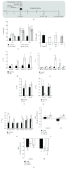
M1 promotes cardiac differentiation of human iPSCs (iPSC-Foreskin-2 cell line). (a) Schematic of embryoid body- (EB-) based cardiac differentiation protocol with 5 μM of M1 treatment regimen. (b) Effect of M1 on the percentage of beating EBs (n = 8). (c) Percentage of cardiac troponin T-positive (cTnT+) cells in individual beating EBs at day 10 postplating (n = 8). (d–h) mRNA expression of mesodermal cardiac transcription factors (d), cardiac-specific muscle proteins (e), endoderm lineage markers (f), ectoderm lineage markers (g), and mitochondrial fission and fusion markers (h) in human iPSCs treated with DMSO (control) or 5 μM M1 for 6 days during EB formation (n = 4). (i, j) Changes in the beating rate of cardiomyocytes derived from control or M1 groups following treatment with isoproterenol hydrochloride (isoprenaline: 1–100 nM (i)) or carbamylcholine (carbachol: 1–100 nM (j)) (n = 10). Data are expressed as mean ± SEM. P < 0.05, ∗∗P < 0.01, and ∗∗∗P < 0.001 vs. control by one-way ANOVA with Dunnett's post hoc test (b, c, i, j) and by paired Student's t-test (d–h).
Figure 4
Figure 4
M1 does not alter ATP synthase subunit expression or interact with any of the 468 human protein kinases screened. (a) mRNA (n = 7) and (b) protein (n = 6) expression of ATP synthase subunits in human iPSCs (iPSC-Foreskin-2 cell line) cultured in TeSR-E8 and treated with M1 for 48 hours. (c) Kinase interaction map of M1 at 10 μM with 468 human protein kinases. Zero interactions with a binding score of ≥35% relative to DMSO were identified. Data are expressed as mean ± SEM and analyzed by one-way paired ANOVA with Dunnett's post hoc test.

References

    1. Verma V., Purnamawati K., Manasi, Shim W. Steering signal transduction pathway towards cardiac lineage from human pluripotent stem cells: a review. Cellular Signalling. 2013;25(5):1096–1107. doi: 10.1016/j.cellsig.2013.01.027. - DOI - PubMed
    1. Chen H., Chan D. C. Mitochondrial dynamics in regulating the unique phenotypes of cancer and stem cells. Cell Metabolism. 2017;26(1):39–48. doi: 10.1016/j.cmet.2017.05.016. - DOI - PMC - PubMed
    1. Lees J. G., Gardner D. K., Harvey A. J. Pluripotent stem cell metabolism and mitochondria: beyond ATP. Stem Cells International. 2017;2017:17. doi: 10.1155/2017/2874283.2874283 - DOI - PMC - PubMed
    1. Seo B., Yoon S., Do J. Mitochondrial dynamics in stem cells and differentiation. International Journal of Molecular Sciences. 2018;19(12):p. 3893. doi: 10.3390/ijms19123893. - DOI - PMC - PubMed
    1. Wanet A., Arnould T., Najimi M., Renard P. Connecting mitochondria, metabolism, and stem cell fate. Stem Cells and Development. 2015;24(17):1957–1971. doi: 10.1089/scd.2015.0117. - DOI - PMC - PubMed

LinkOut - more resources