. 2020 Jan;24(1):126-138.
doi: 10.1111/jcmm.14663. Epub 2019 Oct 23.

Nuclear localization of the tyrosine kinase BMX mediates VEGFR2 expression

Affiliations

Nuclear localization of the tyrosine kinase BMX mediates VEGFR2 expression

Tingting Liu et al. J Cell Mol Med. 2020 Jan.

Abstract

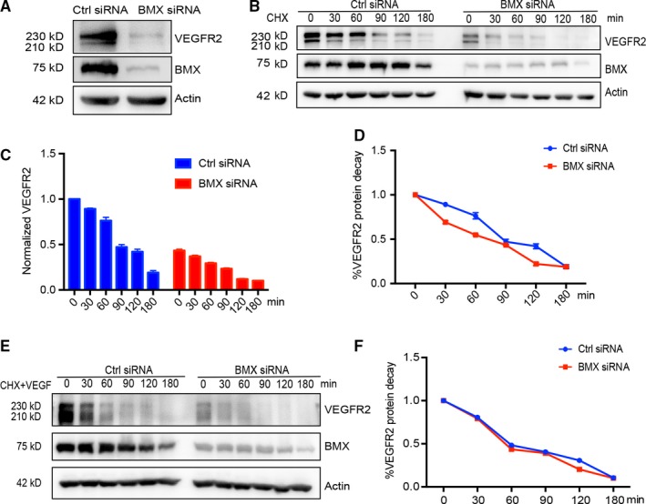
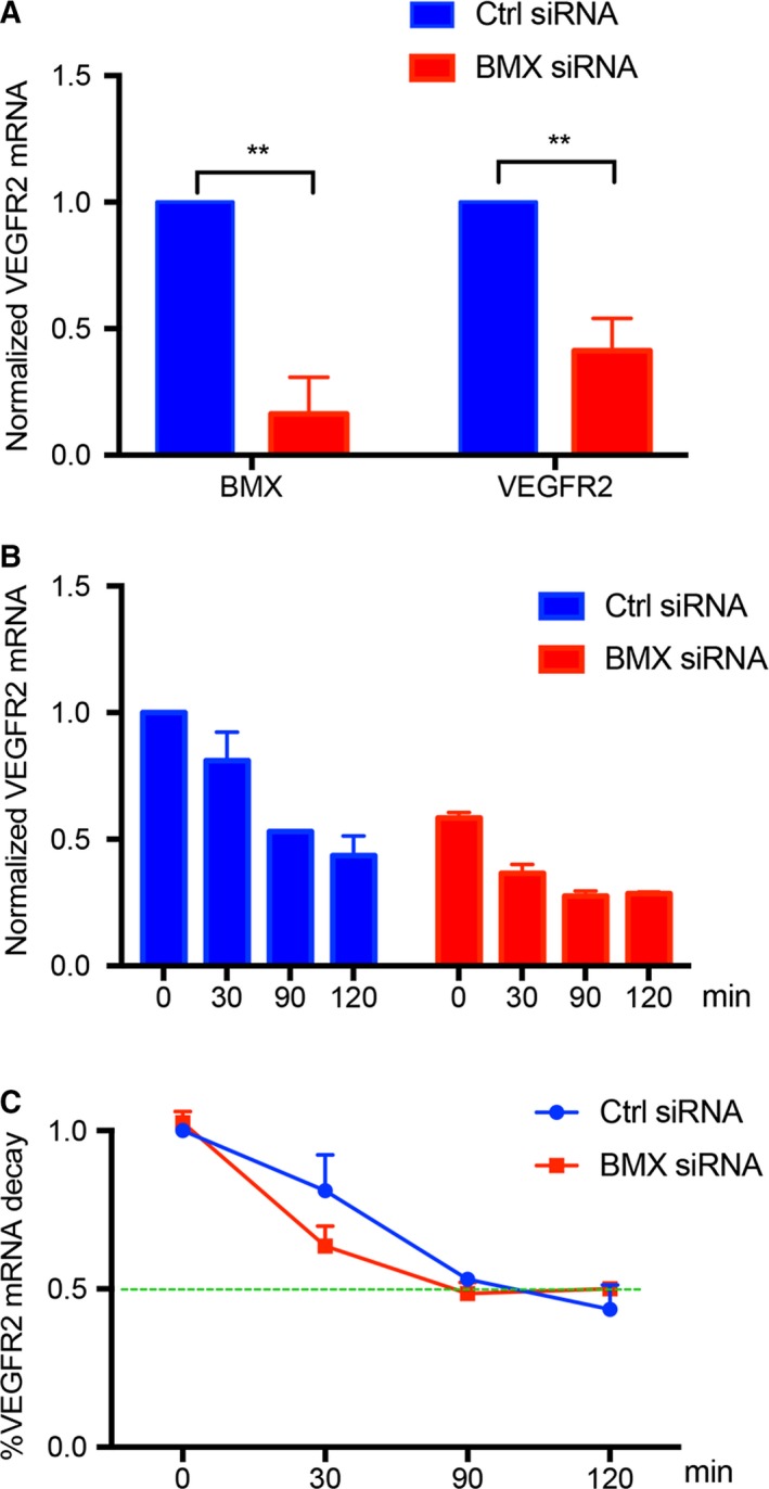
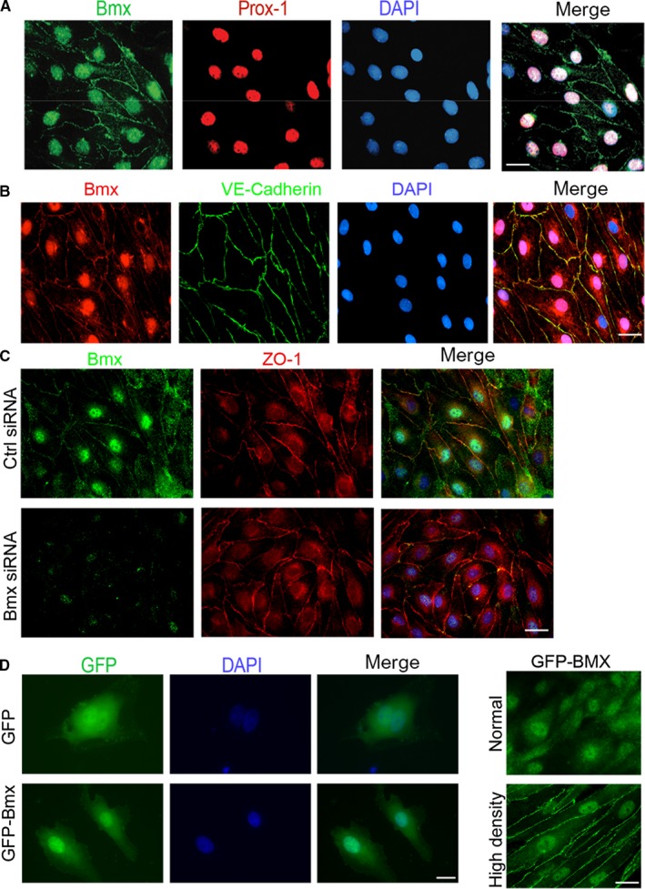
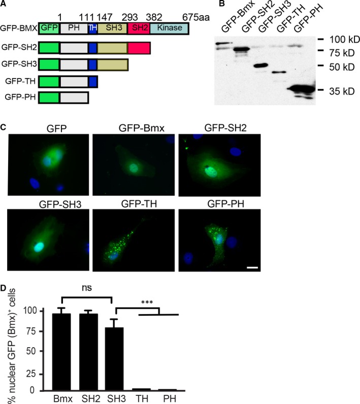
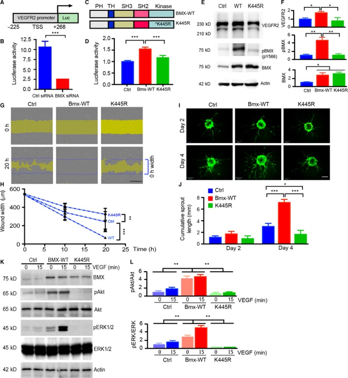
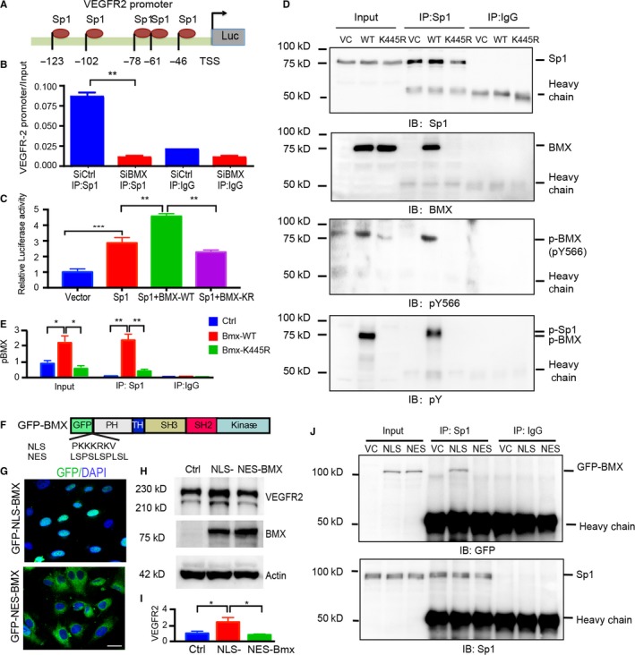
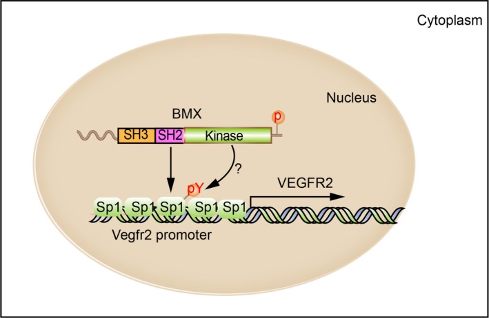
Vascular endothelial growth factor receptors (VEGFRs) are major contributors to angiogenesis and lymphangiogenesis through the binding of VEGF ligands. We have previously shown that the bone marrow tyrosine kinase BMX is critical for inflammatory angiogenesis via its direct transactivation of VEGFR2. In the present study, we show that siRNA-mediated silencing of BMX led to a significant decrease in the total levels of VEGFR2 mRNA and protein, without affecting their stability, in human endothelial cells (ECs). Interestingly, BMX was detected in the nuclei of ECs, and the SH3 domain of BMX was necessary for its nuclear localization. Luciferase assays showed a significant decrease in the Vegfr2 (kdr) gene promoter activity in ECs after BMX silencing, indicating that BMX is necessary for Vegfr2 transcription. In addition, we found that wild-type BMX, but not a catalytic inactive mutant BMX-K445R, promoted Vegfr2 promoter activity and VEGF-induced EC migration and tube sprouting. Mechanistically, we show that the enhancement of Vegfr2 promoter activity by BMX was mediated by Sp1, a transcription factor critical for the Vegfr2 promoter. Loss of BMX significantly reduced Sp1 binding to the Vegfr2 promoter as assayed by chromatin immunoprecipitation assays. Wild-type BMX, but not a kinase-inactive form of BMX, associated with and potentially phosphorylated Sp1. Moreover, a nuclear-targeted BMX (NLS-BMX), but not cytoplasm-localized form (NES-BMX), bound to Sp1 and augmented VEGFR2 expression. In conclusion, we uncovered a novel function of nuclear-localized BMX in regulating VEGFR2 expression and angiogenesis, suggesting that BMX is a therapeutic target for angiogenesis-related diseases.

Keywords: Bmx; Sp1; VEGFR2; angiogenesis; promoter; transcription.


Conflict of interest statement

The authors confirm that there are no conflicts of interest.

Figures

Figure 1
Figure 1
BMX regulates VEGFR2 expression but not VEGFR2 protein stability in ECs. HUVECs were transfected with human BMX siRNA or control siRNA (20 nmol/L) for 48 h. A, Total VEGFR2 and BMX proteins were determined by Western blotting with specific antibodies. β‐Actin was used as a loading control. B‐D, BMX siRNA on VEGFR2 stability. siRNA‐transfected HUVECs were incubated with cycloheximide (CHX, 10 μg/mL) for the indicated time‐points. Total VEGFR2 and BMX proteins were determined by Western blotting with specific antibodies. β‐Actin was used as a loading control (B). The protein bands in B were quantified by densitometry, and the relative VEGFR2 levels were presented by setting untreated control siRNA as 1.0 (C). Normalized VEGFR2 expression shows its decay rate (D). E‐F, BMX siRNA on VEGFR2 stability in the presence of VEGF‐A. siRNA‐transfected HUVECs were incubated with cycloheximide (CHX, 10 μg/mL) in the presence of VEGF‐A (50 ng/mL) for the indicated time‐points. VEGFR2 protein detection (E) and normalization (F) as described in B‐D. The data are means ± SEM from three independent experiments
Figure 2
Figure 2
BMX regulates VEGFR2 expression at the transcriptional level in ECs. HUVECs were transfected with human BMX siRNA or control siRNA (20 nmol/L) for 48 h. A, siRNA‐transfected HUVECs were incubated with actinomycin D (10 μg/mL) for the indicated time‐points. VEGFR2 mRNA was assessed at the indicated time‐points by qRT‐PCR with normalization to β‐actin mRNA. B, The % decrease of VEGFR2 mRNA due to actinomycin D was quantified normalized to Ctrl siRNA at 0 min, using the time 0 of each respective group as the reference point. C, Normalized VEGFR2 mRNA levels. The data are means ± SEM from three independent experiments. **, P < .01
Figure 3
Figure 3
Localization of BMX in HDLECs. A, Confluent HDLECs were co‐stained with anti‐BMX and anti‐Prox‐1, followed by Alexa Fluor 488‐ and 594‐conjugated secondary antibodies (donkey anti‐goat and donkey anti‐rabbit, respectively). The merged images of BMX, Prox‐1 and DAPI are shown on the right. B. Confluent HDLECs were co‐stained with anti‐BMX and anti‐VE‐cadherin, followed by Alexa Fluor 594‐ and 488‐conjugated secondary antibodies (donkey anti‐goat and donkey antimouse, respectively). The merged images of BMX, VE‐cadherin and DAPI are shown on the right. 63× magnification images are shown for all images. C, HDLECs were transfected with human BMX siRNA or control siRNA (20 nmol/L) for 48 h. Confluent HDLECs were co‐stained with anti‐BMX and anti‐ZO‐1, followed by Alexa Fluor 488‐ and 594‐conjugated secondary antibodies (donkey anti‐goat and donkey antimouse, respectively). The merged images of BMX, ZO‐1 and DAPI from the control and BMX siRNA groups are shown on the right. D‐E, HDLECs were infected with lentivirus expressing GFP or GFP‐BMX. IF microscopy was used to visualize GFP or GFP‐BMX. Single cells (D), normal‐density (E, top) and high‐density cells (E, bottom) are shown. All experiments were repeated at least three times. Scale bar: 25 μm
Figure 4
Figure 4
The SH3 domain of BMX is critical for BMX nuclear localization. A, Schematic diagram for BMX structural domains and expression constructs. GFP indicates green fluorescent protein; PH, pleckstrin homology domain; TH, TEC homology domain; K: kinase domain; aa, amino acids. Western blotting is shown in (B) using an anti‐GFP antibody to detect proteins. C, HDLECs were transfected with either 1.0 μg of GFP plasmid, GFP‐BMX full‐length plasmid (human BMX‐WT fused to an N‐terminal GFP tag), GFP‐SH2 plasmid (human BMX cDNA lacking the kinase domain fused to an N‐terminal GFP tag), GFP‐SH3 plasmid (human BMX PH, TH and SH3 domains domain fused to an N‐terminal GFP tag), GFP‐TH plasmid (human BMX PH and TH domains fused to an N‐terminal GFP tag) or GFP‐PH plasmid (human BMX PH domain fused to an N‐terminal GFP tag). IF microscopy was used to visualize the GFP tagged proteins. D, Quantification of nuclear GFP‐BMX+ cells. The data are means ± SEM from three independent experiments. One hundred GFP‐positive cells were counted for each group. ***, P < .0001. ns: non‐significant. Scale bar: 25 μm
Figure 5
Figure 5
BMX regulates VEGFR2 transcription and VEGF‐induced angiogenesis in a kinase‐dependent manner. A, Schematic diagram for the Vegfr2 promoter‐luciferase reporter gene. −225 and +258 are the positions related to the transcription start site (TSS; +1). B, HDLECs were cotransfected with the Vegfr2 luciferase reporter, Renilla luciferase plasmid and either human BMX siRNA or control siRNA (20 nmol/L) for 48 h, and then, the dual‐luciferase assay was performed. For the dual‐luciferase assay, the firefly luciferase readout was normalized to that of Renilla luciferase. C, Schematic diagram of BMX‐WT and kinase‐dead mutant BMX‐K445R. D, HDLECs were cotransfected with the Vegfr2 luciferase reporter, Renilla luciferase plasmid and either vector control (Vector), BMX‐WT or kinase‐dead K445R‐BMX for 48 h, and then, the dual‐luciferase assay was performed. E, Overexpression of BMX‐WT, but not the kinase‐dead mutant BMX‐K445R in HUVECs, increases VEGFR2 expression. HUVECs were infected by lentivirus expressing vector control (Ctrl), BMX‐WT or BMX‐K445R. VEGFR2, BMX and phosphor‐BMX (pY566) were determined by Western blotting with indicated antibodies. F‐G, BMX‐WT promotes, while BMX‐K445R inhibits, VEGF‐induced EC migration. HUVECs were infected by lentivirus expressing vector control (Ctrl), BMX‐WT or BMX‐K445R. Cells were subjected to wound injury, followed by incubation for 10 h and 20 h in the presence of VGF‐A (50 ng/mL). F, Representative images of cell migration are shown. The initial wound gap (0 h) is indicated by blue lines at 20 h images, while the remaining gaps are indicated by yellow areas. G, Quantitation of EC migration. The average wound width was quantified, n = 3. Scale bar: 1 mm. H‐I, 3D spheroid sprouting assay. GFP, GFP‐BMX‐WT‐ or GFP‐BMX‐KR‐expressing HUVECs were coated with microbeads, embedded in fibrin gels and grown in EGM‐2 medium for 4 d. A representative image of 10 beads for each sample is shown (H). Scale bar: 100 μm. Quantifications of sprout number, sprout length are shown in I, n = 10. J, BMX‐WT promotes, while BMX‐K445R inhibits, VEGF‐induced signalling. HUVECs were infected by lentivirus expressing vector control (Ctrl), BMX‐WT or BMX‐K445R. Cells were treated with VEGF‐A (50 ng/mL) for 15 min, and VEGFR2 downstream signalling was determined by Western blotting with indicated antibodies, n = 3. Normalized p‐Akt:total Akt and p‐ERK1/2:total ERK1/2 were quantified by taking untreated Ctrl as 1.0. All data are means ± SEM from three independent experiments. *P < .05, **P < .01 and ***P < .001 by unpaired two‐tailed Student's t test
Figure 6
Figure 6
Active BMX interacts with Sp1 in the nucleus and facilitates Sp1 binding to the Vegfr2 promoter. A, Schematic diagram for the Sp1 binding sites located on the Vegfr2 promoter. −123 to −46 are positions related to the transcription start site (TSS; +1). B, BMX promotes Sp1 binding to the Vegfr2 promoter. HDLECs were transfected with human BMX siRNA or control siRNA (20 nmol/L) for 48 h. ChIP assay was then performed with Sp1 antibody. An Sp1 binding region of the Vegfr2 promoter was used as a primer for quantitative PCR. C, HUVECs were cotransfected with a Vegfr2 reporter (−123 to +1), Renilla luciferase plasmid and either vector control (Vector), BMX‐WT or kinase‐dead K445R‐BMX alone or together with Sp1 for 48 h. The dual‐luciferase assay was then performed. The firefly luciferase readout was normalized to that of Renilla luciferase. The data are means ± SEM from three independent experiments. **, P < .01; ***, P < .0001. D‐E, HUVECs were infected by BMX‐WT or BMX‐K445R lentivirus. The nuclear fractions were isolated, and the association of BMX with Sp1 was determined by immunoprecipitation with anti‐Sp1 antibody, followed by immunoblotting (IB) with anti‐Sp1, anti‐BMX, anti‐BMXpY566 and anti‐phosphor‐tyrosine (anti‐pY). IgG was used to a control. Quantification of p‐BMX was presented in (E). F‐G, Location of NLS‐BMX and NES‐BMX. F, Schematic diagram of GFP‐NLS‐BMX and GFP‐NES‐BMX. Insertion of a NLS (PKKKRKV) or NES (LSPSLSPLSL) is indicated. G, HUVECs were infected by lentivirus expressing GFP‐NLS‐BMX or GFP‐NES‐BMX, and location of BMX was visualized under fluorescence microscope. H‐J, NLS‐BMX, but not NES‐BMX, increases VEGFR2 and binds to Sp1. HUVECs were infected by lentivirus expressing vector control, GFP‐NLS‐BMX or GFP‐NES‐BMX. VEGFR2 expression was determined by Western blotting (H) with quantification in (I), and association of BMX with Sp1 was determined by co‐immunoprecipitation (J)
Figure 7
Figure 7
Schematic model of BMX regulates VEGFR2 transcription by interaction with Sp1. The SH2/SH3 domain of BMX confers its nuclear localization in ECs. Nuclear BMX in an active form interacts with (and possibly phosphorylates) Sp1 to facilitate the recruitment of Sp1 to the VEGFR2 promoter and its transcription in ECs

References

    1. Pober JS, Min W, Bradley JR. Mechanisms of endothelial dysfunction, injury, and death. Annu Rev Pathol. 2009;4:71‐95. - PubMed
    1. Alitalo K. The lymphatic vasculature in disease. Nat Med. 2011;17:1371‐1380. - PubMed
    1. Karpanen T, Alitalo K. Molecular biology and pathology of lymphangiogenesis. Annu Rev Pathol. 2008;3:367‐397. - PubMed
    1. Jones D, Min W. An overview of lymphatic vessels and their emerging role in cardiovascular disease. J Cardiovasc Dis Res. 2011;2:141‐152. - PMC - PubMed
    1. Ferrara N, Gerber HP, LeCouter J. The biology of VEGF and its receptors. Nat Med. 2003;9:669‐676. - PubMed

Publication types

MeSH terms

Substances