

The quantitative assessment of interstitial lung disease with positron emission tomography scanning in systemic sclerosis patients
- PMID: 31642912
- PMCID: PMC7244784
- DOI: 10.1093/rheumatology/kez483
The quantitative assessment of interstitial lung disease with positron emission tomography scanning in systemic sclerosis patients
Abstract
Objectives: The reversibility of interstitial lung disease (ILD) in SSc is difficult to assess by current diagnostic modalities and there is clinical need for imaging techniques that allow for treatment stratification and monitoring. 18F-Fluorodeoxyglucose (FDG) PET/CT scanning may be of interest for this purpose by detection of metabolic activity in lung tissue. This study aimed to investigate the potential role of 18F-FDG PET/CT scanning for the quantitative assessment of SSc-related active ILD.
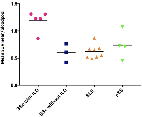
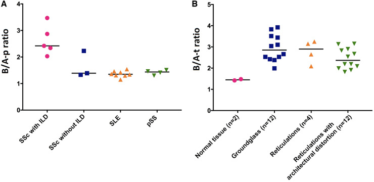
Methods: 18F-FDG PET/CT scans and high resolution CT scans of eight SSc patients, including five with ILD, were analysed. For comparison, reference groups were included: eight SLE patients and four primary Sjögren's syndrome (pSS) patients, all without ILD. A total of 22 regions of interest were drawn in each patient at apical, medial and dorsobasal lung levels. 18F-FDG uptake was measured as mean standardized uptake value (SUVmean) in each region of interest. Subsequently, basal/apical (B/A) and medial/apical (M/A) ratios were calculated at patient level (B/A-p and M/A-p) and at tissue level (B/A-t and M/A-t).
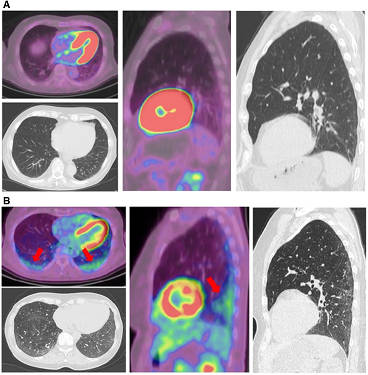
Results: SUVmean values in dorsobasal ROIs and B/A-p ratios were increased in SSc with ILD compared with SSc without ILD (P = 0.04 and P = 0.07, respectively), SLE (P = 0.003 and P = 0.002, respectively) and pSS (P = 0.03 and P = 0.02, respectively). Increased uptake in the dorsobasal lungs and increased B/A-t ratios corresponded to both ground glass and reticulation on high resolution CT.
Conclusion: Semi-quantitative assessment of 18F-FDG PET/CT is able to distinguish ILD from non-affected lung tissue in SSc, suggesting that it may be used as a new biomarker for SSc-ILD disease activity.
Keywords: 18F-FDG PET/CT; Systemic sclerosis; interstitial lung disease; lung fibrosis; positron emission tomography.
© The Author(s) 2019. Published by Oxford University Press on behalf of the British Society for Rheumatology.
Figures
Comment in
-
The race against time to disclose lung inflammation in interstitial lung disease in systemic sclerosis: is PET scan the winning solution?Rheumatology (Oxford). 2020 Jun 1;59(6):1198-1199. doi: 10.1093/rheumatology/keaa077. Rheumatology (Oxford). 2020. PMID: 32159796 Free PMC article. No abstract available.
References
-
- Morales-Cardenas A, Perez-Madrid C, Arias L. et al. Pulmonary involvement in systemic sclerosis. Autoimmun Rev 2016;15:1094–108. - PubMed
-
- Goh NS, Desai SR, Veeraraghavan S. et al. Interstitial lung disease in systemic sclerosis: a simple staging system. Am J Respir Crit Care Med 2008;177:1248–54. - PubMed
-
- Denton CP, Khanna D.. Systemic sclerosis. Lancet 2017;390:1685–99. - PubMed
-
- Antoniou KM, Margaritopoulos G, Economidou F, Siafakas NM.. Pivotal clinical dilemmas in collagen vascular diseases associated with interstitial lung involvement. Eur Respir J 2009;33:882–96. - PubMed
