. 2020 Feb 1;318(2):L407-L418.
doi: 10.1152/ajplung.00561.2018. Epub 2019 Oct 23.

Perinatal maternal antibiotic exposure augments lung injury in offspring in experimental bronchopulmonary dysplasia

Affiliations

Perinatal maternal antibiotic exposure augments lung injury in offspring in experimental bronchopulmonary dysplasia

Kent A Willis et al. Am J Physiol Lung Cell Mol Physiol. .

Abstract

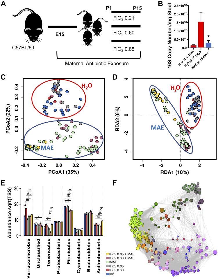
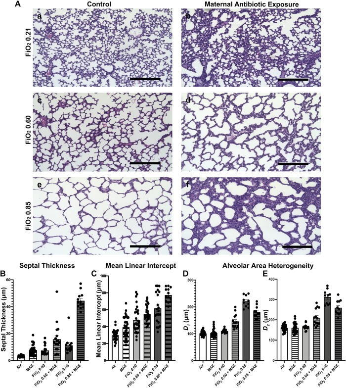
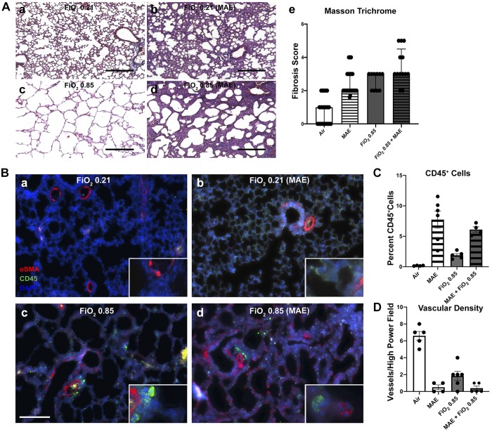
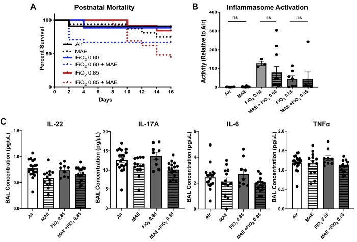
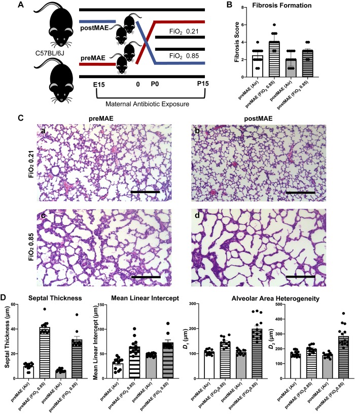
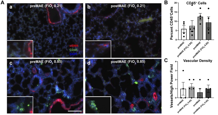
During the newborn period, intestinal commensal bacteria influence pulmonary mucosal immunology via the gut-lung axis. Epidemiological studies have linked perinatal antibiotic exposure in human newborns to an increased risk for bronchopulmonary dysplasia, but whether this effect is mediated by the gut-lung axis is unknown. To explore antibiotic disruption of the newborn gut-lung axis, we studied how perinatal maternal antibiotic exposure influenced lung injury in a hyperoxia-based mouse model of bronchopulmonary dysplasia. We report that disruption of intestinal commensal colonization during the perinatal period promotes a more severe bronchopulmonary dysplasia phenotype characterized by increased mortality and pulmonary fibrosis. Mechanistically, metagenomic shifts were associated with decreased IL-22 expression in bronchoalveolar lavage and were independent of hyperoxia-induced inflammasome activation. Collectively, these results demonstrate a previously unrecognized influence of the gut-lung axis during the development of neonatal lung injury, which could be leveraged to ameliorate the most severe and persistent pulmonary complication of preterm birth.

Keywords: gut-lung axis; interleukin-22; microbiome; mucosal immunology; pulmonary fibrosis.


Conflict of interest statement

No conflicts of interest, financial or otherwise, are declared by the authors.

Figures

Fig. 1.
Fig. 1.
Maternal antibiotic exposure induces a characteristic commensal disruption of the neonatal gut microbiome at 15 days of life. A: experimental schema. C57Bl/6J mice were cohoused to normalize the microbiome and then time-mated to produce multiple simultaneous litters exposed to either maternal antibiotic exposure (MAE) or purified water (controls). After birth, neonatal mice were exposed to either normoxia (air) or hyperoxia for 14 days. Neonatal mice were humanely euthanized for stool and tissue collection at 15 days of life. E15, embryonic day 15; P15, postnatal day 15. B: as quantified by qRT-PCR, MAE reduced the absolute abundance of the bacterial 16S rRNA gene in the neonatal colon at 15 days to a level similar to a specific pathogen-free 5-day-old pup (one-way ANOVA, P = 0.0374). C: structured principal coordinates analysis [PCoA, permutational multivariate ANOVA (PERMANOVA) with 999 permutations of Bray–Curtis dissimilarity, R2 = 0.302, P = 0.00033]. D: unstructured redundancy analysis (RDA, variance 87.29, f = 8.46, P = 0.001). C and D demonstrate that the composition of the neonatal colonic microbiome at 15 days of life differs by exposure to MAE and not hyperoxia. Centroids added for emphasis. E: differences in relative abundance at the phylum level. Sqrt(TSS), square root total sum normalization (Hellinger Transformation). F: Spearman network analysis. Positive correlations with false discovery rate-adjusted P values < 0.05 are displayed as an edge, and relative size represents significance of the edge to the surrounding network. n = 5–8 pups/group in 3 independent experiments; data points represent individual animals. One-way ANOVA, Bonferroni, *P < 0.05, **P < 0.01, ***P < 0.001. FIO2, fraction of inspired oxygen; P0, day of birth.
Fig. 2.
Fig. 2.
Commensal disruption produces a more severe phenotype of bronchopulmonary dysplasia. A: representative micrographs of hematoxylin and eosin-stained lung sections that were gravity inflation-fixed from 15-day-old neonatal mice exposed to either normoxia (air) (Aa), maternal antibiotic exposure (MAE) and normoxia (Ab), fraction of inspired oxygen (FIO2) 0.60 (Ac), MAE and FIO2 0.60 (Ad), FIO2 0.85 (Ae), or MAE and FIO2 0.85 (Af). Scale bars represent 200 μm. B: septal thickness, two-way ANOVA interaction, MAE and hyperoxia, P < 0.001. C: mean linear intercept, two-way ANOVA interaction not significant (NS), MAE, P = 0.0004; hyperoxia, P < 0.001. D and E: airway heterogeneity (weighted means of airway diameter, D1 and D2) demonstrates only an interactive effect of MAE (two-way ANOVA interaction, P < 0.0001; MAE NS, hyperoxia, P < 0.0001); n = 5–8 pups/group in 3 independent experiments; data points represent individual animals and error bars display mean ± SE.
Fig. 3.
Fig. 3.
Commensal disruption increases fibrosis and immune cell recruitment and decreases vascular density. ad: for each group of representative micrographs: normoxia alone (a), maternal antibiotic exposure (MAE) alone (b), hyperoxia exposure at fraction of inspired oxygen (FIO2) 0.85 alone (c), and the combined exposure of MAE and hyperoxia (d). A: Masson trichrome staining with data presented as median ± interquartile range of a pathology score (Ae) (Friedman rank sum test, P < 0.0001). Black scale bars represent 200 μm. Data points represent individual animals; n = 3–5 pups/group in 3 independent experiments. B: representative immunohistochemistry for CD45 (Cy5, pseudocolored green channel) and α-smooth muscle actin (αSMA, red channel). C: quantification of the percentage of CD45-positive cells (two-way ANOVA interaction and hyperoxia exposure not significant; MAE, P < 0.001). D: quantification of the number of αSMA-positive vessels <50 μm in diameter per ×20 microscopic field (two-way ANOVA interaction, P = 0.001; hyperoxia exposure and MAE, P < 0.001). White scale bar represents 100 μm. n = 3–5 pups/group; data points represent individual animals, and error bars display mean ± SE.
Fig. 4.
Fig. 4.
Commensal disruption increases mortality and produces changes in some bronchoalveolar lavage (BAL) cytokine levels. A: maternal antibiotic exposure (MAE) leads to increased mortality during hyperoxia exposure [fraction of inspired oxygen (FIO2) 0.85 compared with MAE and FIO2 0.85, log-rank test, P = 0.002]. Normoxia (black), intermediate hyperoxia (blue), and elevated hyperoxia (red), with MAE exposure indicated by a dotted line. B: inflammasome activity is increased by hyperoxia but not by the addition of MAE [two-way ANOVA hyperoxia exposure, P < 0.0001; MAE, not significant (NS)]. C: low-level BAL cytokine concentrations with a limited effect of MAE but not hyperoxia (IL-22: two-way ANOVA interaction, NS; MAE, P = 0.0042; hyperoxia exposure, NS; IL-17A: two-way ANOVA interaction, NS; MAE, P = 0.005; hyperoxia exposure, NS; IL-6: two-way ANOVA interaction, NS; MAE, P = 0.0217; hyperoxia exposure, NS; TNF-α: two-way ANOVA interaction, NS; MAE, P = 0.0396; hyperoxia exposure, NS). Except where noted, error bars represent mean ± SE; n = 7–20 pups/group; data points represent individual animals. Significance was determined by two-way ANOVA, and P < 0.05 was considered significant.
Fig. 5.
Fig. 5.
Prenatal exposure to maternal antibiotics enhances fibrosis, while postnatal exposure accentuates changes in lung architecture from hyperoxia exposure. A: experimental schema showing the birth of simultaneous litters to either purified water- or antibiotic-exposed dams. The resulting offspring were then fostered to dams in the opposite study arm at birth, before exposure to hyperoxia or normoxia, producing prenatal maternal antibiotic-exposed (preMAE, red) and postnatal maternal antibiotic-exposed (postMAE, blue) cohorts. B: preMAE enhances fibrosis (Friedman rank sum test, P = 0.00016). C: representative micrographs of hematoxylin and eosin-stained lung sections that were gravity inflation-fixed from 15-day-old neonatal mice exposed to either preMAE and normoxia (air, a), preMAE and fraction of inspired oxygen (FIO2) 0.85 (b), postMAE and normoxia (c), or postMAE and FIO2 0.85 (d). D: septal thickness (two-way ANOVA interaction, P = 0.0092; antibiotics and hyperoxia, P < 0.0001) is increased in preMAE, but the mean linear intercept (two-way ANOVA interaction, not significant; antibiotics, P = 0.0013; hyperoxia, P < 0.0001) and weighted means of airway diameter (D1: two-way ANOVA interaction, P = 0.0002; antibiotics, P = 0.0003; hyperoxia, P < 0.0001 and D2: two-way ANOVA interaction, P < 0.0001; antibiotics, P = 0.0002; hyperoxia, P < 0.0001) are increased in postMAE. n = 5–8 pups/group in 2 independent experiments; datapoints represent individual animals, and error bars represent mean ± SE. E15, embryonic day 15; P0, day of birth; P15, postnatal day 15.
Fig. 6.
Fig. 6.
Prenatal and postnatal maternal antibiotic exposure produce equivalent alterations in pulmonary immune cell recruitment and vascular density. A: representative micrographs for CD45 (Cy5, pseudocolored green channel) and α-smooth muscle actin (αSMA, red channel). a: prenatal maternal antibiotic exposure (preMAE) at normoxia (air). b: postnatal maternal antibiotic exposure (postMAE) at normoxia. c: preMAE at fraction of inspired oxygen (FIO2) 0.85. d: postMAE at FIO2 0.85. B: quantification of the percentage of CD45-positive cells [two-way ANOVA, not significant (NS)]. C: quantification of the number of αSMA-positive vessels <50 μm in diameter per ×20 microscopic field (two-way ANOVA, NS). White scale bar represents 100 μm. n = 3–5 pups/group; datapoints represent individual animals, and error bars display mean ± SE.

Comment in

References

    1. Alvira CM. Aberrant pulmonary vascular growth and remodeling in bronchopulmonary dysplasia. Front Med (Lausanne) 3: 21, 2016. doi:10.3389/fmed.2016.00021. - DOI - PMC - PubMed
    1. Ambalavanan N, Carlo WA, D’Angio CT, McDonald SA, Das A, Schendel D, Thorsen P, Higgins RD; Eunice Kennedy Shriver National Institute of Child Health and Human Development Neonatal Research Network . Cytokines associated with bronchopulmonary dysplasia or death in extremely low birth weight infants. Pediatrics 123: 1132–1141, 2009. doi:10.1542/peds.2008-0526. - DOI - PMC - PubMed
    1. Arboleya S, Sánchez B, Milani C, Duranti S, Solís G, Fernández N, de los Reyes-Gavilán CG, Ventura M, Margolles A, Gueimonde M. Intestinal microbiota development in preterm neonates and effect of perinatal antibiotics. J Pediatr 166: 538–544, 2015. doi:10.1016/j.jpeds.2014.09.041. - DOI - PubMed
    1. Arrieta MC, Stiemsma LT, Dimitriu PA, Thorson L, Russell S, Yurist-Doutsch S, Kuzeljevic B, Gold MJ, Britton HM, Lefebvre DL, Subbarao P, Mandhane P, Becker A, McNagny KM, Sears MR, Kollmann T, Mohn WW, Turvey SE, Finlay BB; CHILD Study Investigators . Early infancy microbial and metabolic alterations affect risk of childhood asthma. Sci Transl Med 7: 307ra152, 2015. doi:10.1126/scitranslmed.aab2271. - DOI - PubMed
    1. Baker CD, Abman SH. Impaired pulmonary vascular development in bronchopulmonary dysplasia. Neonatology 107: 344–351, 2015. doi:10.1159/000381129. - DOI - PMC - PubMed

Publication types

MeSH terms