. 2019 Oct 22;29(4):844-859.e3.
doi: 10.1016/j.celrep.2019.09.042.

Cutaneous Immune Cell-Microbiota Interactions Are Controlled by Epidermal JunB/AP-1

Affiliations

Cutaneous Immune Cell-Microbiota Interactions Are Controlled by Epidermal JunB/AP-1

Özge Uluçkan et al. Cell Rep. .

Abstract

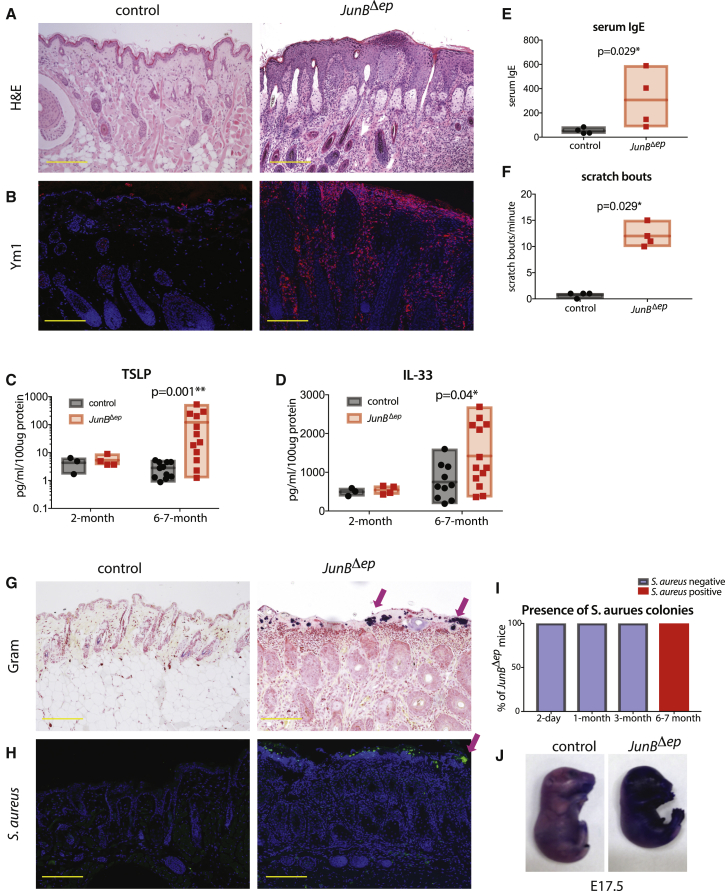
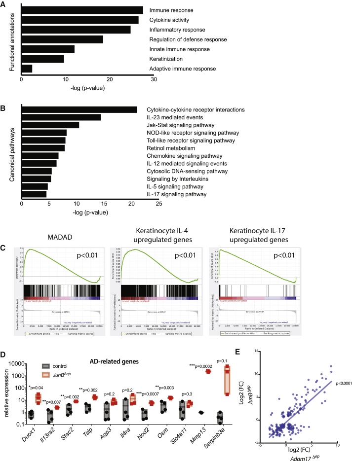
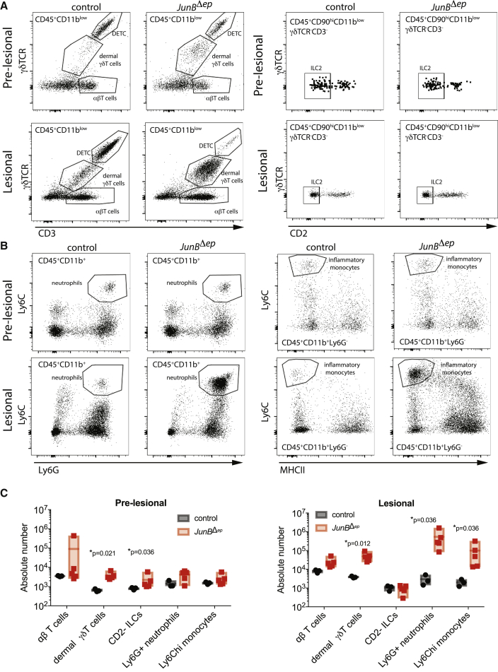
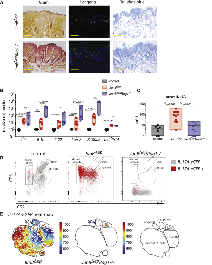
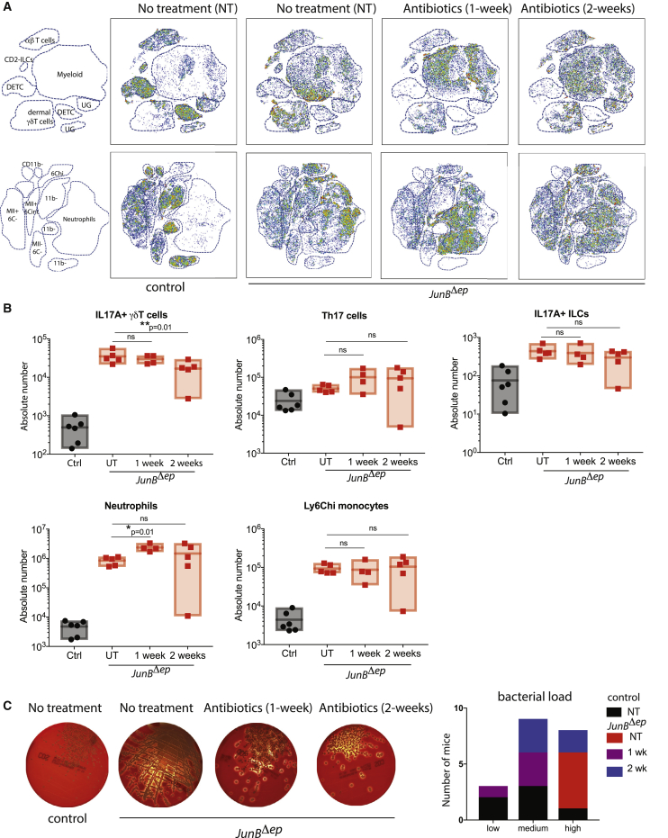
Atopic dermatitis (AD) is a multi-factorial skin disease with a complex inflammatory signature including type 2 and type 17 activation. Although colonization by S. aureus is common in AD, the mechanisms rendering an organism prone to dysbiosis, and the role of IL-17A in the control of S. aureus-induced skin inflammation, are not well understood. Here, we show several pathological aspects of AD, including type 2/type 17 immune responses, elevated IgE, barrier dysfunction, pruritus, and importantly, spontaneous S. aureus colonization in JunBΔep mice, with a large transcriptomic overlap with AD. Additionally, using Rag1-/- mice, we demonstrate that adaptive immune cells are necessary for protection against S. aureus colonization. Prophylactic antibiotics, but not antibiotics after established dysbiosis, reduce IL-17A expression and skin inflammation, examined using Il17a-eGFP reporter mice. Mechanistically, keratinocytes lacking JunB exhibit higher MyD88 levels in vitro and in vivo, previously shown to regulate S. aureus colonization. In conclusion, our data identify JunB as an upstream regulator of microbiota-immune cell interactions and characterize the IL-17A response upon spontaneous dysbiosis.

Keywords: AP-1; JunB; atopic dermatitis; dysbiosis; microbiota; skin inflammation; type 2 immunity.


Conflict of interest statement

Ö.U. and B.R. are presently employees of the Novartis Institutes for Biomedical Research. Novartis did not fund the study, nor did it play any role in the study design, data collection and analysis, decision to publish, or preparation of the manuscript. All other authors declare no competing interests.

Figures

None
Graphical abstract
Figure 1
Figure 1
Spontaneous S. aureus Colonization with AD Features Is Observed in JunBΔep Mice (A) Representative images of H&E staining of skin from control and JunBΔep mice at 6–7 months of age (n > 10 per genotype). The yellow scale bar indicates 200 μm. (B) Representative images of Ym1 staining of skin from control and JunBΔep mice at 6–7 months (n = 4, 4). (C) TSLP levels in skin lysates of control and JunBΔep mice at 2 (n = 3, 4) and 6–7 months (n = 11, 12). (D) IL-33 levels in skin lysates of control and JunBΔep mice at 2 (n = 3, 4) and 6–7 months (n = 11, 12). (E) Serum IgE levels of control and JunBΔep mice at 6–7 months of age (n = 4, 4). (F) Scratch bouts of control and JunBΔep mice at 6–7 months of age (n = 4, 4). (G) Representative images of Gram staining of skin from control and JunBΔep mice at 6–7 months (n > 10 per genotype). Arrows indicate Gram-positive colonies of bacteria. The yellow scale bar indicates 200 μm. (H) Representative images of S. aureus immunofluorescence staining of skin from control and JunBΔep mice at 6–7 months (n > 10 per genotype). Arrow indicates S. aureus colonies. (I) Percentage of JunBΔep mice positive for S. aureus colonization, as assessed by IF for S. aureus. (J) Outside-in barrier assay using toluidine blue dye at embryonic day 17.5 (n = 6, 6).
Figure 2
Figure 2
Lesional Skin from JunBΔep Mice Shows Overlapping Transcriptomic Signature with Human AD, with Both Type 2 and Type 17 Immune Activation (A) Functional annotations derived from RNA sequencing (RNA-seq) analysis of skin from control (n = 3) and JunBΔep mice (n = 3). (B) Canonical pathways derived from RNA-seq analysis of skin from control (n = 3) and JunBΔep mice (n = 3). (C) Gene set enrichment analysis (GSEA) showing positive correlations with MADAD (meta-analysis-derived AD) signature, IL-4- and IL-17-responsive genes in keratinocytes. (D) Expression levels of genes implicated in AD pathology, as assessed by qPCR. (E) Correlation of transcriptomic changes of commonly expressed genes in JunBΔep mice versus Adam17Δep mice (Woodring et al., 2018).
Figure 3
Figure 3
T Cells and ILC2s Infiltrate Skin before the Onset of Overt Inflammation (A and B) Representative flow cytometry plots showing (A) T cell subsets, ILCs, and (B) neutrophils, inflammatory monocytes of pre-lesional (3 months of age; n = 3, 5) and lesional (6 months of age; n = 3, 5) skin in control and JunBΔep mice. (C) Absolute numbers of immune cell subsets as analyzed using flow cytometry of pre-lesional (3 months of age) and lesional (6 months of age) skin in control and JunBΔep mice. Markers defining each cell subset are as follows: αβT cells: CD45+CD11b−/ lowCD3+γδTCR; γδT cells: CD45+CD11b−/lowCD3+gdTCRint; DETC: CD45+CD11b−/lowCD3higdTCRhi; CD2− ILCs: CD45+CD90hiCD11bCD3γδTCRCD2; Ly6G+ neutrophils: CD45+CD11bhiCD3gdTCRLy6Ghi; Ly6Chi monocytes: CD45+CD11b+CD3gdTCRLy6GLy6ChiMHCII−.
Figure 4
Figure 4
Skin Inflammation and S. aureus Colonization Are Exacerbated in the Absence of Adaptive Immunity (A) Representative images of Gram, langerin, and toluidine blue of skin in JunBΔep and JunBΔepRag1–/– mice at 3 months of age (n = 6, 6). The yellow scale bar indicates 200 μm. (B) Relative expression levels of pro-inflammatory cytokines in the skin of JunBΔep and JunBΔepRag1–/– mice at 3 months of age compared with controls (n = 3, 5, 6). (C) Serum IL-17A levels in JunBΔep and JunBΔepRag1–/– mice at 3 months of age compared with controls (n = 7, 11, 9). (D) Representative flow cytometry plots with GFP+ cells overlaid in the skin of JunBΔep and JunBΔepRag1–/– mice at 4 months of age compared with controls. The cells shown are gated on CD45+CD90+CD11b. (E) Heatmaps showing IL-17A expression on t-SNE plots from total IL-17A-eGFP+ cells. The map on the right is created using conventional gating, as described in Figure 2.
Figure 5
Figure 5
Prophylactic Antibiotics Reduce Skin Inflammation (A) Representative H&E and Gram staining images of control and JunBΔep mice with or without prophylactic antibiotics (UT: n = 4, 3; Ab: n = 2, 5). The blue scale bar indicates 200 μm. (B) Blood agar plates from swabs from control and JunBΔep skin with or without prophylactic antibiotic treatment and quantification of total bacterial load on skin. (C) Number of infiltrating immune cells into the skin of control and JunBΔep mice with or without prophylactic antibiotics as analyzed using flow cytometry. Gating strategy as described in Figure 2. (D) Representative flow cytometry images of IL-17A expressing T cells of control and JunBΔep mice with or without prophylactic antibiotics. (E) Representative flow cytometry images of IL-17A expressing ILCs of control and JunBΔep mice with or without prophylactic antibiotics.
Figure 6
Figure 6
Antibiotic Treatment Post-development of Skin Inflammation Does Not Provide Therapeutic Benefit (A) t-SNE plots showing the distribution of lymphoid (top) and myeloid (bottom) cells in JunBΔep mice with or without 1 and 2 weeks of antibiotics compared with controls as analyzed using flow cytometry. Maps on the left were made using conventional gating as described in Figure 2. UG, ungated (control [Ctrl], n = 6; JunBΔep UT, n = 5; 1 week, n = 4; 2 weeks, n = 5). (B) Number of infiltrating immune cells into the skin of JunBΔep mice with or without 1 and 2 weeks of antibiotics compared with controls as analyzed by conventional flow cytometry. (C) Blood agar plates from swabs from JunBΔep mice with or without 1 and 2 weeks of antibiotics compared with controls as analyzed using flow cytometry and quantification of total bacterial load on lesional skin.
Figure 7
Figure 7
MyD88 Signaling Is Upregulated in JunBΔep Skin, and MyD88 Is Upregulated in JunB-Deficient Keratinocytes (A) Relative expression levels of il1f6 (Il36α), Il1f8 (Il36β), and Il1f9 (Il36γ) in the lesional skin of JunBΔep mice compared with controls (n = 5, 6). (B) Schematic diagram depicting the experimental design for (C) and (E). (C) Relative expression levels of chemokines and alarmins upon JunB deletion in JunBlox/lox keratinocytes using adenoviruses expressing Cre recombinase with or without supernatant (S/N) derived from swabs from lesional skin of JunBΔep mice (n = 3, 3, repeated three separate times). (D) MyD88 and its downstream genes are significantly regulated in the transcriptome of JunBΔep skin. (E) Western blot using control or JunBΔep keratinocytes upon S/N administration (as described in B) for indicated time points. MyD88 levels and vinculin (as loading control) are shown. (F) Representative images of MyD88 levels by immunohistochemistry in skin from control or JunBΔep mice. Arrows indicate examples of MyD88-positive keratinocytes. The yellow scale bar indicates 100 μm. (G) Upon loss of keratinocyte expression of JunB, MyD88 levels are increased. Together with microbial products, increased MyD88 levels lead to increased alarmin/chemokine expression, starting a cascade of inflammatory response, including increased IL-17A production from Th17, γδT cells, and ILC3s. In a Rag1−/− background, IL-17A is expressed by ILC3s, but skin inflammation is exacerbated in the absence of the adaptive immune system.

Similar articles

Cited by

References

    1. Bieber T. Atopic dermatitis. N. Engl J. Med. 2008;358:1483–1494. - PubMed
    1. Bin L., Leung D.Y.M. Genetic and epigenetic studies of atopic dermatitis. Allergy Asthma Clin. Immunol. 2016;12:52. - PMC - PubMed
    1. Bird L. Asthma and allergy: TSLP: key role in allergic responses confirmed. Nat. Rev. Immunol. 2005;5:747.
    1. Bitschar K., Wolz C., Krismer B., Peschel A., Schittek B. Keratinocytes as sensors and central players in the immune defense against Staphylococcus aureus in the skin. J. Dermatolog. Sci. 2017;87:215–220. - PubMed
    1. Brunner P.M., Israel A., Zhang N., Leonard A., Wen H.C., Huynh T., Tran G., Lyon S., Rodriguez G., Immaneni S. Early-onset pediatric atopic dermatitis is characterized by TH2/TH17/TH22-centered inflammation and lipid alterations. J. Allergy Clin. Immunol. 2018;141:2094–2106. - PubMed

Publication types

MeSH terms