Prevalence and Clinicopathological Significance of MET Overexpression and Gene Amplification in Patients with Gallbladder Carcinoma
- PMID: 31645095
- PMCID: PMC7176974
- DOI: 10.4143/crt.2019.370
Prevalence and Clinicopathological Significance of MET Overexpression and Gene Amplification in Patients with Gallbladder Carcinoma
Abstract
Purpose: Mesenchymal epithelial transition (MET) is a proto-oncogene that encodes a heterodimeric transmembrane receptor tyrosine kinase for the hepatocyte growth factor. Aberrant MET signaling has been described in several solid tumors-especially non-small cell lung cancer- and is associated with tumor progression and adverse prognosis. As MET is a potential therapeutic target, information regarding its prevalence and clinicopathological relevance is crucial.
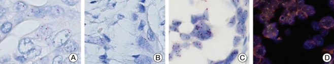
Materials and methods: We investigated MET expression and gene amplification in 113 gallbladder cancers using tissue microarray. Immunohistochemistry was used to evaluate MET overexpression, and silver/fluorescence in situ hybridization (ISH) was used to assess gene copy number.
Results: MET overexpression was found in 37 cases of gallbladder carcinoma (39.8%), and gene amplification was present in 17 cases (18.3%). MET protein expression did not correlate with MET amplification. MET amplification was significantly associated with aggressive clinicopathological features, including high histological grade, advanced pT category, lymph node metastasis, and advanced American Joint Committee on Cancer stage. There was no significant correlation between any clinicopathological factors and MET overexpression. No difference in survival was found with respect to MET overexpression and amplification status.
Conclusion: Our data suggested that MET might be a potential therapeutic target for targeted therapy in gallbladder cancer, because MET amplification was found in a subset of tumors associated with adverse prognostic factors. Detection of MET amplification by ISH might be a useful predictive biomarker test for anti-MET therapy.
Keywords: Gallbladder cancer; Gene amplification; Gene copy number; Immunohistochemistry; In situ hybridization; MET.
Conflict of interest statement
Conflict of interest relevant to this article was not reported.
Figures


References
-
- Li M, Zhang Z, Li X, Ye J, Wu X, Tan Z, et al. Whole-exome and targeted gene sequencing of gallbladder carcinoma identifies recurrent mutations in the ErbB pathway. Nat Genet. 2014;46:872–6. - PubMed
MeSH terms
Substances
Grants and funding
LinkOut - more resources
Full Text Sources
Medical
Research Materials
Miscellaneous