. 2019 Dec;47(8):913-953.
doi: 10.1177/0192623319876929. Epub 2019 Oct 23.

Proceedings of the 2019 National Toxicology Program Satellite Symposium

Affiliations

Proceedings of the 2019 National Toxicology Program Satellite Symposium

Susan A Elmore et al. Toxicol Pathol. 2019 Dec.

Abstract

The 2019 annual National Toxicology Program Satellite Symposium, entitled "Pathology Potpourri," was held in Raleigh, North Carolina, at the Society of Toxicologic Pathology's 38th annual meeting. The goal of this symposium was to present and discuss challenging diagnostic pathology and/or nomenclature issues. This article presents summaries of the speakers' talks along with select images that were used by the audience for voting and discussion. Various lesions and topics covered during the symposium included aging mouse lesions from various strains, as well as the following lesions from various rat strains: rete testis sperm granuloma/fibrosis, ovarian cystadenocarcinoma, retro-orbital schwannoma, periductal cholangiofibrosis of the liver and pancreas, pars distalis hypertrophy, chronic progressive nephropathy, and renal tubule regeneration. Other cases included polyovular follicles in young beagle dogs and a fungal blood smear contaminant. One series of cases challenged the audience to consider how immunohistochemistry may improve the diagnosis of some tumors. Interesting retinal lesions from a rhesus macaque emphasized the difficulty in determining the etiology of any particular retinal lesion due to the retina's similar response to vascular injury. Finally, a series of lesions from the International Harmonization of Nomenclature and Diagnostic Criteria Non-Rodent Fish Working Group were presented.

Keywords: INHAND; NTP satellite symposium; aging mouse lesions; ovarian cystadenoma; periductal cholangiofibrosis; polyovular follicles; sperm granuloma.


Conflict of interest statement

Declaration of Conflicting Interests Statement

The author(s) declared no potential, real, or perceived conflicts of interest with respect to the research, authorship, and/or publication of this article.

Figures
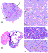
Figure 1.
Figure 1.
Adrenal gland from an adult C3H/HeJ male mouse (A-C) and seminal vesicle from an adult NOD.B10Sn-H2b/J male mouse, both from the National Toxicology Program Aging Mouse Study (D-F). (A) The adrenal gland is diffusely enlarged and infiltrated by a non-encapsulated neoplasm that is effacing and replacing the normal tissue architecture. Note that the neoplasm appears to be arising from the subcapsular surface (B) Neoplastic cells arranged in nests and packets are characterized by basophilic cytoplasm and fusiform (type A) morphology (“arrow B” region in Figure A). (C) In a different area of the neoplasm (“arrow C” region in Figure A), neoplastic cells are polygonal (type B) with abundant eosinophilic cytoplasm and are arranged in islands or sheets. (D) The seminal vesicle is markedly dilated and infiltrated by an unencapsulated neoplasm. (E) In some areas (arrow “E” in figure D), the neoplasm appears epithelial in origin, with neoplastic cells arranged in nests, islands and trabeculae. (F) In other areas of the neoplasm (arrow “F” in Figure D), the neoplastic cells are more elongated and spindle-shaped with pale, eosinophilic cytoplasm and are arranged in bundles and streams.
Figure 2.
Figure 2.
Pituitary (A-C) and pancreatic (D) lesions from adult 129S1/Svlmj mice from the National Toxicology Program Aging Mouse Study. (A) Photomicrograph of the pituitary gland showing a poorly circumscribed mass (arrows) expanding the pars intermedia and compressing the adjacent pars distalis in more than one quadrant (arrowheads). (B) Photomicrograph of the pars intermedia showing characteristic nests of pleomorphic cells (center). (C) Photomicrograph showing neoplastic pars intermedia cells extending into the pars nervosa (arrows). (D) Photomicrograph of the pancreas. Inset shows higher magnification image of the peri-insular acinar hypertrophy characterized by intracytoplasmic accumulation of abundant zymogen granules within acinar cells surrounding the islets of Langerhans.
Figure 3.
Figure 3.
Bone marrow lesions (A-D) and lymph node lesions (E-F) from adult female B6C3F1/J mice from the National Toxicology Program Aging Mouse Study (Case 5). Increased adipocytes in the bone marrow (A-B) were only noted in females from this particular strain of aged mouse. Note that hematopoietic elements of the bone marrow, particularly in the metasphyseal region, are almost entirely replaced by abundant numbers of adipocytes, as compared to the normal bone marrow (C-D). Infiltration of adipocytes in the medullary sinuses of the mesenteric (E) and mandibular (F) lymph nodes in adult female B6C3F1/J mice from the National Toxicology Program Aging Mouse Study. This finding occasionally accompanied incidences of increased adipocytes in the bone marrow in this strain. Chronic active inflammation of the epicardium from an adult female NZO/HILtJ mouse from the National Toxicology Program Aging Mouse Study (G-J) (Case 6) and lung lesions from Cast/EiJ mice (K-L) (Case 7) from the National Toxicology Program Aging Mouse Study. In the NZO/HILtJ mouse strain, inflammation was only present in females and was always associated with mediastinal lymphoma (G). In the heart (H-J), circumferential thickening of the epicardium was characterized by infiltration of lymphocytes, plasma cells, lesser numbers of neutrophils, fibrosis, and mesothelial hyperplasia. Eosinophilic crystals (K) were a common finding in both males and females from the Cast/EiJ strain of mouse and were typically not associated with any additional pathology in the lung, except for rare diagnoses of proteinosis (L).
Figure 3.
Figure 3.
Bone marrow lesions (A-D) and lymph node lesions (E-F) from adult female B6C3F1/J mice from the National Toxicology Program Aging Mouse Study (Case 5). Increased adipocytes in the bone marrow (A-B) were only noted in females from this particular strain of aged mouse. Note that hematopoietic elements of the bone marrow, particularly in the metasphyseal region, are almost entirely replaced by abundant numbers of adipocytes, as compared to the normal bone marrow (C-D). Infiltration of adipocytes in the medullary sinuses of the mesenteric (E) and mandibular (F) lymph nodes in adult female B6C3F1/J mice from the National Toxicology Program Aging Mouse Study. This finding occasionally accompanied incidences of increased adipocytes in the bone marrow in this strain. Chronic active inflammation of the epicardium from an adult female NZO/HILtJ mouse from the National Toxicology Program Aging Mouse Study (G-J) (Case 6) and lung lesions from Cast/EiJ mice (K-L) (Case 7) from the National Toxicology Program Aging Mouse Study. In the NZO/HILtJ mouse strain, inflammation was only present in females and was always associated with mediastinal lymphoma (G). In the heart (H-J), circumferential thickening of the epicardium was characterized by infiltration of lymphocytes, plasma cells, lesser numbers of neutrophils, fibrosis, and mesothelial hyperplasia. Eosinophilic crystals (K) were a common finding in both males and females from the Cast/EiJ strain of mouse and were typically not associated with any additional pathology in the lung, except for rare diagnoses of proteinosis (L).
Figure 4.
Figure 4.
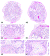
Rete testis lesions in Hsd:Sprague Dawley SD rats (A-F) from a National Toxicology Program perinatal chronic bioassay. All were exposed in utero to a phthalate. (A, C, E) The rete testis (arrow) is expanded by a sperm granuloma and fibrosis. (E) Numerous spermatids in the rete testis are surrounded by a rim of epithelioid and multinucleated macrophages. Within the granuloma there are smaller aggregates of macrophages and cholesterol clefts. (B, D, F) Fibrosis in the rete testis (arrow). (F) The rete testis is expanded by fibrosis with smaller aggregates of foamy macrophages, suggesting progression from a sperm granuloma.
Figure 5.
Figure 5.
Ovarian tumor (A-I) from a control nearly 20-week-old Sprague Dawley rat. A low magnification image (A) of the cauliflower-like tumor with an exophytic growth pattern on the surface of one of the ovaries. Higher magnification (B) shows the mass is derived from the ovarian surface epithelium (OSE), which is relatively well demarcated from the adjacent areas. The mass is composed of solid or papillary structures of pleomorphic, cuboidal to low columnar cells that are occasionally ciliated. Higher magnification (C) shows the neutrophilic infiltration and neovascularization in some solid regions of the tumor with a delicate stromal compartment. (D-F) Immunohistochemical characterization of the tumor. Neoplastic cells are positive for vimentin (~60%) (D) and CK-7 (~10%) (E) and negative for CK-WS (F). (G-I) Immunohistochemical characterization of the OSE in the SD rat. The normal OSE stains positively for vimentin (G), CK-7 (H) and CK-WS (I). Immunohistochemical characterization of the ovarian surface epithelium (OSE) in the SD rat (G-I). The normal OSE stains positively for vimentin (G), CK-7 (H) and CK-WS (I).
Figure 5.
Figure 5.
Ovarian tumor (A-I) from a control nearly 20-week-old Sprague Dawley rat. A low magnification image (A) of the cauliflower-like tumor with an exophytic growth pattern on the surface of one of the ovaries. Higher magnification (B) shows the mass is derived from the ovarian surface epithelium (OSE), which is relatively well demarcated from the adjacent areas. The mass is composed of solid or papillary structures of pleomorphic, cuboidal to low columnar cells that are occasionally ciliated. Higher magnification (C) shows the neutrophilic infiltration and neovascularization in some solid regions of the tumor with a delicate stromal compartment. (D-F) Immunohistochemical characterization of the tumor. Neoplastic cells are positive for vimentin (~60%) (D) and CK-7 (~10%) (E) and negative for CK-WS (F). (G-I) Immunohistochemical characterization of the OSE in the SD rat. The normal OSE stains positively for vimentin (G), CK-7 (H) and CK-WS (I). Immunohistochemical characterization of the ovarian surface epithelium (OSE) in the SD rat (G-I). The normal OSE stains positively for vimentin (G), CK-7 (H) and CK-WS (I).
Figure 6.
Figure 6.
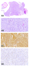
Retroorbital Schwannoma (A-D) from a 2-year-old male F344/N rat from a National Toxicology Program chronic bioassay that was treated with 1,2-Epoxybutane via inhalation (high dose group). (A) The unencapsulated, multinodular mass expands the retroorbital region. (B) Bundles and streams of spindle cells with indistinct borders dissect a fibrovascular stroma. Cells have low amounts of eosinophilic, fibrillar cytoplasm and ovoid to spindle nuclei with coarsely granular chromatin and indistinct nucleoli. Cellular and nuclear pleomorphism is low, and mitoses are not observed. There is robust diffuse nuclear immunoreactivity for S-100 (C) and diffuse nuclear immunoreactivity for Sox-10 (D). (E-F) There are numerous, multifocal, small to large pulmonary tumors similar to the retroorbital mass. There is robust nuclear immunoreactivity for S-100 (G) and lack of immunoreactivity for EMA (H).
Figure 6.
Figure 6.
Retroorbital Schwannoma (A-D) from a 2-year-old male F344/N rat from a National Toxicology Program chronic bioassay that was treated with 1,2-Epoxybutane via inhalation (high dose group). (A) The unencapsulated, multinodular mass expands the retroorbital region. (B) Bundles and streams of spindle cells with indistinct borders dissect a fibrovascular stroma. Cells have low amounts of eosinophilic, fibrillar cytoplasm and ovoid to spindle nuclei with coarsely granular chromatin and indistinct nucleoli. Cellular and nuclear pleomorphism is low, and mitoses are not observed. There is robust diffuse nuclear immunoreactivity for S-100 (C) and diffuse nuclear immunoreactivity for Sox-10 (D). (E-F) There are numerous, multifocal, small to large pulmonary tumors similar to the retroorbital mass. There is robust nuclear immunoreactivity for S-100 (G) and lack of immunoreactivity for EMA (H).
Figure 7.
Figure 7.
Examples of spontaneous periductal cholangiofibrosis (A-D) in 2-year-old Hsd:Sprague Dawley SD rats rats from a National Toxicology Program chronic bioassay. Spontaneous periductal cholangiofibrosis in the liver of a female rat as was presented in Case 1 (A-B). Low magnification (A) shows a focal, nodular, extracapsular mass immediately adjacent to the main hepatic bile duct. At higher magnification (B) the mass is characterized by dilated and/or tortuous bile ductule-like structures with mucous-like intraluminal material, cuboidal to columnar lining epithelium with intestinal metaplasia, and abundant supporting fibrous stroma with variable inflammatory cell infiltrates. Spontaneous periductal cholangiofibrosis in the pancreas of a female rat as presented in Case 2 (C-D). Low magnification (C) shows a focal finding in the parenchyma of the pancreas. Note the close approximation to the common bile duct. Higher magnification (D) shows the mass is characterized by dilated and/or tortuous bile ductule-like structures with mucous-like intraluminal material, cuboidal to columnar lining epithelium with intestinal metaplasia, and abundant supporting fibrous stroma with variable inflammatory cell infiltrates.
Figure 8.
Figure 8.
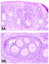
Polyovular follicles in Beagle dogs (A-B). (A) shows an ovary of a young untreated Beagle dog with multiple polyovular follicles containing up to 6 intact ova. (B) is a high magnification of a polyovular follicle from a second dog showing that each ovum is surrounded by granulosa cells.
Figure 9.
Figure 9.
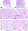
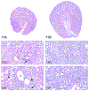
Hypertrophy of pars distalis endocrine cells from an adult male Hsd:Sprague Dawley SD rat from a 2-year National Toxicology Program perinatal chronic bioassay presented as Case 1 (A-F). At low magnification (B - original objective 4x), there are numerous pale-staining cells scattered throughout the pars distalis in the pituitary gland from a male rat exposed to the highest dose of a phthalate, when compared to a control male rat (A). At high magnification (D - original magnification 40x), the pale-staining cells are enlarged with expanded cytoplasm, some are finely vacuolated, some have large vacuoles that do not peripheralize the nuclei (arrowheads), and some have a single large vacuole that peripheralizes the nucleus (“signet ring” cells; arrow), when compared to a control (C). The treated animal with pars distalis hypertrophy in B and D had testicular lesions, including atrophy and Leydig cell hyperplasia (F, compare to control rat in E). Hypertrophy of pars distalis endocrine cells from an adult male Fischer 344/N rat from a 13-week National Toxicology Program study presented as Case 2 (G-L). The rat treated with ethylene thiourea (H, J) had enlarged endocrine cells scattered throughout the pars distalis (compared to a corresponding control rat G, I). These cells had pale cytoplasm that was frequently finely vacuolated (H, J). When compared to control animals at the same magnification, there were increased incidences of diffuse follicular cell hyperplasia in the thyroid glands of ethylene thiourea-treated rats (L, compare to control rat in K).
Figure 9.
Figure 9.
Hypertrophy of pars distalis endocrine cells from an adult male Hsd:Sprague Dawley SD rat from a 2-year National Toxicology Program perinatal chronic bioassay presented as Case 1 (A-F). At low magnification (B - original objective 4x), there are numerous pale-staining cells scattered throughout the pars distalis in the pituitary gland from a male rat exposed to the highest dose of a phthalate, when compared to a control male rat (A). At high magnification (D - original magnification 40x), the pale-staining cells are enlarged with expanded cytoplasm, some are finely vacuolated, some have large vacuoles that do not peripheralize the nuclei (arrowheads), and some have a single large vacuole that peripheralizes the nucleus (“signet ring” cells; arrow), when compared to a control (C). The treated animal with pars distalis hypertrophy in B and D had testicular lesions, including atrophy and Leydig cell hyperplasia (F, compare to control rat in E). Hypertrophy of pars distalis endocrine cells from an adult male Fischer 344/N rat from a 13-week National Toxicology Program study presented as Case 2 (G-L). The rat treated with ethylene thiourea (H, J) had enlarged endocrine cells scattered throughout the pars distalis (compared to a corresponding control rat G, I). These cells had pale cytoplasm that was frequently finely vacuolated (H, J). When compared to control animals at the same magnification, there were increased incidences of diffuse follicular cell hyperplasia in the thyroid glands of ethylene thiourea-treated rats (L, compare to control rat in K).
Figure 10.
Figure 10.
An example of a fungal spore that is an incidental contamination artifact of peripheral blood smears that can occur with air or staining solutions. This smear shows a single, encapsulated (arrow), coiled structure consistent with a fungal spore (i.e., the conidia of a helicosporous hyphomycete).
Figure 11.
Figure 11.
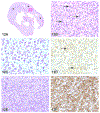
Selected renal lesions (A-F) from a two-year National Toxicology Program study in B6C3F1 mouse presented as Cases 1 and 2. Case 1, Chronic Progressive Nephropathy (CPN) (A,C,E). (A) End-stage CPN characterized by a shrunken, fibrotic kidney with an undulating capsular surface. (C) CPN features include hyaline casts (asterisks), interstitial fibrosis, glomerulosclerosis, tubular hypertrophy and hyperplasia, as well as inflammatory infiltrates (arrow) and pigment. (E) Higher magnification of areas of interstitial fibrosis and glomerulosclerosis (arrow), tubular dilation (asterisk), tubular degeneration and hypertrophy (arrowhead). Case 2, Renal Tubule – Regeneration (B, D, F). (B) Renal cortex is diffusely highlighted by increased basophilia. (D) Within the cortex, the interstitium is compressed by wall-to-wall basophilic tubules. (F) Higher magnification reveals additional features of karyomegaly, hypertrophy and hyperplasia as well as regeneration and increased mitoses. Fibrotic changes are notably absent as well as prominent thickened membranes.
Figure 12.
Figure 12.
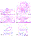
Mast cell tumor in a kidney of a female F344/N rat (A-F) from a National Toxicology Program two-year carcinogenicity bioassay presented as Case 1. H&E stained section of a kidney (A) with a neoplasm (asterisks) effacing the normal architecture of the kidney. Magnified view (B) of the neoplasm shown in (A). The neoplasm has effaced the normal architecture of the kidney and is composed of round to polygonal cells containing cytoplasmic granules (arrows). Toluidine blue stained section (C) demonstrates metachromatic granules in the neoplastic cells. Immunohistochemistry with a CKIT antibody (D) demonstrates membranous staining on the neoplastic cells (arrows). Diaminobenzidine (DAB) chromogen and hematoxylin counterstain. Periodic acid–Schiff (PAS) (E) staining shows no PAS-positive granules in the neoplastic cells. Neoplastic cells are negative for IBA1 (F), a macrophage marker. However, there are large numbers of IBA1-positive tumor associated macrophages. DAB chromogen and hematoxylin counterstain. Natural Killer (NK) cell tumor from a female Wistar Han rat (G-L) from a National Toxicology Program two-year carcinogenesis bioassay presented as Case 2. H&E stained section (G) of a subcutaneous mass showing a very large neoplasm (asterisk). Magnified view (H) of the neoplasm shown in (G) shows large round neoplastic cells with granular cytoplasm. PAS stained section (I) shows PAS-positive cytoplasmic pink granules in the neoplastic cells. The neoplastic cells are diffusely positive for cytoplasmic perforin (J) and CD3 (K). Compare the cytoplasmic staining for CD3 in the inset associated with (K) to the membranous CD3 staining in the inset associated with Figure R. The neoplastic cells in (L) are negative for IBA1 but the tumor associated macrophages are positive (arrows). Diaminobenzidine (DAB) chromogen and hematoxylin counterstain (J,K,L). Histiocytic sarcoma from a male F344/N rat (M-R) from a National Toxicology Program two-year carcinogenesis bioassay presented as Case 3. Section of a pancreatic lymph node (M) which is enlarged due to the presence of a neoplasm. Magnified view (N) of the neoplasm demonstrates variably sized round to polygonal cells. Some of the cells show granules in their cytoplasm (arrows). The remnant lymphocytes are admixed with the neoplastic cells. The neoplastic cells contain no metachromatic granules in the toluidine blue stained section (O) and are negative for perforin (P) and positive for IBA1 (Q). Most of the unstained cells in between the neoplastic cells in (Q) are remnant lymphocytes which stain for CD3 (R). The inset in (R) highlights the CD3 staining in the membrane as opposed to the cytoplasmic staining seen in the NK cell tumor (K). Diaminobenzidine (DAB) chromogen and hematoxylin counterstain (Q,R). A simplified algorithm (S) for diagnosing a tumor with cytoplasmic granules in the neoplastic cells.
Figure 12.
Figure 12.
Mast cell tumor in a kidney of a female F344/N rat (A-F) from a National Toxicology Program two-year carcinogenicity bioassay presented as Case 1. H&E stained section of a kidney (A) with a neoplasm (asterisks) effacing the normal architecture of the kidney. Magnified view (B) of the neoplasm shown in (A). The neoplasm has effaced the normal architecture of the kidney and is composed of round to polygonal cells containing cytoplasmic granules (arrows). Toluidine blue stained section (C) demonstrates metachromatic granules in the neoplastic cells. Immunohistochemistry with a CKIT antibody (D) demonstrates membranous staining on the neoplastic cells (arrows). Diaminobenzidine (DAB) chromogen and hematoxylin counterstain. Periodic acid–Schiff (PAS) (E) staining shows no PAS-positive granules in the neoplastic cells. Neoplastic cells are negative for IBA1 (F), a macrophage marker. However, there are large numbers of IBA1-positive tumor associated macrophages. DAB chromogen and hematoxylin counterstain. Natural Killer (NK) cell tumor from a female Wistar Han rat (G-L) from a National Toxicology Program two-year carcinogenesis bioassay presented as Case 2. H&E stained section (G) of a subcutaneous mass showing a very large neoplasm (asterisk). Magnified view (H) of the neoplasm shown in (G) shows large round neoplastic cells with granular cytoplasm. PAS stained section (I) shows PAS-positive cytoplasmic pink granules in the neoplastic cells. The neoplastic cells are diffusely positive for cytoplasmic perforin (J) and CD3 (K). Compare the cytoplasmic staining for CD3 in the inset associated with (K) to the membranous CD3 staining in the inset associated with Figure R. The neoplastic cells in (L) are negative for IBA1 but the tumor associated macrophages are positive (arrows). Diaminobenzidine (DAB) chromogen and hematoxylin counterstain (J,K,L). Histiocytic sarcoma from a male F344/N rat (M-R) from a National Toxicology Program two-year carcinogenesis bioassay presented as Case 3. Section of a pancreatic lymph node (M) which is enlarged due to the presence of a neoplasm. Magnified view (N) of the neoplasm demonstrates variably sized round to polygonal cells. Some of the cells show granules in their cytoplasm (arrows). The remnant lymphocytes are admixed with the neoplastic cells. The neoplastic cells contain no metachromatic granules in the toluidine blue stained section (O) and are negative for perforin (P) and positive for IBA1 (Q). Most of the unstained cells in between the neoplastic cells in (Q) are remnant lymphocytes which stain for CD3 (R). The inset in (R) highlights the CD3 staining in the membrane as opposed to the cytoplasmic staining seen in the NK cell tumor (K). Diaminobenzidine (DAB) chromogen and hematoxylin counterstain (Q,R). A simplified algorithm (S) for diagnosing a tumor with cytoplasmic granules in the neoplastic cells.
Figure 13.
Figure 13.
Retinal degeneration from a 10-year-old male rhesus macaque (A-G) that received 8.05 Gray whole-body irradiation and had a history of type II diabetes mellitus and systemic hypertension. The sections are stained with H&E (A-D) and perioidic acid-Schiff (PAS; E and F). (A-B) There is diffuse retinal degeneration with marked cystoid edema of the nuclear layers, and the presumed remains of a damaged vessel surrounded by eosinophilic debris in the nerve fiber layer. At higher magnification (C), the destroyed vessel is seen as accumulations of eosinophilic debris with no discernible architecture and a mild gliosis. Another region of the nerve fiber layer (D), shows an accumulation of eosinophilic bodies, interpreted as cytoid bodies and are the result of axonal obstruction. (E) shows a tortuous, redundant vessel with plump endothelial cells in the ganglion cell layer. When stained with PAS (E and F), basement membrane splitting of an arteriole in the nerve fiber layer can be appreciated, as well as a damaged capillary leaking PAS positive material.
Figure 14.
Figure 14.
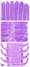
Examples(A-Q) of morphologic findings that might be encountered during evaluations of toxicological studies that utilize fish models. Low magnification (A) of tilapia (Oreochromis spp.) gills from Case 1. High magnification (B) of tilapia gills to further demonstrate diagnoses of diffuse lamellar epithelial hyperplasia, diffuse lamellar fusion, and mononuclear cell infiltrates. High magnification (C) of comparatively normal (actually mildly affected) tilapia gill from the same study, included for comparison. Low magnification (D) view of ovary from a female fathead minnow (Pimephales promelas) from Case 2. High magnification (E) view of ovary to demonstrate granulomatous inflammation, characterized by sheets of epithelioid cells in this case. Granulomatous inflammation in fish can resemble dysplastic or neoplastic squamous epithelium. Another high magnification (F) of the same fathead minnow ovary. The ring of irregular dense pink material is the degenerating zona pellucida of an oocyte (compare with normal oocyte in H). The ooplasm is filled with microsporidia spores. Oil magnification (G) of microsporidia, which have the typical “bedroom slipper” appearance. Normal cortical alveolar stage oocyte (H) in the ovary of a fathead minnow, included for comparison. Here, the ooplasm is filled with cortical alveoli, which are yolk precursor granules. Microsporidia spores (I) as viewed using polarized light. Not all developmental stages are refractile, thus polarized light does not always detect the organisms. H&E, polarized. Low magnification (J) of liver from an adult female white sucker (Catostomus commersonii) from Case 3. Note the scalloped edge of the mass-like lesion, and slight peripheral compression of the surrounding liver. Medium magnification (K) of white sucker liver. The lesion occupies the approximate upper half of the image, and the unaffected liver is in the lower half. In this area, finger-like projections of the lesion encircle thick cords of viable-appearing hepatocytes. Medium magnification (L) of white sucker liver. Area from the center of the lesion, which is characterized by streaming cells and whorls reminiscent of a perivascular or nerve sheath tumor. At high magnification (M), resemblance to hemangiopericytoma or nerve sheath tumor vanishes. Here it is evident that the whorls are formed by plump cells that have eccentric nuclei, marginated and clumped chromatin, and faintly granular translucent cytoplasm. Another high magnification (N) image. Here the plump cells either have eosinophilic granular cytoplasm, or relatively clear cytoplasm with fine spicules. The latter type are consistent with mature rodlet cells. It is proposed that the cells with eosinophilic granular cytoplasm are rodlet cell precursors, while the cells in (M) appear to be an intermediate stage. Periodic acid Schiff (PAS) staining (O) highlights the rodlet cell spicules, granular cytoplasm of the precursor cells, membranous extensions of the tumor cell cytoplasm, and possibly extracellular rodlet-like material.. Masson’s trichrome staining (P) illustrates the relative absence of collagen within the lesion, while the wall of a blood vessel serves as an internal control. The lack of fibrosis further suggests that this is not a chronic inflammatory lesion. Transmission electron micrograph (Q) demonstrates a rodlet cell (mid-left) and a putative precursor cell with granular cytoplasm (lower right). Note the presence of extracellular rodlet-like material near the top of the image (arrows).
Figure 14.
Figure 14.
Examples(A-Q) of morphologic findings that might be encountered during evaluations of toxicological studies that utilize fish models. Low magnification (A) of tilapia (Oreochromis spp.) gills from Case 1. High magnification (B) of tilapia gills to further demonstrate diagnoses of diffuse lamellar epithelial hyperplasia, diffuse lamellar fusion, and mononuclear cell infiltrates. High magnification (C) of comparatively normal (actually mildly affected) tilapia gill from the same study, included for comparison. Low magnification (D) view of ovary from a female fathead minnow (Pimephales promelas) from Case 2. High magnification (E) view of ovary to demonstrate granulomatous inflammation, characterized by sheets of epithelioid cells in this case. Granulomatous inflammation in fish can resemble dysplastic or neoplastic squamous epithelium. Another high magnification (F) of the same fathead minnow ovary. The ring of irregular dense pink material is the degenerating zona pellucida of an oocyte (compare with normal oocyte in H). The ooplasm is filled with microsporidia spores. Oil magnification (G) of microsporidia, which have the typical “bedroom slipper” appearance. Normal cortical alveolar stage oocyte (H) in the ovary of a fathead minnow, included for comparison. Here, the ooplasm is filled with cortical alveoli, which are yolk precursor granules. Microsporidia spores (I) as viewed using polarized light. Not all developmental stages are refractile, thus polarized light does not always detect the organisms. H&E, polarized. Low magnification (J) of liver from an adult female white sucker (Catostomus commersonii) from Case 3. Note the scalloped edge of the mass-like lesion, and slight peripheral compression of the surrounding liver. Medium magnification (K) of white sucker liver. The lesion occupies the approximate upper half of the image, and the unaffected liver is in the lower half. In this area, finger-like projections of the lesion encircle thick cords of viable-appearing hepatocytes. Medium magnification (L) of white sucker liver. Area from the center of the lesion, which is characterized by streaming cells and whorls reminiscent of a perivascular or nerve sheath tumor. At high magnification (M), resemblance to hemangiopericytoma or nerve sheath tumor vanishes. Here it is evident that the whorls are formed by plump cells that have eccentric nuclei, marginated and clumped chromatin, and faintly granular translucent cytoplasm. Another high magnification (N) image. Here the plump cells either have eosinophilic granular cytoplasm, or relatively clear cytoplasm with fine spicules. The latter type are consistent with mature rodlet cells. It is proposed that the cells with eosinophilic granular cytoplasm are rodlet cell precursors, while the cells in (M) appear to be an intermediate stage. Periodic acid Schiff (PAS) staining (O) highlights the rodlet cell spicules, granular cytoplasm of the precursor cells, membranous extensions of the tumor cell cytoplasm, and possibly extracellular rodlet-like material.. Masson’s trichrome staining (P) illustrates the relative absence of collagen within the lesion, while the wall of a blood vessel serves as an internal control. The lack of fibrosis further suggests that this is not a chronic inflammatory lesion. Transmission electron micrograph (Q) demonstrates a rodlet cell (mid-left) and a putative precursor cell with granular cytoplasm (lower right). Note the presence of extracellular rodlet-like material near the top of the image (arrows).

References

    1. goRENI. https://www.goreni.org/ Accessed July 15, 2019.
    1. Brandli-Baiocco A, Balme E, Bruder M, et al. Nonproliferative and Proliferative Lesions of the Rat and Mouse Endocrine System. J Toxicol Pathol 2018;31(3 Suppl):1S–95S. - PMC - PubMed
    1. Nolte T, Brander-Weber P, Dangler C, et al. Nonproliferative and Proliferative Lesions of the Gastrointestinal Tract, Pancreas and Salivary Glands of the Rat and Mouse. J Toxicol Pathol 2016;29(1 Suppl):1S–125S. - PMC - PubMed
    1. Cora M, Travlos G. Bone Marrow—Hypocellularity. National Toxicology Program Nonneoplastic Lesion Atlas 2014; https://ntp.niehs.nih.gov/nnl/hematopoietic/bone_marrow/hypocell/index.htm Accessed July 10, 2019.
    1. Barlow NJ, McIntyre BS, Foster PM. Male reproductive tract lesions at 6, 12, and 18 months of age following in utero exposure to di(n-butyl) phthalate. Toxicol Pathol 2004;32(1):79–90. - PubMed

Publication types

LinkOut - more resources