Case Reports
. 2019 Oct 23;5(5):a004440.
doi: 10.1101/mcs.a004440. Print 2019 Oct.

Novel PDGFRB rearrangement in multifocal infantile myofibromatosis is tumorigenic and sensitive to imatinib

Affiliations
Case Reports

Novel PDGFRB rearrangement in multifocal infantile myofibromatosis is tumorigenic and sensitive to imatinib

Mohammed Hassan et al. Cold Spring Harb Mol Case Stud. .

Abstract

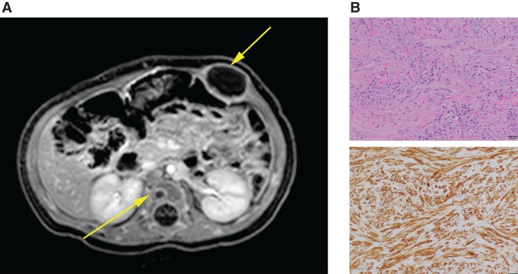
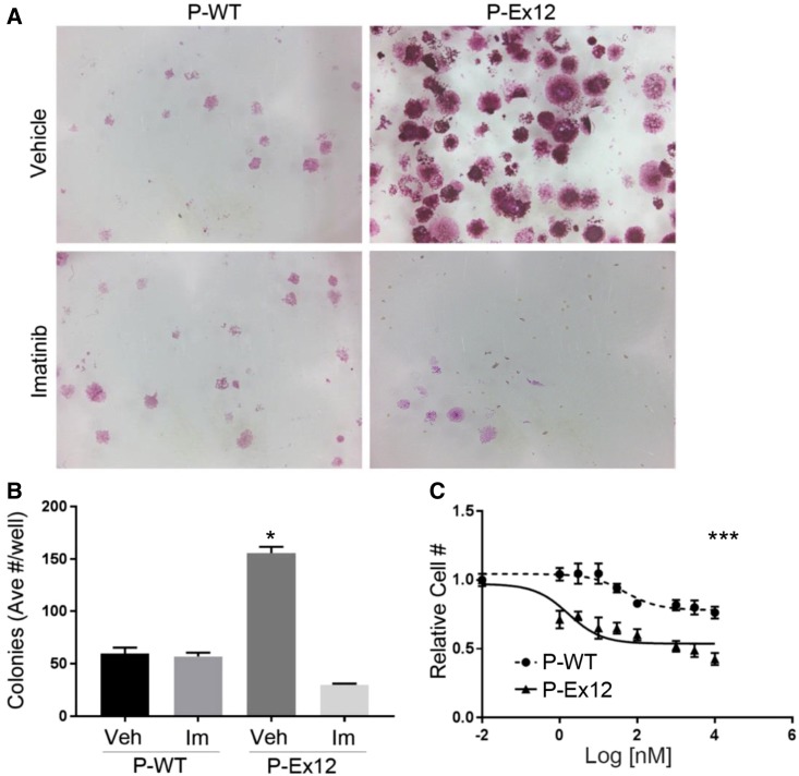
Infantile myofibromatosis (IM) is an aggressive neoplasm composed of myofibroblast-like cells in children. Although typically localized, it can also present as multifocal disease, which represents a challenge for effective treatment. IM has previously been linked to activating somatic and germline point mutations in the PDGFRβ tyrosine kinase encoded by the PDGFRB gene. Clinical panel-based targeted tumor sequencing of a tumor from a newborn with multifocal IM revealed a novel PDGFRB rearrangement, which was reported as being of unclear significance. Additional sequencing of cDNA from tumor and germline DNA confirmed a complex somatic/mosaic PDGFRB rearrangement with an apparent partial tandem duplication disrupting the juxtamembrane domain. Ectopic expression of cDNA encoding the mutant form of PDGFRB markedly enhanced cell proliferation of mouse embryo fibroblasts (MEFs) compared to wild-type PDGFRB and conferred tumor-forming capacity on nontumorigenic 10T1/2 fibroblasts. The mutated protein enhanced MAPK activation and retained sensitivity to the PDGFRβ inhibitor imatinib. Our findings reveal a new mechanism by which PDGFRB can be activated in IM, suggest that therapy with tyrosine kinase inhibitors including imatinib may be beneficial, and raise the possibility that this receptor tyrosine kinase might be altered in a similar fashion in additional cases that would similarly present annotation challenges in clinical DNA sequencing analysis pipelines.

Keywords: neoplasm of the skin.


Figures

Figure 1.
Figure 1.
Tumor analysis and classification. (A) Postcontrast T1-weighted MRI demonstrates multiple rim-enhancing masses in the abdominal wall musculature and vertebral body. (B) Histopathologic examination of a biopsy from the abdominal wall mass shows a myofibroma. (Top) Hematoxylin and eosin, 200× magnification: (bottom) smooth muscle actin immunostain, 200× magnification.
Figure 2.
Figure 2.
Sequencing and mapping of the gene rearrangement. (A) Complex PDGFRB rearrangement is detected by next-generation sequencing (NGS). The rearrangement was detected in DNA and RNA by NGS but is difficult to ascertain because of event complexity. (B) PDGFRB rearrangement predicted by NGS is confirmed by Sanger sequencing of genomic DNA (gDNA) and RNA (cDNA) from tumor. (C) PDGFRB is highly expressed in RNA-seq data from the patient's tumor (circled) compared to other cases of IM with and without activating PDGFRB point mutations. (D) Rearrangement results in the replacement of a portion of the juxtamembrane domain of PDGFRβ by a novel amino acid sequence derived from a portion of exon 15 read out-of-frame. The wild-type kinase domain is retained.
Figure 3.
Figure 3.
Functional evaluation of the rearrangement using PDGFRB-null mouse embryonic fibroblasts. (A) Diagram shows primers amplification for both wild-type PDGFRB (WT) and PDGFRB bearing the rearrangement identified in this patient's tumor (P-Ex12). (B) RT-PCR of PDGFRB–null mouse embryonic fibroblasts (MEFs) transduced with lentivirus expressing GFP only (CTL), P-WT, or P-Ex12. (C) A representative western blot of PDGFRB–null mouse embryonic fibroblasts that demonstrates expression of PDGFRB in P-WT- and P-Ex12-transduced cells and increased expression of phospho–p44/42 MAPK of cells transduced with P-Ex12 as compared to P-WT- and CTL-expressing mouse embryonic fibroblasts. (D) Photographs of colony formation assay of MEFs plated at low density show increased colony formation in P-Ex12-expressing cells compared to P-WT- and CTL-expressing MEFS. (E) Quantification of cell plate area covered by transduced MEFS shown in D.
Figure 4.
Figure 4.
P-Ex12 is tumorigenic in 10T1/2 fibroblasts. (A) RT-PCR of 10T1/2 cells transduced with P-Ex12, P-WT, and CTL lentiviral vectors. (B) Representative western blot of 10T1/2 cells confirms expression of PDGFRB and increased expression of phospho-p44/42 MAPK of cells transduced with lentivirus bearing P-Ex12 as compared to P-WT- and CTL-expressing fibroblasts. (C) Kaplan–Meier plot displays decreased overall survival of mice bearing P-Ex12-transduced 10T1/2 cells compared to both CTL- and P-WT-transduced cells. (D) Histopathological examination of processed tumor samples show signs of tumor infiltration through nerve cells, blood vessels, and muscle cells and areas of necrosis (hematoxylin and eosin, 200× and 400× magnification).
Figure 5.
Figure 5.
Analysis of tumors formed by 10T1/2 fibroblasts expressing the rearranged PDGFRB cDNA. (A) RT-PCR of tumor samples confirms expression of P-Ex12 in the tumor (Tu); plasmid (Pl) used as control in PCR. (B) Immunohistochemistry staining of tumor samples shows positive staining for Ki67 (α-Ki67) and phospho-histone H3 (α-PH3).
Figure 6.
Figure 6.
Treatment of transduced mouse embryonic fibroblasts. (A) Representative photographs of transduced PDGFRB-null mouse embryonic fibroblasts show increased colony formation with P-Ex12 expression that is blunted with imatinib treatment (1 µM). (B) Quantification of colony formation in treated and untreated P-WT- and P-Ex12-transduced MEFs. (C) MEFs transduced with P-Ex12 demonstrate increased sensitivity to imatinib as compared to P-WT MEFs.

References

    1. Abe A, Emi N, Tanimoto M, Terasaki H, Marunouchi T, Saito H. 1997. Fusion of the platelet-derived growth factor receptor β to a novel gene CEV14 in acute myelogenous leukemia after clonal evolution. Blood 90: 4271–4277. - PubMed
    1. Agaimy A, Bieg M, Michal M, Geddert H, Märkl B, Seitz J, Moskalev EA, Schlesner M, Metzler M, Hartmann A, et al. 2017. Recurrent somatic PDGFRB mutations in sporadic infantile/solitary adult myofibromas but not in angioleiomyomas and myopericytomas. Am J Surg Pathol 41: 195–203. 10.1097/PAS.0000000000000752 - DOI - PubMed
    1. Alaggio R, Barisani D, Ninfo V, Rosolen A, Coffin CM. 2008. Morphologic overlap between infantile myofibromatosis and infantile fibrosarcoma: a pitfall in diagnosis. Pediatr Dev Pathol 11: 355–362. 10.2350/07-09-0355.1 - DOI - PubMed
    1. Andrae J, Gallini R, Betsholtz C. 2008. Role of platelet-derived growth factors in physiology and medicine. Genes Dev 22: 1276–1312. 10.1101/gad.1653708 - DOI - PMC - PubMed
    1. Arts FA, Chand D, Pecquet C, Velghe AI, Constantinescu S, Hallberg B, Demoulin JB. 2016. PDGFRB mutants found in patients with familial infantile myofibromatosis or overgrowth syndrome are oncogenic and sensitive to imatinib. Oncogene 35: 3239–3248. 10.1038/onc.2015.383 - DOI - PubMed

Publication types

MeSH terms

Substances

Supplementary concepts

LinkOut - more resources