. 2019 Dec 12;13(2):dmm041319.
doi: 10.1242/dmm.041319.

Transgenic minipig model of Huntington's disease exhibiting gradually progressing neurodegeneration

Affiliations

Transgenic minipig model of Huntington's disease exhibiting gradually progressing neurodegeneration

Taras Ardan et al. Dis Model Mech. .

Abstract

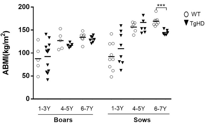
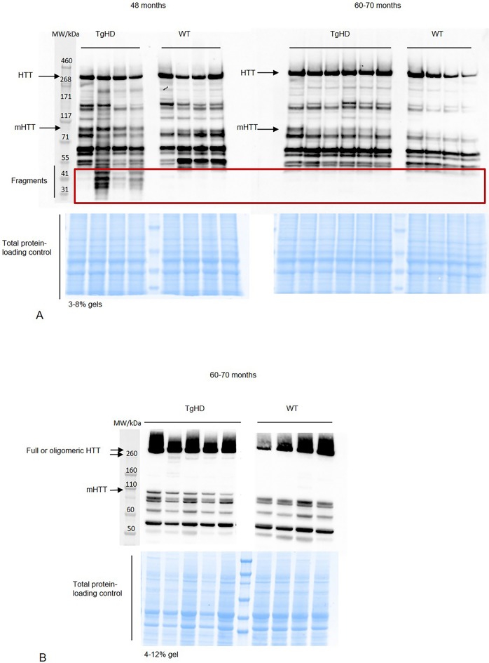
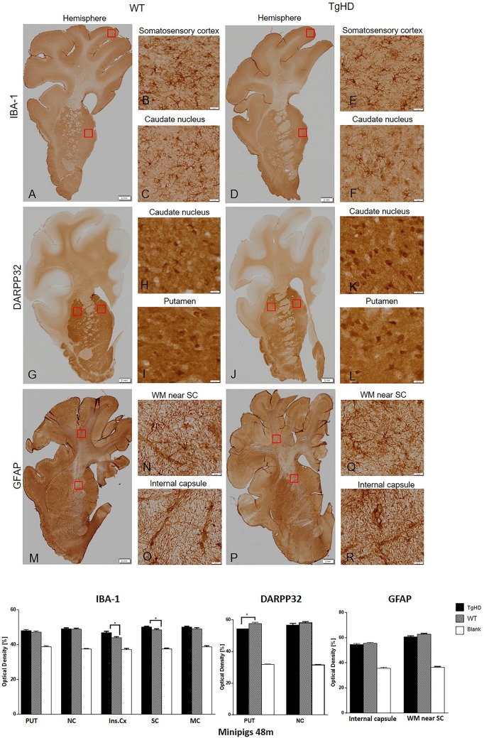
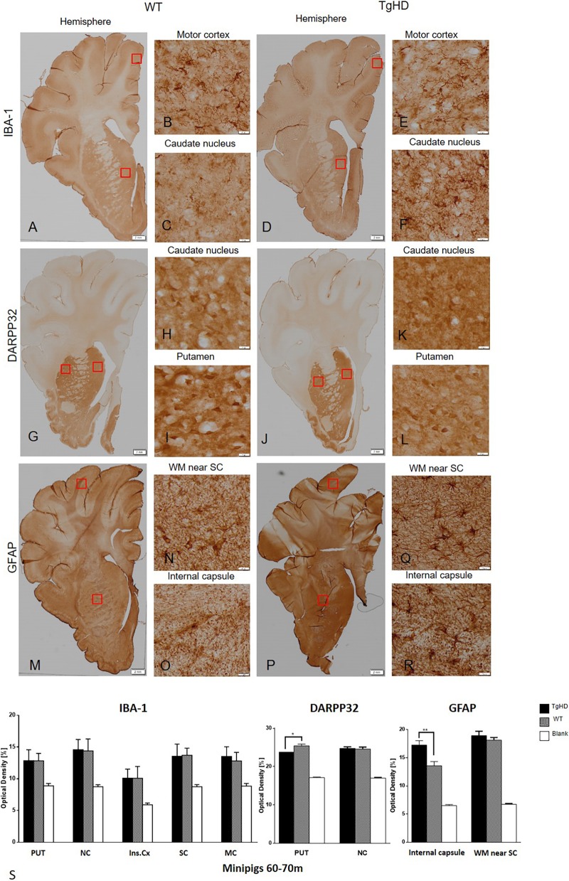
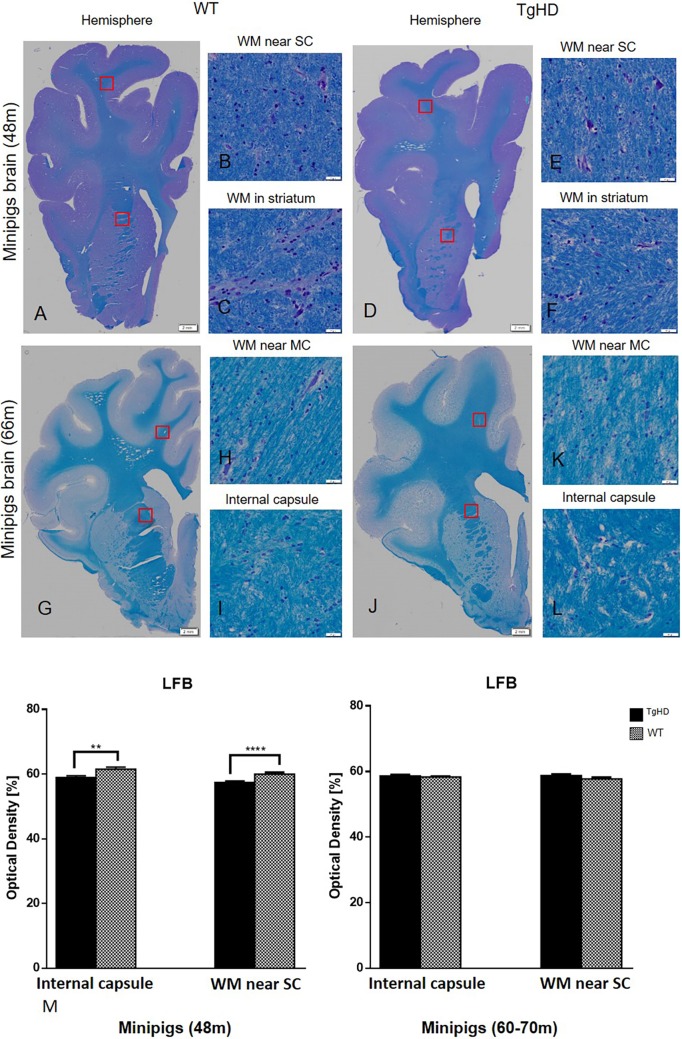
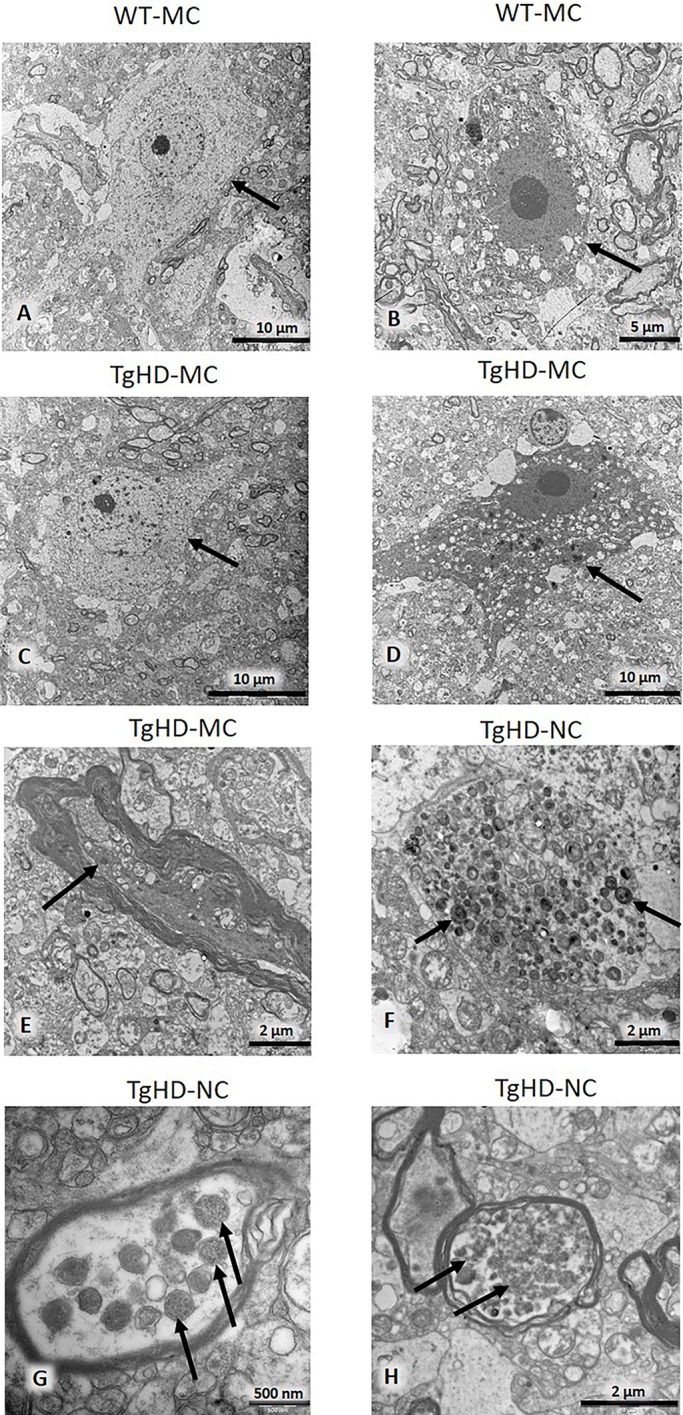
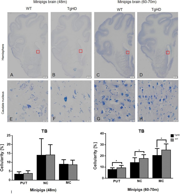
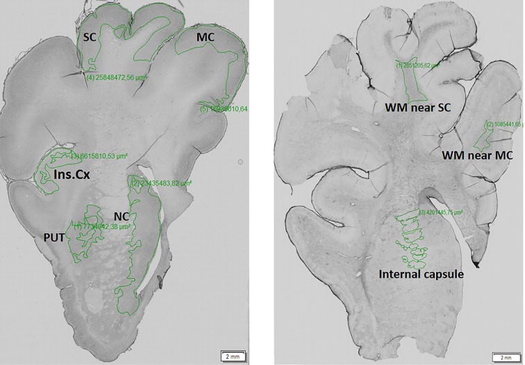
Recently developed therapeutic approaches for the treatment of Huntington's disease (HD) require preclinical testing in large animal models. The minipig is a suitable experimental animal because of its large gyrencephalic brain, body weight of 70-100 kg, long lifespan, and anatomical, physiological and metabolic resemblance to humans. The Libechov transgenic minipig model for HD (TgHD) has proven useful for proof of concept of developing new therapies. However, to evaluate the efficacy of different therapies on disease progression, a broader phenotypic characterization of the TgHD minipig is needed. In this study, we analyzed the brain tissues of TgHD minipigs at the age of 48 and 60-70 months, and compared them to wild-type animals. We were able to demonstrate not only an accumulation of different forms of mutant huntingtin (mHTT) in TgHD brain, but also pathological changes associated with cellular damage caused by mHTT. At 48 months, we detected pathological changes that included the demyelination of brain white matter, loss of function of striatal neurons in the putamen and activation of microglia. At 60-70 months, we found a clear marker of neurodegeneration: significant cell loss detected in the caudate nucleus, putamen and cortex. This was accompanied by clusters of structures accumulating in the neurites of some neurons, a sign of their degeneration that is also seen in Alzheimer's disease, and a significant activation of astrocytes. In summary, our data demonstrate age-dependent neuropathology with later onset of neurodegeneration in TgHD minipigs.

Keywords: Brain; Huntingtin; Large animal model; Neuropathology; TgHD.


Conflict of interest statement

Competing interestsThe authors declare no competing or financial interests.

Figures

Fig. 1.
Fig. 1.
The animal body mass index (ABMI) measurement of TgHD and WT minipigs of F1 and F2 generations at different ages. A graph shows ABMIs for sows and boars within three age groups: 1- to 3-year-old (1-3 Y) boars (TgHD N=12, WT N=5) and sows (TgHD N=8, WT N=9), 4-5 Y boars (TgHD N=5, WT N=5) and sows (TgHD N=7, WT N=6) and 6-7 Y boars (TgHD N=7, WT N=7) and sows (TgHD N=6, WT N=8). Student's t-test with Mann–Whitney test. ***P<0.001.
Fig. 2.
Fig. 2.
Western blot analysis of HTT forms. (A) Detection of fragmented HTT in putamen from 48- and 60- to 70-month-old minipigs using 3-8% gels and EPR-5526 anti-HTT antibody. (B) Detection of oligomeric forms of HTT in putamen from 60- to 70-month-old minipigs using 4-12% gels and EPR-5526 anti-HTT antibody. Representative blots from different TgHD and WT animals are shown.
Fig. 3.
Fig. 3.
Immunohistochemical investigation of expression of IBA-1, DARPP32 and GFAP in the brain sections of 48-month-old animals. IBA-1 (A-F); DARPP32 (G-L); GFAP (M-R). The graph below shows that image analysis of the immunohistochemical staining demonstrated significantly increased IBA-1 expression in the insular and somatosensory cortex, and significantly decreased DARPP32 expression in putamen of TgHD animals. **P<0.05; PUT, putamen; NC, caudate nucleus; Ins.Cx, insular cortex; SC, somatosensory cortex; MC, motor cortex; WM, white matter; Blank, staining without primary antibody. Scale bars: hemispheres, 2 mm; enlargements of brain structures, 50 µm.
Fig. 4.
Fig. 4.
Immunohistochemical investigation of expressions of IBA-1, DARPP32 and GFAP in the brain sections of 60- to 70-month-old animals. IBA-1 (A-F); DARPP32 (G-L); GFAP (M-R). (S) Image analysis of the immunohistochemical staining demonstrated significantly increased GFAP expression in the internal capsule and significantly decreased DARPP32 expression in putamen of TgHD animals. *P<0.05; **P≤0.01; PUT, putamen; NC, caudate nucleus; Ins.Cx, insular cortex; SC, somatosensory cortex; MC, motor cortex; WM, white matter; Blank, staining witout primary antibody. Scale bars: hemispheres, 2 mm; enlargements of brain structures, 50 µm.
Fig. 5.
Fig. 5.
Histochemical staining of pig brains and quantification of myelinization in white matter. (A-L) Luxol Fast Blue (LFB) histochemical staining of pig brains. (M) Quantification of myelinization in white matter on minipig coronal brain sections of 48- and 60- to 70-month-old animals. Significantly decreased intensity of myelin staining was detected in the internal capsule (E) and somatosensory cortex (F) of 48-month-old TgHD animals. **P≤0.01; ****P≤0.001. No changes of myelinization were detected in 60- to 70-month-old minipigs (G-M). WM, white matter; SC, somatosensory cortex; MC, motor cortex. Scale bars: hemispheres, 2 mm; enlargements of brain structures, 50 µm.
Fig. 6.
Fig. 6.
Electron microscopy of motor cortex and caudate nucleus. Arrows indicate light (A,C) and dark (B,D) neurons. Dystrophic neurite (E). Accumulation of multilamellar bodies in unmyelinated neuronal process (F). Dense bodies in the myelinated process are probably remnants of degenerated mitochondria (G). Autophagic vacuoles in a myelinated process (H). MC, motor cortex; NC, caudate nucleus.
Fig. 7.
Fig. 7.
Toluidine Blue histochemical staining and quantification of cellularity. Hemispheres (A-D); caudate nucleus (E-H). (I) Quantification of cellularity in striatum and motor cortex of minipig brain sections of both 48- and 60- to 70-month-old animals using image analysis methods. Significantly decreased cellularity was detected in the putamen (PUT), caudate nucleus (NC) and motor cortex (MC) of TgHD 66-month-old animals. *P≤0.05. TB, Toluidine Blue. Scale bars: hemispheres, 2 mm; enlargements of brain structures, 50 µm.
Fig. 8.
Fig. 8.
Brain regions that underwent densitometric measurement of the intensity staining. The manually selected areas of the porcine brain hemisphere (left) such as motor cortex (MC), somatosensory cortex (SC), insular cortex (Ins.Cx), putamen (PUT) and nucleus caudate (NC), and white matter (WM; right) internal capsule, and WM near the SC and MC, which underwent a densitometric measurement of the intensity staining. The evaluated regions of interest of these brain areas were marked out by a green line.

References

    1. Askeland G., Rodinova M., Štufková H., Dosoudilova Z., Baxa M., Smatlikova P., Bohuslavova B., Klempir J., Nguyen T. D., Kuśnierczyk A. et al. (2018). A transgenic minipig model of Huntington's disease shows early signs of behavioral and molecular pathologies. Dis. Model. Mech. 11, dmm035949 10.1242/dmm.035949 - DOI - PMC - PubMed
    1. Aziz N. A., van der Burg J. M. M., Tabrizi S. J. and Landwehrmeyer G. B. (2018). Overlap between age-at-onset and disease-progression determinants in Huntington disease. Neurology 90, e2099-e2106. 10.1212/WNL.0000000000005690 - DOI - PMC - PubMed
    1. Bartzokis G., Lu P. H., Tishler T. A., Fong S. M., Oluwadara B., Finn J. P., Huang D., Bordelon Y., Mintz J. and Perlman S. (2007). Myelin breakdown and iron changes in Huntington's disease: pathogenesis and treatment implications. Neurochem. Res. 32, 1655-1664. 10.1007/s11064-007-9352-7 - DOI - PubMed
    1. Bates G. P., Dorsey R., Gusella J. F., Hayden M. R., Kay C., Leavitt B. R., Nance M., Ross C. A., Scahill R. I., Wetzel R. et al. (2015). Huntington disease. Nat. Rev. Dis. 1, 15005 10.1038/nrdp.2015.5 - DOI - PubMed
    1. Baxa M., Hruska-Plochan M., Juhas S., Vodicka P., Pavlok A., Juhasova J., Miyanohara A., Nejime T., Klima J., Macakova M. et al. (2013). A transgenic minipig model of Huntington's disease. J. Huntingtons Dis. 2, 47-68. 10.1136/jnnp.2010.222570.26 - DOI - PubMed

Publication types

MeSH terms