. 2019 Nov 29;294(48):18451-18464.
doi: 10.1074/jbc.RA119.011010. Epub 2019 Oct 23.

Synergistic mutations in soluble guanylyl cyclase (sGC) reveal a key role for interfacial regions in the sGC activation mechanism

Affiliations

Synergistic mutations in soluble guanylyl cyclase (sGC) reveal a key role for interfacial regions in the sGC activation mechanism

Kenneth C Childers et al. J Biol Chem. .

Abstract

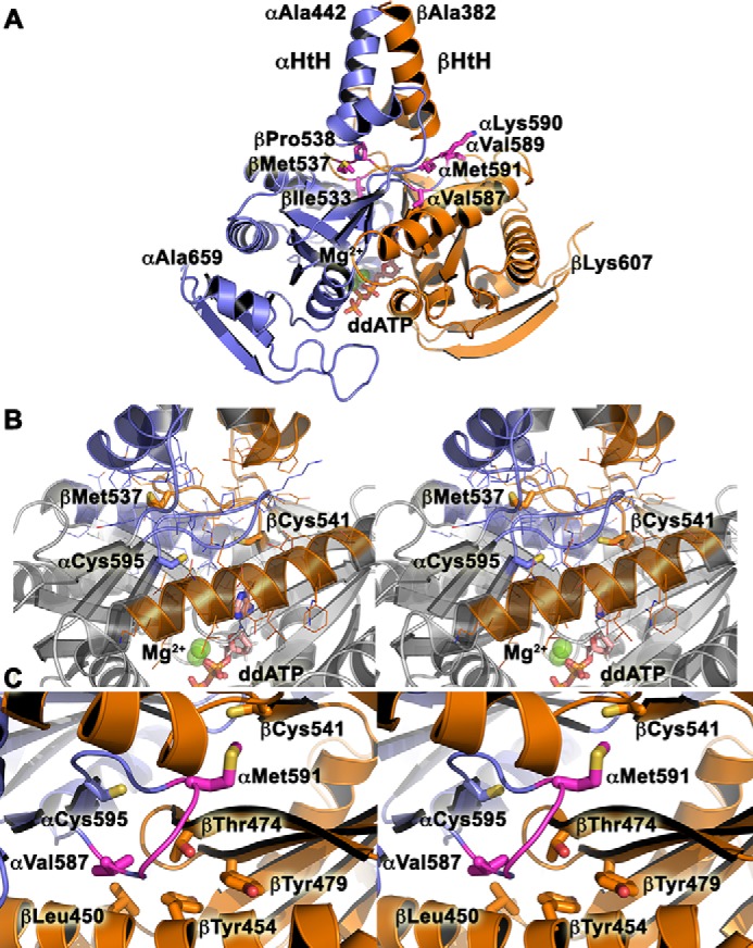
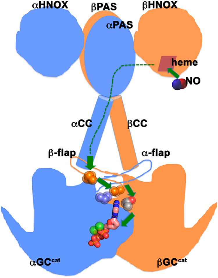
Soluble guanylyl cyclase (sGC) is the main receptor for nitric oxide (NO) and a central component of the NO-cGMP pathway, critical to cardiovascular function. NO binding to the N-terminal sensor domain in sGC enhances the cyclase activity of the C-terminal catalytic domain. Our understanding of the structural elements regulating this signaling cascade is limited, hindering structure-based drug design efforts that target sGC to improve the management of cardiovascular diseases. Conformational changes are thought to propagate the NO-binding signal throughout the entire sGC heterodimer, via its coiled-coil domain, to reorient the catalytic domain into an active conformation. To identify the structural elements involved in this signal transduction cascade, here we optimized a cGMP-based luciferase assay that reports on heterologous sGC activity in Escherichia coli and identified several mutations that activate sGC. These mutations resided in the dorsal flaps, dimer interface, and GTP-binding regions of the catalytic domain. Combinations of mutations from these different elements synergized, resulting in even greater activity and indicating a complex cross-talk among these regions. Molecular dynamics simulations further revealed conformational changes underlying the functional impact of these mutations. We propose that the interfacial residues play a central role in the sGC activation mechanism by coupling the coiled-coil domain to the active site via a series of hot spots. Our results provide new mechanistic insights not only into the molecular pathway for sGC activation but also for other members of the larger nucleotidyl cyclase family.

Keywords: activation; allosteric regulation; cyclic nucleotide; enzyme catalysis; enzyme mechanism; enzyme mutation; guanylate cyclase (guanylyl cyclase); luciferase assay; molecular dynamics; nitric oxide.


Conflict of interest statement

The authors declare that they have no conflicts of interest with the contents of this article

Figures

Figure 1.
Figure 1.
Residues in the GC-1 catalytic domains targeted for mutagenesis. A, ventral (left) and dorsal (right) sides of the GC-1 catalytic domains (αβGCcat) in the modeled activated conformation with bound Mg2+ and dideoxy-ATP (ddATP) (13). B, residues αCys-595 and βAsn-548 were mutated to inactivate GC-1. C, dorsal flaps residues were mutated. D, residues αCys-595, αGlu-526, and βThr-474 were predicted to form a hydrogen-bond triad (13). E, substrate-binding residue βCys-541 was proposed to modulate substrate specificity (38).
Figure 2.
Figure 2.
Alignment of guanylyl cyclase and adenylyl cyclase dorsal flaps. A, alignment generated with CLUSTAL Omega (71–73) and visualized with ESPript (74, 75). Numbering corresponds to the dorsal flap sequence for αGC-1 (Homo sapiens). Similar residues (red letters) are in blue boxes and invariant residues are highlighted in red. Residues in H. sapiens GC-1 that were mutated in this study are highlighted as follows: αVal-587 (green), αVal-587 (yellow), αLys-590 (pale green), αMet591 (pink), βIle-533 (blue), βMet-537 (purple), and βPro-538 (teal). Uniprot accession codes are indicated for each sequence. B, the graphic representation of conserved residues was generated with WebLogo (76).
Figure 3.
Figure 3.
Molecular dynamics simulations reveal distinct backbone conformations among WT and mutant αβGCcat that are related to GC-1 activation. A, RMSD of backbone atoms, with respect to the crystallographic structure (PDB code 4NI2), derived from MD simulations for WT and αβGCcat mutants. B, PCA performed on Cartesian coordinates of backbone atoms from the simulations. Simulation-generated conformational snapshots are projected as shaded areas in the subspace spanned by the two principal components capturing the largest structural variance (PC1 and PC2; the number in the axis label indicates the percentage of variance captured by the corresponding PC). Contour lines represent probability density distributions of conformational samples, where the outmost line indicates the boundary of sampled space. The crystallographic structure representing the inactive conformation (PDB code 4NI2) and the active structural model are also mapped. C and D, collective motions represented by PC1 and PC2, respectively. Two extreme interpolated structures of αβGCcat along each PC are superimposed and represented as cartoons. C, blue and green are structures at −100 and 120, respectively, along PC1. D, blue and white represent −70 and 70, respectively, along PC2. The substrate-binding site is indicated by a yellow star.
Figure 4.
Figure 4.
Function-related rearrangements of interfacial residue-residue contacts/interactions upon mutation. Contact probability changes (denoted by df) from WT αβGCcat to a specific mutant (column) are mapped to the crystallographic structure of αβGCcat (PDB code 4NI2, white cartoon). Only contacts between subunits are shown for clarity. Blue and red cylinders represent contacts with df ≥ 0.1 and df ≤−0.1, respectively, where the cylinder radius is proportional to |df|. The substrate-binding site is indicated by a yellow star.
Figure 5.
Figure 5.
Model for WT CC-GCcat. A, the model for αGC-1 (442–659, blue) and βGC-1 (383–607, orange) was generated with SWISSMODEL (77, 78). Key residues from the dorsal flaps mutated in this study are shown as sticks and labeled. The helix-turn-helix (αHtH and βHtH) motifs of the penultimate coiled-coil domain are indicated. Magnesium ions (green balls) and dideoxy-ATP (sticks, ddATP) are included in the model. B, stereoview of the central positioning for the dorsal flaps, which are sandwiched between the helix-turn-helix motif and the active site of the catalytic domain. The view is the same as in A. Key residues are shown in sticks and colored in blue (αGC) and orange (βGC). C, stereoview of the hydrophobic pocket around residue βThr-474.
Figure 6.
Figure 6.
Proposed mechanism for sGC activation. Schematic representation of full-length sGC with the N-terminal HNOX domains, the dimerization domains (PAS), the coiled-coil domains (CC), and the C-terminal catalytic domains (GCcat). Upon NO binding to the HNOX heme, the activation signal gets transmitted through all the domains (dashed green lines). Hot spot residues (spheres) belonging to coupled networks in the dorsal flaps, the intersubunit interface, and the active site couple the preceding CC domains to the GCcat domains to promote activation.

References

    1. Ignarro L. J., and Freeman B. (eds) (2017) Nitric Oxide: Biology and Pathobiology, 3rd Ed., Academic Press, San Diego
    1. Montfort W. R., Wales J. A., and Weichsel A. (2017) Structure and activation of soluble guanylyl cyclase, the nitric oxide sensor. Antioxid. Redox Signal. 26, 107–121 10.1089/ars.2016.6693 - DOI - PMC - PubMed
    1. Bian K., and Murad F. (2003) Nitric oxide (NO)–biogeneration, regulation, and relevance to human diseases. Front. Biosci. 8, d264–78 10.2741/997 - DOI - PubMed
    1. Childers K. C., and Garcin E. D. (2018) Structure/function of the soluble guanylyl cyclase catalytic domain. Nitric Oxide 77, 53–64 10.1016/j.niox.2018.04.008 - DOI - PMC - PubMed
    1. Ma X., Sayed N., Beuve A., and van den Akker F. (2007) NO and CO differentially activate soluble guanylyl cyclase via a heme pivot-bend mechanism. EMBO J. 26, 578–588 10.1038/sj.emboj.7601521 - DOI - PMC - PubMed

Publication types

MeSH terms

LinkOut - more resources