. 2019 Oct 23;11(515):eaax5866.
doi: 10.1126/scitranslmed.aax5866.

Characterization of orally efficacious influenza drug with high resistance barrier in ferrets and human airway epithelia

Affiliations

Characterization of orally efficacious influenza drug with high resistance barrier in ferrets and human airway epithelia

Mart Toots et al. Sci Transl Med. .

Abstract

Influenza viruses constitute a major health threat and economic burden globally, frequently exacerbated by preexisting or rapidly emerging resistance to antiviral therapeutics. To address the unmet need of improved influenza therapy, we have created EIDD-2801, an isopropylester prodrug of the ribonucleoside analog N 4-hydroxycytidine (NHC, EIDD-1931) that has shown broad anti-influenza virus activity in cultured cells and mice. Pharmacokinetic profiling demonstrated that EIDD-2801 was orally bioavailable in ferrets and nonhuman primates. Therapeutic oral dosing of influenza virus-infected ferrets reduced group pandemic 1 and group 2 seasonal influenza A shed virus load by multiple orders of magnitude and alleviated fever, airway epithelium histopathology, and inflammation, whereas postexposure prophylactic dosing was sterilizing. Deep sequencing highlighted lethal viral mutagenesis as the underlying mechanism of activity and revealed a prohibitive barrier to the development of viral resistance. Inhibitory concentrations were low nanomolar against influenza A and B viruses in disease-relevant well-differentiated human air-liquid interface airway epithelia. Correlating antiviral efficacy and cytotoxicity thresholds with pharmacokinetic profiles in human airway epithelium models revealed a therapeutic window >1713 and established dosing parameters required for efficacious human therapy. These data recommend EIDD-2801 as a clinical candidate with high potential for monotherapy of seasonal and pandemic influenza virus infections. Our results inform EIDD-2801 clinical trial design and drug exposure targets.


Conflict of interest statement

Competing interests: G.R.B., M.G.N. and G.R.P. hold patent 20190022116, “N4-Hydroxycytidine and Derivatives and Anti-Viral Uses Related Thereto”, covering composition of matter and method of use of EIDD-2801 for influenza therapy. This study could affect their personal financial status. All other authors declare no competing interests.

Figures

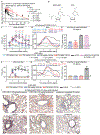
Fig. 1.
Fig. 1.. Anti-influenza efficacy of EIDD-2801 in ferrets.
(A) Plasma PK profiles of NHC and therapeutic candidate EIDD-2801 in cynomolgus macaques (N = 8) after 100 mg/kg oral dose. F indicates oral bioavailability. (B) 2D structures of EIDD-2801 and its hydrolysis product, NHC. (C-I) Ferrets infected intranasally with Ca/09 (C-E, I; N = 3-6) or Wi/05 (F-H; N = 3-7) were treated orally with 100 or 20 mg/kg EIDD-2801 b.i.d. for 3.5 days post-infection. Shed (C, F) and nasal turbinate (E, H) virus titers are shown. (D, G) Body temperature was continuously monitored telemetrically. Lowes analysis of data obtained from all animals/group. Prophylactic treatment of Ca/09-infected animals with oseltamivir is shown for comparison. Symbols in (A-H) represent biological repeats, graphs indicate medians ± SD; 2-way ANOVA (shed titers) or 1-way ANOVA (nasal turbinate titers) with Dunnett’s post hoc test. Differences in resolve of fever were assessed through time-to-event Mantel-Cox test. LoD – limit of detection; MTFR – median time to fever resolve. (I) Representative images of lung sections.
Fig. 2.
Fig. 2.. Resistance profiling.
(A) Virus titers after passages shown in fig. S7 (N = 3). (B) Adaptation of influenza virus to baloxavir marboxil and NHC. Adaptation profile and amino acid frequencies of resistance mutations after deep-sequencing of baloxavir marboxil-experienced (N = 2) viruses (top panel). Deep-sequencing to identify mutation frequency (cut-off 5%) in influenza virus polymerase components after ten passages at 1 or 2 μM NHC or vehicle (lower panels). (C) C-to-U and G-to-A transition events after five and ten passages at 1 or 2 μM NHC relative to vehicle. Symbols show biological repeats, columns are means and error bars represent SDs. Statistical significance was explored by 2-way ANOVA and Dunnett’s post hoc test. (D) Re-passaging of NHC-experienced virus populations from (A) at 4 μM NHC. Symbols represent biological repeats, lines connect means. N = 4 for all NHC and vehicle-experienced virus populations in B-D.
Fig. 3.
Fig. 3.. Anti-IAV and IBV efficacy in well-differentiated human airway epithelium cultures.
(A) Confocal microscopy 21 days post-ALI induction, showing hallmarks of airway epithelia: tight junctions (anti-ZO-1), adherens junctions (anti-E-Cadherin), goblet cells (anti-Muc5AC), and ciliated cells (anti-β-tubulin). Nuclei stained with DAPI. (B) TEER measurements throughout the 21-day differentiation at ALI. Symbols represent individual transwells (N = 10), line shows mean; 2-way ANOVA with Dunnett’s post hoc test. (C) Ca/09 or B/Brisbane/60/2008-infected airway epithelia cultures, treated basolaterally with 1.8 μM NHC or DMSO volume equivalents. Stained are viral antigens (Ca/09: anti-NS1; B/Brisbane/60/2008: anti-IBV), tight junctions, and nuclei. (D) NHC dose-response curves against IAV and IBV in 3D epithelia cultures. Oseltamivir tested against Ca/09 only (N = 3-6/concentration point). Apically shed virus was harvested three days post-infection, EC50 calculations through four-parameter variable-slope regression modeling. Symbols show biological repeats, lines connect means. (E) TEER after 3-day exposure to 50 μM NHC or vehicle (N = 3). Line connects means. (F) Confocal microscopy of samples from (E), showing tight junctions and nuclei. Enlarged immunofluorescence images in fig. S9 and fig. S16–17.
Fig. 4.
Fig. 4.. Simulation of influenza therapy in well-differentiated human airway epithelium models.
(A) Transitions in HBTEC nuclear (SDH-A) and mitochondrial (COX1) mRNAs after 3-day NHC treatment; ≥5 clones and ≥5,000 nucleotides/concentration examined; Fisher’s exact test was applied. (B) Transitions in ferret lung nuclear (TNFα) and mitochondrial (COX15) mRNAs after 7 oral b.i.d. EIDD-2801 doses at 100 mg/kg; 11 clones and ≥7,000 nucleotides were examined. (C) Recapitulation of 128 mg/kg EIDD-2801 oral ferret NHC plasma PK profile in the basolateral chamber of well-differentiated human airway epithelium cultures. NHC concentrations applied is shown in gray columns, corresponding NHC-TP concentrations were measured by LC-MS/MS after 4, 12, and 24 hours. Symbols show biological repeats (N = 3/time point). (D) NHC-TP concentrations in human airway epithelia recapitulating oral EIDD-2801 b.i.d. treatment at 128, 20 or 7 mg/kg in ferrets. Solid lines connect measured NHC-TP from (C) and fig. S13, dashed lines extrapolate b.i.d. treatment, dotted lines mark robust efficacy (EC99), sterilizing antiviral activity, and cytotoxicity thresholds. (E-G) Ca/09-infected ferrets treated therapeutically with 7 mg/kg oral EIDD-2801 b.i.d. after a single 20 mg/kg loading dose (LD) (E) Shed viral load. Symbols (N = 3) show biological repeats, lines connect medians; 2-way ANOVA with Sidak’s post hoc test. (F) Lowes analysis of continuously monitored body temperature. Differences in resolve of fever were assessed through time-to-event Mantel-Cox test. MTFR – median time to fever resolve. (G) Select cytokine and chemokine mRNA induction in nasal turbinates 2.5 days post-infection; values are relative to uninfected animals, symbols show biological repeats (N = 3), columns means ± SD; Welch’s unequal variances t-test was applied.
Fig. 5.
Fig. 5.. Experimental validation of simulation results.
Ca/09-infected ferrets treated therapeutically with 7 mg/kg EIDD-2801 b.i.d for 3.5 days. (A) Shed viral load. Biological repeats (N = 5) are shown, lines connect medians; 2-way ANOVA with Sidak’s post hoc test. (B) Lowes analysis of body temperature. Differences in resolve of fever were assessed through time-to-event Mantel-Cox test. MTFR – median time to fever resolve. (C) Total number of cells in nasal lavages. (D) Total white blood cell counts. Symbols in (C) and (D) show biological repeats (N = 5), lines connect means; 2-way ANOVA with Sidak’s post hoc test. (E) Virus titers in upper and lower respiratory tracts 3.5 days after infection. Symbols show biological repeats (N = 3); graphs indicate medians ± SD; individual statistical assessments of titers in the distinct respiratory tract compartments with unpaired t-tests. NT: nasal turbinates; BALF: bronchioalveolar lavage fluid. (F) Virus distribution in individual lung lobes. Symbols show titers in caudal and cranial lung lobes for individual animals; graphs indicate medians ± SD. (G) Immunohistochemistry of nasal turbinates from vehicle and EIDD-2801-treated animals. Specific detection with anti-IAV HA antiserum and DAB staining, hematoxylin counterstain. Red arrows mark isolated DAB-positive cells detected in treated animals. Scale bars represent 100 μm (overview microphotographs) and 25 μm (inserts), representative fields of view are shown. (H) Immunohistochemistry of lung sections from vehicle and EIDD-2801-treated animals. Staining and size bars as in (G).

References

    1. Palese P, Shaw ML, in Fields Virology, Knipe DM, Howley PM, Eds. (Wolters Kluwer/Lippincott Williams & Wilkins, Philadelphia, 2007), vol. 2, chap. 47, pp. 1647–1690.
    1. Neumann G, Kawaoka Y, The first influenza pandemic of the new millennium. Influenza Other Respir Viruses 5, 157–166 (2011). - PMC - PubMed
    1. Neumann G, Noda T, Kawaoka Y, Emergence and pandemic potential of swine-origin H1N1 influenza virus. Nature 459, 931–939 (2009). - PMC - PubMed
    1. World Health Organization fact sheets influenza (seasonal) https://www.who.int/news-room/fact-sheets/detail/influenza-(seasonal). (2019).
    1. Garten R, Blanton L, Elal AIA, Alabi N, Barnes J, Biggerstaff M, Brammer L, Budd AP, Burns E, Cummings CN, Davis T, Garg S, Gubareva L, Jang Y, Kniss K, Kramer N, Lindstrom S, Mustaquim D, O’Halloran A, Sessions W, Taylor C, Xu X, Dugan VG, Fry AM, Wentworth DE, Katz J, Jernigan D, Update: Influenza Activity in the United States During the 2017-18 Season and Composition of the 2018-19 Influenza Vaccine. MMWR Morb Mortal Wkly Rep 67, 634–642 (2018). - PMC - PubMed

Publication types