. 2019 Dec 2;218(12):4007-4016.
doi: 10.1083/jcb.201904148. Epub 2019 Oct 23.

Mitochondrial stress causes neuronal dysfunction via an ATF4-dependent increase in L-2-hydroxyglutarate

Affiliations

Mitochondrial stress causes neuronal dysfunction via an ATF4-dependent increase in L-2-hydroxyglutarate

Rachel J Hunt et al. J Cell Biol. .

Abstract

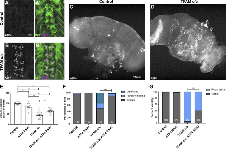
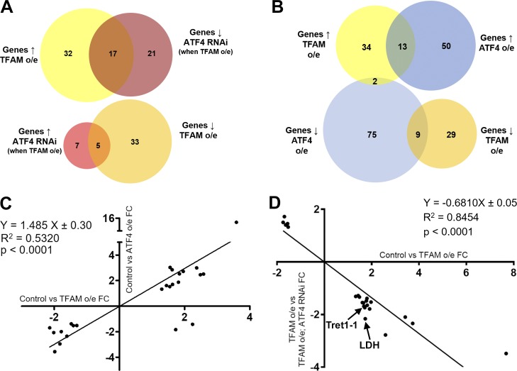
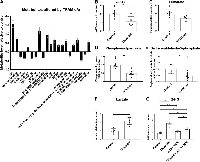
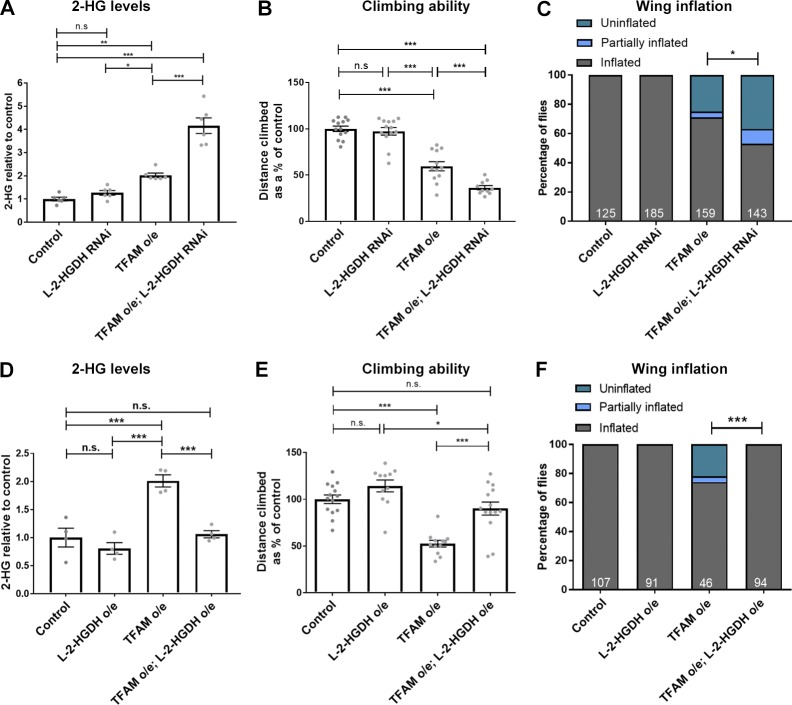
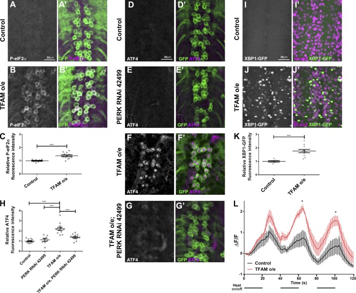
Mitochondrial stress contributes to a range of neurological diseases. Mitonuclear signaling pathways triggered by mitochondrial stress remodel cellular physiology and metabolism. How these signaling mechanisms contribute to neuronal dysfunction and disease is poorly understood. We find that mitochondrial stress in neurons activates the transcription factor ATF4 as part of the endoplasmic reticulum unfolded protein response (UPR) in Drosophila We show that ATF4 activation reprograms nuclear gene expression and contributes to neuronal dysfunction. Mitochondrial stress causes an ATF4-dependent increase in the level of the metabolite L-2-hydroxyglutarate (L-2-HG) in the Drosophila brain. Reducing L-2-HG levels directly, by overexpressing L-2-HG dehydrogenase, improves neurological function. Modulation of L-2-HG levels by mitochondrial stress signaling therefore regulates neuronal function.


Figures

Figure 1.
Figure 1.
Mitochondrial stress signaling via ATF4 in neurons. (A–B′) ATF4 is not expressed in control larval neurons (A and A′), but its expression is activated in motor neurons overexpressing (o/e) TFAM (B and B) using OK371-Gal4. ATF4 in white in A and B and magenta in A and B. CD8-GFP (green) marks motor neurons. (C and D) ATF4 is not expressed in the adult brain (C) but is activated in neurons overexpressing TFAM with nSyb-Gal4 (D). (E and F) Knockdown of ATF4 alleviates the climbing (E) and wing inflation (F) defects caused by TFAM overexpression in motor neurons using D42-Gal4. Numbers of flies for wing inflation are in white. (G) Knockdown of ATF4 alleviates the pupal lethality caused by pan-neuronal TFAM overexpression with nSyb-Gal4. Number of pupal cases are in white. Controls are Gal4 hemizygotes. Data in E are presented as mean ± SEM. n = 20 for all genotypes. ***, P < 0.001.
Figure 2.
Figure 2.
Neuronal mitochondrial stress activates a transcriptional response via ATF4. (A) Venn diagrams showing the number of significantly misregulated genes in the control versus TFAM overexpression (o/e) and TFAM overexpression versus TFAM overexpression combined with ATF4 RNAi conditions as determined by RNA sequencing. ↑ = up-regulated; ↓ = down-regulated. (B) Venn diagrams showing the number of significantly misregulated genes in the control versus TFAM overexpression and control versus ATF4 overexpression conditions. (C) Gene expression correlations between genes significantly misregulated in both the control versus TFAM overexpression and control versus ATF4 overexpression conditions. (D) Gene expression correlations between genes significantly misregulated in both the control versus TFAM overexpression and TFAM overexpression versus TFAM overexpression combined with ATF4 RNAi conditions. FC, fold change. See Materials and methods and Tables S1, S2, S3, S4, and S5 for details.
Figure 3.
Figure 3.
Mitochondrial stress signaling increases 2-HG levels via ATF4. (A) Metabolites whose levels are significantly changed (5% false discovery rate) in adult heads from flies with pan-neuronal TFAM overexpression (o/e) using nSyb-Gal4. (B–F) Levels of individual TCA cycle and glycolytic metabolites in adult heads from flies with pan-neuronal TFAM overexpression using nSyb-Gal4. See Table S6 for details. (G) Knockdown of ATF4 reduces the increase in 2-HG levels caused by pan-neuronal TFAM overexpression using nSyb-Gal4. Controls are nSyb-Gal4 hemizygotes. Data are represented as mean ± SEM. n = 5 for all genotypes. ns, not significant; *, P ≤ 0.05; **, P ≤ 0.01; ***, P ≤ 0.001.
Figure 4.
Figure 4.
Mitochondrial stress–induced L-2-HG regulates neuronal function. (A) The increase in 2-HG levels caused by pan-neuronal TFAM overexpression (o/e) is exacerbated by L-2-HGDH knockdown. n = 6 for all genotypes. (B) L-2-HGDH knockdown enhances the climbing defect caused by pan-neuronal TFAM overexpression. Control n = 13, L-2-HGDH RNAi n = 13, TFAM overexpression n = 12, TFAM overexpression + L-2-HGDH RNAi n = 10. (C) L-2-HGDH knockdown enhances the wing inflation defect caused by pan-neuronal TFAM overexpression. Numbers of flies are shown in white. (D) L-2-HGDH overexpression prevents the increase in 2-HG levels caused by TFAM overexpression. n = 4 for all genotypes. (E) L-2-HGDH overexpression alleviates the climbing defect caused by pan-neuronal TFAM overexpression. Control n = 14, L-2-HGDH overexpression n = 11, TFAM overexpression n = 12, TFAM overexpression + L-2-HGDH overexpression n = 14. (F) L-2-HGDH overexpression alleviates the wing inflation defect caused by pan-neuronal TFAM overexpression. Numbers of flies are shown in white. nSyb-Gal4 was used for pan-neuronal expression. Controls are nSyb-Gal4 hemizygotes. Data are represented as mean ± SEM. n.s., not significant; *, P ≤ 0.05; **, P ≤ 0.01; ***, P ≤ 0.001.
Figure 5.
Figure 5.
Mitochondrial stress–induced activation of ATF4 via the ER UPR. (A–B′) Compared with controls (A and A′), overexpression of TFAM in larval motor neurons causes increased expression of phospho-eIF2α (P-eIF2α; B and B′). P-eIF2α in white in A and B and magenta in A′ and B′. CD8-GFP (green) marks motor neurons. (C) Quantification of P-eIF2α expression. Control n = 8, TFAM overexpression n = 12. (D–G′) Knockdown of PERK (RNAi line 42499) prevents the increase in ATF4 expression (white in D–G and magenta in D′–G′) caused by overexpression (o/e) of TFAM in larval motor neurons using OK371-Gal4. CD8-GFP expression (green) marks motor neurons. (H) Quantification of ATF4 expression. Control n = 12, PERK RNAi n = 8, TFAM overexpression n = 12, TFAM overexpression + PERK RNAi n = 12. (I–J′) Compared with controls (I and I′), overexpression of TFAM in larval motor neurons using OK371-Gal4 causes increased expression of XBP1-GFP (J and J). XBP1-GFP in white in I and J and green in I′ and J′. nLacZ (magenta) marks motor neurons. (K) Quantification of XBP1-GFP expression. Control n = 6, TFAM overexpression n = 11. (L) TFAM overexpression in larval motor neurons disrupts activity-dependent Ca2+ buffering. Mean of traces for GCaMP6m fluorescence in motor neuron cell bodies (using OK371-Gal4) as TrpA1 channels were activated by 20-s on/off pulses of 25°C heat, as indicated by the horizontal black lines below the graph (line = heat on, space = heat off). Control = black trace (n = 5), TFAM overexpression = red trace (n = 5). Controls are Gal4 hemizygotes. Data are represented as mean ± SEM. *, P ≤ 0.05; ***, P ≤ 0.001.

References

    1. Afgan E., Baker D., Batut B., van den Beek M., Bouvier D., Cech M., Chilton J., Clements D., Coraor N., Grüning B.A., et al. . 2018. The Galaxy platform for accessible, reproducible and collaborative biomedical analyses: 2018 update. Nucleic Acids Res. 46(W1):W537–W544. 10.1093/nar/gky379 - DOI - PMC - PubMed
    1. Ansó E., Weinberg S.E., Diebold L.P., Thompson B.J., Malinge S., Schumacker P.T., Liu X., Zhang Y., Shao Z., Steadman M., et al. . 2017. The mitochondrial respiratory chain is essential for haematopoietic stem cell function. Nat. Cell Biol. 19:614–625. 10.1038/ncb3529 - DOI - PMC - PubMed
    1. Arnould T., Michel S., and Renard P.. 2015. Mitochondria Retrograde Signaling and the UPR mt: Where Are We in Mammals? Int. J. Mol. Sci. 16:18224–18251. 10.3390/ijms160818224 - DOI - PMC - PubMed
    1. Bao X.R., Ong S.E., Goldberger O., Peng J., Sharma R., Thompson D.A., Vafai S.B., Cox A.G., Marutani E., Ichinose F., et al. . 2016. Mitochondrial dysfunction remodels one-carbon metabolism in human cells. eLife. 5:e10575 10.7554/eLife.10575 - DOI - PMC - PubMed
    1. Benjamini Y., Krieger A.M., and Yekutieli D.. 2006. Adaptive linear step-up procedures that control the false discovery rate. Biometrika. 93:491–507. 10.1093/biomet/93.3.491 - DOI

Publication types