Randomized Controlled Trial
. 2020 Mar;34(3):735-745.
doi: 10.1038/s41375-019-0604-8. Epub 2019 Oct 23.

A six-gene leukemic stem cell score identifies high risk pediatric acute myeloid leukemia

Affiliations
Randomized Controlled Trial

A six-gene leukemic stem cell score identifies high risk pediatric acute myeloid leukemia

Abdelrahman H Elsayed et al. Leukemia. 2020 Mar.

Erratum in

Abstract

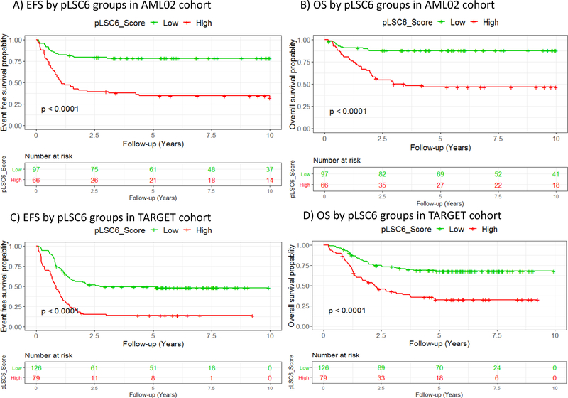
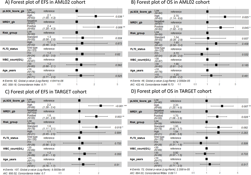
Recently, mRNA-expression signature enriched in LSCs was used to create a 17-gene leukemic stem cell (LSC17) score predictive of prognosis in adult AML. By fitting a Cox-LASSO regression model to the clinical outcome and gene-expression levels of LSC enriched genes in 163 pediatric participants of the AML02 multi-center clinical trial (NCT00136084), we developed a six-gene LSC score of prognostic value in pediatric AML (pLSC6). In the AML02 cohort, the 5-year event-free survival (EFS) of patients within low-pLSC6 group (n = 97) was 78.3 (95% CI = 70.5-86.9%) as compared with 34.5(95% CI = 24.7-48.2 %) in patients within high-pLSC6 group (n = 66 subjects), p < 0.00001. pLSC6 remained significantly associated with EFS and overall survival (OS) after adjusting for induction 1-MRD status, risk-group, FLT3-status, WBC-count at diagnosis and age. pLSC6 formula developed in the AML02 cohort was validated in the pediatric AML-TARGET project data (n = 205), confirming its prognostic value in both single-predictor and multiple-predictor Cox regression models. In both cohorts, pLSC6 predicted outcome of transplant patients, suggesting it as a useful criterion for transplant referrals. Our results suggest that pLSC6 score holds promise in redefining initial risk-stratification and identifying poor risk AML thereby providing guidance for developing novel treatment strategies.


Conflict of interest statement

Conflict of Interest Disclosure: No conflicts to disclose.

Figures

Figure 1:
Figure 1:. Overall schema of study.
Left panel summarizes previous study by Ng et al, 2016 reporting LSC17 and a follow up validation of adult LSC17 in pediatric AML by Duployez et al, 2018. Right panel provides summary of the strategy utilized in the current study to establish a pediatric specific LSC score consisting of 6 genes designated as pLSC6.
Figure 2:
Figure 2:. Pediatric LSC6 (pLSC6) score based on six stem cell genes (DNMT3B, GPR56 and CD34, SOCS2, SPINK2 and FAM30A) predicts clinical outcomes in two independent cohorts of pediatric AML – AML02 and TARGET.
Based on recursive portioning cutoff, Patients were categorized according to their pLSC6 scores into two groups; low (green color; around 60% of AML02, TARGET patients) and high (red color; around 40% of the patients). High pLSC6 scores predicts poor event free survival (A, C) and overall survival (B, D) in AML02 and TARGET cohorts respectively. Number of patients at risk during follow up period of 10 years is given and P-values are based on Cox-hazard models.
Figure 3:
Figure 3:. Pediatric LSC6 (pLSC6) score and MRD status after induction I course of treatment.
Patients found positive for residual leukemic cells after induction 1 course of treatment (MRD-IND1 > 0.1%) had statistically significant higher distribution in the pLSC6 high score group as compared to the low pLSC6 score group in AML02 (A) and TARGET (B) cohorts. P-value based on Chi-square test. Event free survival (C and E) and overall survival (D and F) probabilities by pLSC6 score and MRD status in AML02 (C and D) and TARGET cohorts (E and F) respectively. Green color represents patients with low pLSC6 scores while red represent patients with high pLSC6. Solid lines represent MRD-ve patients and dashed lines for MRD+ve patients.
Figure 4:
Figure 4:. pLSC6 score subclassifies standard risk group patients by clinical outcome.
Kaplan-Meier estimates of EFS by high (red) or low (green) pLSC6 score groups in AML02 (A) and TARGET cohort; (B)
Figure 5:
Figure 5:. Forest plots of multivariable Cox-proportional hazard models showing pLSC6 score as an independent prognostic factor of EFS and OS in AML02 and Target cohorts.
Hazard ratios and 95% confidence intervals Cis are listed next to each variable for EFS (A, C) and OS (B, D) in AML02 and TARGET-AML cohorts, respectively. Within Forest plot, HR for each variable is depicted as a black box and 95% CI are shown as horizontal lines. The vertical line crossing the value of 1 represents non statistically significant effect, odds of less than one indicate better, whereas greater than 1 indicate worse effects.
Figure 6:
Figure 6:
Kaplan-Meier estimates of EFS (A, C) and OS (B, D) by pLSC6 score in standard and high-risk AML patients who did or did not receive hematopoietic stem cell transplantation (HSCT) in AML02 and TARGET cohorts, respectively. Green line: low pLSC6, red line :high pLSC6. Solid lines: HSCT and dashed lines: nonHSCT.

References

    1. Terwijn M, Zeijlemaker W, Kelder A, Rutten AP, Snel AN, Scholten WJ, et al. Leukemic stem cell frequency: a strong biomarker for clinical outcome in acute myeloid leukemia. PLoS One. 2014;9(9):e107587. - PMC - PubMed
    1. van Rhenen A, Feller N, Kelder A, Westra AH, Rombouts E, Zweegman S, et al. High stem cell frequency in acute myeloid leukemia at diagnosis predicts high minimal residual disease and poor survival. Clin Cancer Res. 2005;11(18):6520–7. - PubMed
    1. Ng SW, Mitchell A, Kennedy JA, Chen WC, McLeod J, Ibrahimova N, et al. A 17-gene stemness score for rapid determination of risk in acute leukaemia. Nature. 2016;540(7633):433–7. - PubMed
    1. de Rooij JD, Zwaan CM, van den Heuvel-Eibrink M. Pediatric AML: From Biology to Clinical Management. J Clin Med. 2015;4(1):127–49. - PMC - PubMed
    1. Rubnitz JE, Inaba H, Dahl G, Ribeiro RC, Bowman WP, Taub J, et al. Minimal residual disease-directed therapy for childhood acute myeloid leukaemia: results of the AML02 multicentre trial. Lancet Oncol. 2010;11(6):543–52. - PMC - PubMed

Publication types

Associated data