. 2020 Jun;17(6):631-646.
doi: 10.1038/s41423-019-0300-7. Epub 2019 Oct 23.

Anti-inflammatory mechanisms of the novel cytokine interleukin-38 in allergic asthma

Affiliations

Anti-inflammatory mechanisms of the novel cytokine interleukin-38 in allergic asthma

Xiaoyu Sun et al. Cell Mol Immunol. 2020 Jun.

Abstract

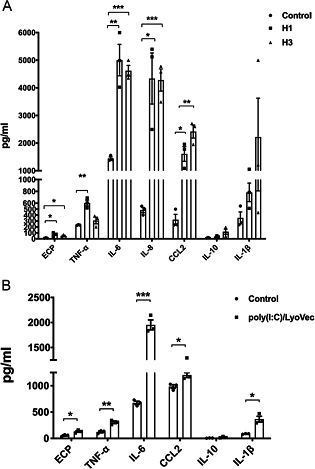
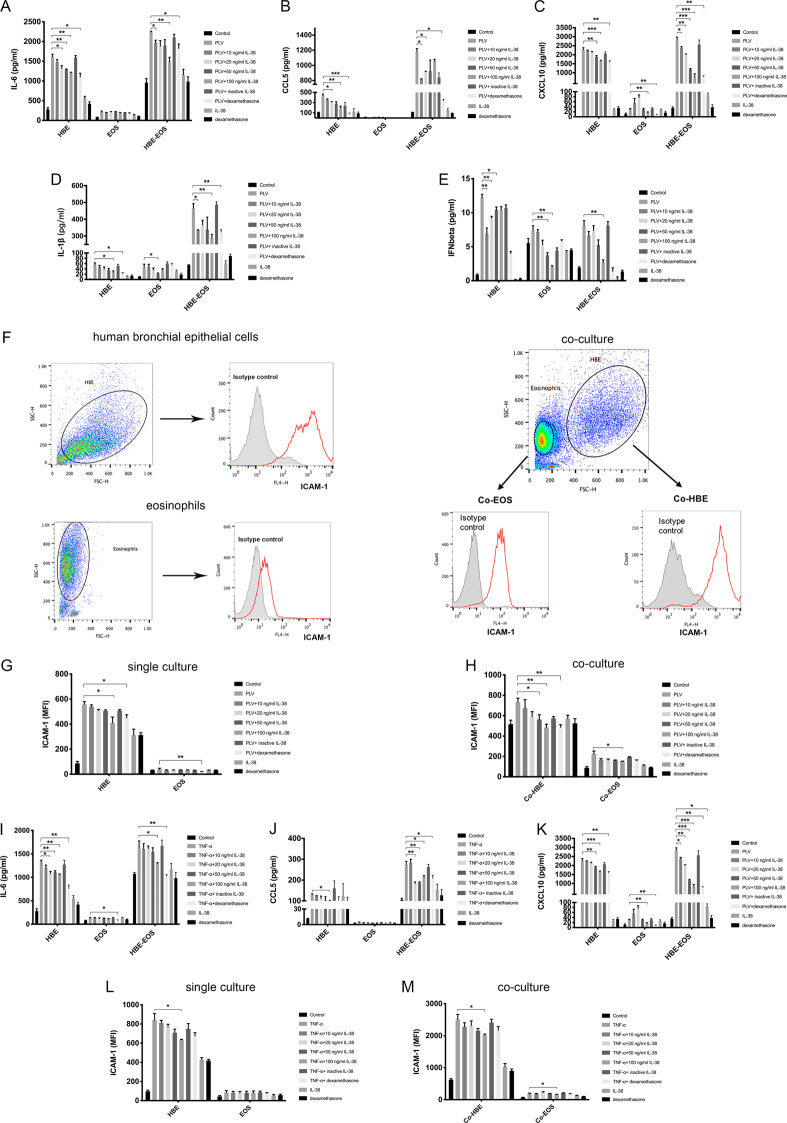
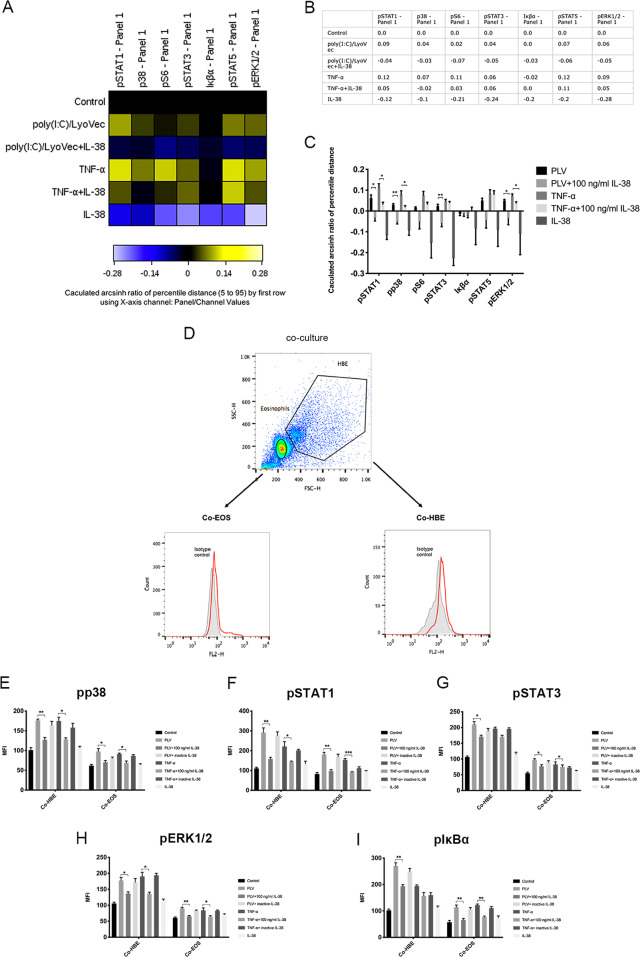
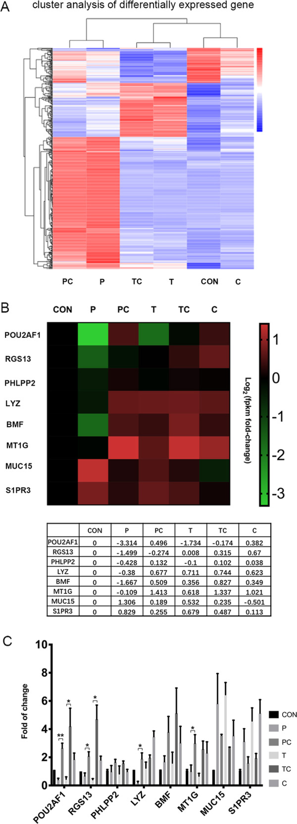
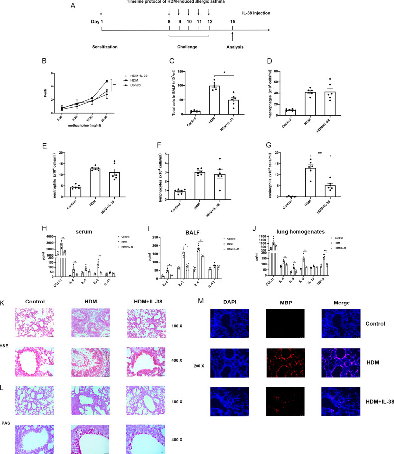
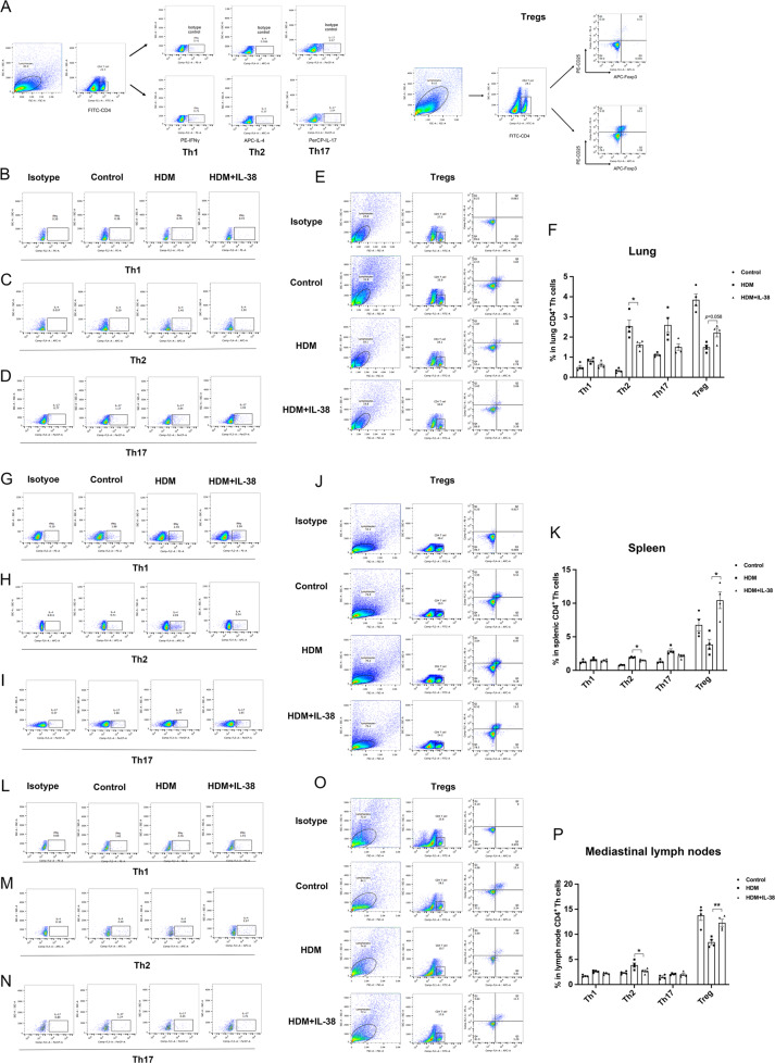
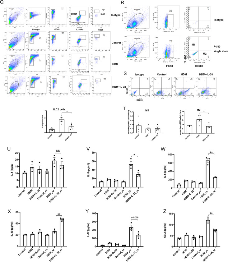
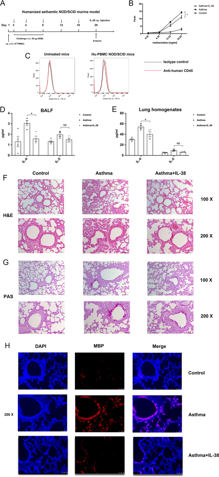
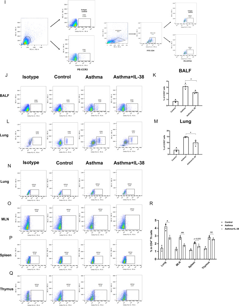
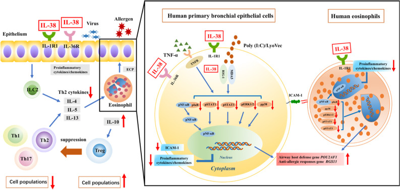
We elucidated the anti-inflammatory mechanisms of IL-38 in allergic asthma. Human bronchial epithelial cells and eosinophils were cocultured upon stimulation with the viral RLR ligand poly (I:C)/LyoVec or infection-related cytokine TNF-α to induce expression of cytokines/chemokines/adhesion molecules. House dust mite (HDM)-induced allergic asthma and humanized allergic asthma NOD/SCID murine models were established to assess anti-inflammatory mechanisms in vivo. IL-38 significantly inhibited induced proinflammatory IL-6, IL-1β, CCL5, and CXCL10 production, and antiviral interferon-β and intercellular adhesion molecule-1 expression in the coculture system. Mass cytometry and RNA-sequencing analysis revealed that IL-38 could antagonize the activation of the intracellular STAT1, STAT3, p38 MAPK, ERK1/2, and NF-κB pathways, and upregulate the expression of the host defense-related gene POU2AF1 and anti-allergic response gene RGS13. Intraperitoneal injection of IL-38 into HDM-induced allergic asthma mice could ameliorate airway hyperreactivity by decreasing the accumulation of eosinophils in the lungs and inhibiting the expression of the Th2-related cytokines IL-4, IL-5, and IL-13 in the bronchoalveolar lavage fluid (BALF) and lung homogenates. Histological examination indicated lung inflammation was alleviated by reductions in cell infiltration and goblet cell hyperplasia, together with reduced Th2, Th17, and innate lymphoid type 2 cell numbers but increased proportions of regulatory T cells in the lungs, spleen, and lymph nodes. IL-38 administration suppressed airway hyperreactivity and asthma-related IL-4 and IL-5 expression in humanized mice, together with significantly decreased CCR3+ eosinophil numbers in the BALF and lungs, and a reduced percentage of human CD4+CRTH2+ Th2 cells in the lungs and mediastinal lymph nodes. Together, our results demonstrated the anti-inflammatory mechanisms of IL-38 and provided a basis for the development of a regulatory cytokine-based treatment for allergic asthma.

Keywords: IL-38; ILC2s; Tregs; allergic asthma; eosinophils.


Conflict of interest statement

The authors declare no competing interests.

Figures

Fig. 1
Fig. 1
Effect of influenza virus H1, influenza virus H3, or poly (I:C)/LyoVec on eosinophils. Eosinophils (5 × 105) were added into tubes and then incubated with 0.5 MOI of H1 or H3 virus, or poly (I:C)/LyoVec (2 μg/ml) for 24 h. The release of cytokines and chemokines into the supernatant of a H1- or H3-treated eosinophils, or b poly (I:C)/LyoVec-treated eosinophils was determined. The results are shown as the mean ± SEM of triplicate independent experiments with a total of three donors. *P < 0.05, **P < 0.01, and ***P < 0.001 when compared between the denoted groups
Fig. 2
Fig. 2
Effect of IL-38 on cytokine/chemokine release in cocultures of human primary bronchial epithelial cells (HBE) and eosinophils (EOS) upon stimulation by poly (I:C)/LyoVec or TNF-α. Primary bronchial epithelial cells (1 × 105) and purified eosinophils (3 × 105) were cocultured with or without human IL-38 pretreatment for 10 min, followed by poly (I:C)/LyoVec (2 μg/ml) or TNF-α (20 ng/ml) treatment for an additional 20 h. Release of a IL-6, b CCL5, c CXCL10, d IL-1β, and e IFN-β into the supernatant of the poly (I:C)/LyoVec-treated coculture was determined. f Gating strategies for bronchial epithelial cells and eosinophils are shown. Bronchial epithelial cells and eosinophils in the cocultures were gated based on the FSC and SSC parameters. The cell surface expression of ICAM-1 on g HBE/EOS single-cultured cells and h cocultured cells was analyzed by flow cytometry. The levels of i IL-6, j CCL5, and k CXCL10 in the supernatant of the TNF-α-treated coculture were measured. The expression of ICAM-1 on l HBE/EOS single-cultured cells and m cocultured cells after stimulation with TNF-α was determined by flow cytometry. A negative control of 56 °C heat-inactivated human IL-38 (100 ng/ml) and a positive control of dexamethasone (1 μM) were included. Abbreviations: HBE, human primary bronchial epithelial cells; PLV, poly (I:C)/LyoVec; EOS, eosinophils; HBE-EOS, coculture of human bronchial epithelial cells and eosinophils; Co-HBE, human primary bronchial epithelial cells in coculture; and Co-EOS, eosinophils in coculture. The results are shown as the mean ± SEM of triplicate independent experiments with a total of three donors. *P < 0.05, **P < 0.01, and ***P < 0.001 when compared between the denoted groups
Fig. 3
Fig. 3
Mass cytometric profiling of intracellular signaling pathways. Cocultured cells were pretreated with human IL-38 (100 ng/ml) for 10 min and then stimulated with poly (I:C)/LyoVec (2 μg/ml) or human TNF-α (20 ng/ml) for an additional 20 min. The phosphorylated signaling molecules in the stimulated cells were stained by using the Maxpar® Phospho Panel Kit. a A heatmap was generated by Cytobank. b, c The change in each phosphoepitope (calculated as the arcsinh difference of the 95th percentile) was quantified for analysis. d Gating strategies for bronchial epithelial cells and eosinophils from cocultures are shown. The intracellular levels of e phosphorylated p38, f phosphorylated STAT1, g phosphorylated STAT3, h phosphorylated ERK1/2, and i phosphorylated IκBα in cells were measured by intracellular staining with specific antibodies and analyzed by flow cytometry. The absolute MFI values for the phosphorylated signals were used for analysis. Heat-inactivated human IL-38 (100 ng/ml) served as a negative control. The results are shown as the mean ± SEM of triplicate independent experiments with a total of three donors. Abbreviations: Co-HBE, human primary bronchial epithelial cells in coculture; Co-EOS, eosinophils in coculture *P < 0.05, **P < 0.01, and ***P < 0.001 when compared between the denoted groups
Fig. 4
Fig. 4
Identification of IL-38 target genes through transcriptional profiling. Cocultured cells were pretreated with human IL-38 (100 ng/ml) for 10 min and then stimulated with poly (I:C)/LyoVec (2 μg/ml) or human TNF-α (20 ng/ml) for 20 h. Total RNA was extracted from each coculture and transcriptome analysis was performed to identify IL-38 target genes. a A heatmap of the DEGs in different groups is shown. b The fold change for differentially expressed genes is displayed in a log2-scale heatmap and the corresponding data are shown in Table. c RNA-seq results were confirmed using real-time qPCR. The GAPDH housekeeping gene was used as the reference gene. The results are shown as the mean ± SEM of triplicate independent experiments with a total of three donors. *P< 0.05, **P < 0.01, and ***P < 0.001 when compared between the denoted groups. Abbreviations: DEG, differentially expressed gene; PC, poly (I:C)/LyoVec + IL-38-treated group; P, poly (I:C)/LyoVec-treated group; TC, TNF-α + IL-38-treated group; T, TNF-α-treated group; CON, unstimulated group; and C, IL-38-treated group
Fig. 5
Fig. 5
In vivo effects of IL-38 on an HDM-induced allergic asthma murine model. a The timeline protocol of the HDM-induced allergic asthma model is shown. b Airway obstruction was measured as Penh values; Penh (% baseline) was calculated by dividing the methacholine-induced Penh value by the PBS baseline value (n = 5). c The total cell number and dg differential cell counts in the bronchoalveolar lavage fluid (BALF) were determined. The release of cytokines and chemokines was measured in h the serum, i the BALF, and j lung homogenates. k, l Representative lung sections stained with H&E or PAS were examined at ×100 or ×400 magnification (n = 5). m Immunostaining analysis of eosinophils in mouse lung tissue samples is shown. Eosinophils were identified using anti-MBP (red) antibodies and nuclei were visualized by DAPI (blue) staining. The lung sections were examined at ×200 magnification. The results are shown as the mean ± SEM of triplicate independent experiments with a total of four to six mice. *P < 0.05, **P < 0.01, and ***P < 0.001 when compared between the denoted groups. Abbreviations: PAS, periodic acid-Schiff; H&E, hematoxylin and eosin
Fig. 6
Fig. 6
Effects of IL-38 on CD4+ Th subsets in mouse lung tissue, splenocyte, and mediastinal lymph node samples. a Th1, Th2, Th17, and Treg cells were gated as indicated in the protocol. The frequencies of Th1, Th2, Th17, and Treg cells in bf lung tissue, gk spleen, and lp mediastinal lymph node samples, as well as q the frequency of ILC2s (LinCD45+CD127+T1/ST2+CD25+ cells) in the mouse spleen were analyzed using flow cytometry (n = 4). q Gating strategies for ILC2s are shown (CD45 single-stained cells were used as a control for staining). r Gating strategies for M1 and M2 macrophages are shown. s, t Representative flow cytometry data for M1 and M2 macrophages in mouse lung tissue samples and the percentages of M1 and M2 macrophages in the lungs are shown. Ex vivo production of u IL-4, v IL-5, w IL-6, x IL-10, y IL-17, and z CCL5 was measured in the supernatant of mouse splenocytes after restimulation with HDM (30 μg/ml) for 72 h. *P < 0.05, **P < 0.01, and ***P < 0.001 when compared between the denoted groups. Control_re, HDM_re, and HDM + IL-38_re indicate splenocytes from the control, HDM, and HDM + IL-38 groups restimulated with HDM, respectively
Fig. 6
Fig. 6
Effects of IL-38 on CD4+ Th subsets in mouse lung tissue, splenocyte, and mediastinal lymph node samples. a Th1, Th2, Th17, and Treg cells were gated as indicated in the protocol. The frequencies of Th1, Th2, Th17, and Treg cells in bf lung tissue, gk spleen, and lp mediastinal lymph node samples, as well as q the frequency of ILC2s (LinCD45+CD127+T1/ST2+CD25+ cells) in the mouse spleen were analyzed using flow cytometry (n = 4). q Gating strategies for ILC2s are shown (CD45 single-stained cells were used as a control for staining). r Gating strategies for M1 and M2 macrophages are shown. s, t Representative flow cytometry data for M1 and M2 macrophages in mouse lung tissue samples and the percentages of M1 and M2 macrophages in the lungs are shown. Ex vivo production of u IL-4, v IL-5, w IL-6, x IL-10, y IL-17, and z CCL5 was measured in the supernatant of mouse splenocytes after restimulation with HDM (30 μg/ml) for 72 h. *P < 0.05, **P < 0.01, and ***P < 0.001 when compared between the denoted groups. Control_re, HDM_re, and HDM + IL-38_re indicate splenocytes from the control, HDM, and HDM + IL-38 groups restimulated with HDM, respectively
Fig. 7
Fig. 7
In vivo effects of IL-38 on humanized HDM-induced allergic asthma mice. a The timeline protocol of the humanized allergic asthma model is shown. b Airway obstruction was measured as Penh values; Penh (% baseline) was calculated by dividing the methacholine-induced Penh value by the PBS baseline value (n = 3). c Human CD45+ cells were assessed on day 20 in the lungs of human PBMC-reconstituted NOD/SCID mice. The secretion of human IL-4 and IL-5 into d the BALF and e lung homogenates was determined. f, g Representative lung sections stained with H&E or PAS were examined at ×100 or ×200 magnification (n = 3). h Immunostaining analysis of eosinophils in mouse lung tissue samples and lung sections was examined at ×200 magnification. i Gating strategies for CCR3+ eosinophils and CD4+CRTH2+ cells are shown. Flow cytometric identification of CCR3+ eosinophils in single-cell suspensions generated from the j, k BALF and l, m lungs is shown. Flow cytometric analysis of CD4+CRTH2+ cells in n the lungs, o MLN, p spleen, and q thymus is shown. r Results are expressed as percentages for CD4+ Th cells. The results are shown as the mean ± SEM of triplicate independent experiments with a total of four mice. *P < 0.05, **P < 0.01, and ***P < 0.001 when compared between the denoted groups. Abbreviation: MLN, mediastinal lymph nodes
Fig. 7
Fig. 7
In vivo effects of IL-38 on humanized HDM-induced allergic asthma mice. a The timeline protocol of the humanized allergic asthma model is shown. b Airway obstruction was measured as Penh values; Penh (% baseline) was calculated by dividing the methacholine-induced Penh value by the PBS baseline value (n = 3). c Human CD45+ cells were assessed on day 20 in the lungs of human PBMC-reconstituted NOD/SCID mice. The secretion of human IL-4 and IL-5 into d the BALF and e lung homogenates was determined. f, g Representative lung sections stained with H&E or PAS were examined at ×100 or ×200 magnification (n = 3). h Immunostaining analysis of eosinophils in mouse lung tissue samples and lung sections was examined at ×200 magnification. i Gating strategies for CCR3+ eosinophils and CD4+CRTH2+ cells are shown. Flow cytometric identification of CCR3+ eosinophils in single-cell suspensions generated from the j, k BALF and l, m lungs is shown. Flow cytometric analysis of CD4+CRTH2+ cells in n the lungs, o MLN, p spleen, and q thymus is shown. r Results are expressed as percentages for CD4+ Th cells. The results are shown as the mean ± SEM of triplicate independent experiments with a total of four mice. *P < 0.05, **P < 0.01, and ***P < 0.001 when compared between the denoted groups. Abbreviation: MLN, mediastinal lymph nodes
Fig. 8
Fig. 8
Graphical summary of the anti-inflammatory activities of IL-38 in allergic airway inflammation. In murine allergic asthma, IL-38 can inhibit the accumulation of eosinophils, ILC2s, and Th2 cells, and the release of CCL11, ECP, and the Th2-related cytokines IL-4, IL-5, and IL-13. Moreover, IL-38 can promote Tregs, which are regulated by IL-10 to maintain immune homeostasis. In our in vitro study, we focused on the interaction between eosinophils and bronchial epithelial cells. The activation of cocultured human primary epithelial cells and eosinophils by the viral mimic dsRNA RLR ligand poly (I:C)/LyoVec or proinflammatory TNF-α could be suppressed by IL-38, and this suppression was mediated by downregulation of the p38, STAT1, STAT3, ERK1/2, and NF-κB pathways, as well as upregulation of the expression of the airway host defense gene POU2AF1 and anti-allergic response gene RGS13 in bronchial epithelial cells, leading to significantly reduced expression of ICAM-1 and proinflammatory cytokines and chemokines. In addition, IL-38 was capable of decreasing the phosphorylation of p38, STAT1, STAT3, ERK1/2, and IκBα in eosinophils in the coculture system, thereby ameliorating allergic airway inflammation

References

    1. Wenzel SE. Asthma: defining of the persistent adult phenotypes. Lancet. 2006;368:804–813. - PubMed
    1. Schatz M, Rosenwasser L. The allergic asthma phenotype. J. Allergy Clin. Immunol. Pract. 2019;2:645–648. - PubMed
    1. Foster PS, et al. Modeling TH2 responses and airway inflammation to understand fundamental mechanisms regulating the pathogenesis of asthma. Immunol. Rev. 2017;278:20–40. - PMC - PubMed
    1. Matucci A, Vultaggio A, Maggi E, Kasujee I. Is IgE or eosinophils the key player in allergic asthma pathogenesis? Are we asking the right question. Respir. Res. 2018;19:1–10. - PMC - PubMed
    1. Mckenzie GJ, et al. Impaired development of Th2 cells in IL-13-deficient mice. Immunity. 1998;9:423–432. - PubMed

Publication types

MeSH terms