. 2019 Oct;574(7780):696-701.
doi: 10.1038/s41586-019-1671-8. Epub 2019 Oct 23.

MHC-II neoantigens shape tumour immunity and response to immunotherapy

Affiliations

MHC-II neoantigens shape tumour immunity and response to immunotherapy

Elise Alspach et al. Nature. 2019 Oct.

Abstract

The ability of the immune system to eliminate and shape the immunogenicity of tumours defines the process of cancer immunoediting1. Immunotherapies such as those that target immune checkpoint molecules can be used to augment immune-mediated elimination of tumours and have resulted in durable responses in patients with cancer that did not respond to previous treatments. However, only a subset of patients benefit from immunotherapy and more knowledge about what is required for successful treatment is needed2-4. Although the role of tumour neoantigen-specific CD8+ T cells in tumour rejection is well established5-9, the roles of other subsets of T cells have received less attention. Here we show that spontaneous and immunotherapy-induced anti-tumour responses require the activity of both tumour-antigen-specific CD8+ and CD4+ T cells, even in tumours that do not express major histocompatibility complex (MHC) class II molecules. In addition, the expression of MHC class II-restricted antigens by tumour cells is required at the site of successful rejection, indicating that activation of CD4+ T cells must also occur in the tumour microenvironment. These findings suggest that MHC class II-restricted neoantigens have a key function in the anti-tumour response that is nonoverlapping with that of MHC class I-restricted neoantigens and therefore needs to be considered when identifying patients who will most benefit from immunotherapy.
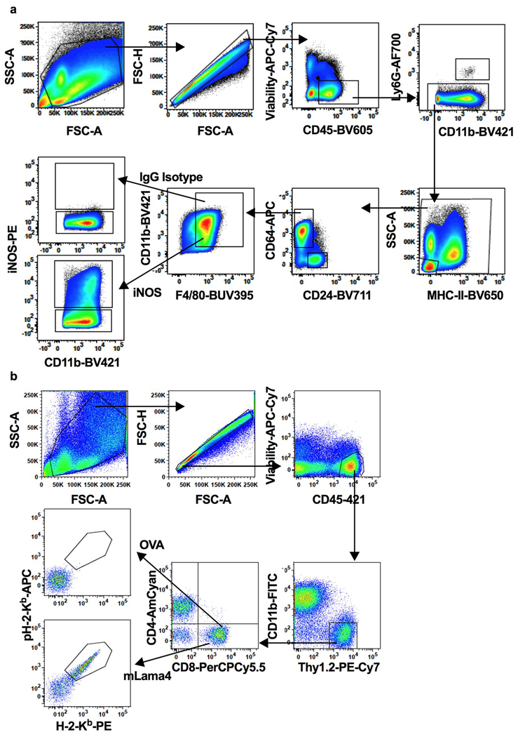

Figures

Extended Data Figure 1:
Extended Data Figure 1:. The hmMHC predictive algorithm and IEDB’18 H2-I-Ab training data set composition
(a) An example of a fully-connected hidden Markov model with 3 hidden states, and emissions corresponding to amino acids. (b-d) Composition of IEDB dataset (MHC full ligand export downloaded on 2018-11-25) represented as number of peptides per binding category and measurement type (b, c) and binding category and peptide length (d). Strong binders: IC50 ≤ 50 nM; binders: 50 nM < IC50 ≤ 500nM; weak binders: 500 nM < IC50 ≤ 5000 nM; non-binders: all remaining peptides. MS: mass spectrometry.
Extended Data Figure 2:
Extended Data Figure 2:. Performance of hmMHC compared to netMHCII-2.3 and netMHCIIpan-3.2
(a) hmMHC (orange stars) underwent 10X cross-validation. In each of the 10 cross-validation partitions, on average there were 4,412 binders in the training set, and 771 binders and 77,086 random natural peptides in the validation set. Performance was compared in terms of AUROC to the performance of netMHCII-2.3 (blue triangles) and netMHCIIpan-3.2 (purple triangles) applied on the same validation sets. For hmMHC, performance for different numbers of hidden states is shown. For netMHCII-2.3 and netMHCIIpan-3.2, performance is shown for both predicted affinity and percentile rank (PR). (b) ROC curves showing performance of hmMHC on H2-I-Ab dataset compared to existing predictors. ROC curves of all peptides and per specific peptide length for every cross-validation partition are shown. (c) Illustration of percentile rank for strong binder classification calibrated on random natural peptides. Red lines indicate the percentile ranks of peptides screened for CD4+ T cell reactivity.
Extended Data Figure 3:
Extended Data Figure 3:. mITGB1 is a major MHC class II-restricted neoantigen in T3 sarcomas.
(a-b) T3 MHC-II neoantigen predictions for all expressed mutations were made using hmMHC (a) and netMHCII-2.3 (b) (netMHCIIpan-3.2 predictions yield very similar results). The predictions are shown as −10 log odds predictor value or logIC50 (smaller values indicate higher likelihood of being presented by I-Ab) and expression level (FPKM). Strong binders are defined as mutations residing in the 2nd percentile of I-Ab binding predictions for random natural peptides for each algorithm (−10logOdds ≤ 26.21 or IC50 ≤ 343.8 nM). The N710Y mutation in Itgb1 met the strong binder threshold in the hmMHC predictions but not in the netMHCII-2.3 predictions. Red dots indicate all mutations that were screened for CD4+ T cell reactivity. Green line denotes high expression cutoff (FPKM=89.1). Blue line indicates strong binder cut off for each algorithm. (c) Two million T3 sarcoma cells were injected subcutaneously into syngeneic mice and CD4+ TIL was isolate on day 12. IFNγ ELISPOT was performed using naïve splenocytes pulsed with 2 μg mL−1 of the indicated peptides. Data is shown as average of three independent experiments ± SEM. (d) Gating strategy for pI-Ab tetramer staining of whole TIL. (e) Quantification of mITGB1-tetramer and CLIP-tetramer staining of CD4+ T cells from whole T3 TIL 12 days post-transplant. Data is shown as average percent tetramer-positive cells of CD4+ cells ± SEM of 3 independent experiments. (f) Syngeneic 129S6 mice were injected subcutaneously with 2x10⁶ T3 sarcoma cells and TIL-derived CD4+ T cells were harvested 12 days post transplant. CD4+ T cells were stimulated with naïve splenocytes pulsed with 2 μg/mL OVA323-339 control or mITGB1697-724 peptide for a flow-based multi-cytokine array. Representative data from one of two independent experiments using pools of 5 tumors each is shown as average of technical triplicate wells from 3 pooled tumors.
Extended Data Figure 4:
Extended Data Figure 4:. T3 TIL-derived CD4+ T cell hybridomas are reactive against mITGB1.
CTLL assay of T3 TIL-derived CD4+ T cell hybridoma lines stimulated with naïve splenocytes pulsed with 2 μg/ml of the individual indicated peptides. Representative data from one of 3 independent experiments is shown as average cpm from technical duplicate wells.
Extended Data Figure 5:
Extended Data Figure 5:. The mITGB1 epitope is presented on I-Ab.
(d) T3 CD4+ T cell hybridomas were stimulated with 2 μg ml−1 mITGB1710Y versus WT Itgb1710N peptide-pulsed splenocytes. Activation was measured by CTLL assay. Representative data from three independent hybridoma lines is shown as average of technical replicate wells. (b) Mapping of the mITGB1 MHC class II binding core was performed using the CD4+ T cell hybridoma line 41 stimulated with naïve splenocytes pulsed with 2 μg/ml of overlapping peptides covering mITGB1697-724. Red denotes the T3-specific mutant amino acid at position p1 of the minimal epitope; underlined portion denotes the validated binding core. Green amino acids represent random residue substitutions used to specifically define valines at residues 715 and 718 as the p6 and p9 MHC-II binding positions and the complete MHC-II binding core. Representative data from 2 independent experiments is shown as the average of technical triplicate wells. (c) MHC-II I-Ab staining of parental T3 cells, IFNγ-stimulated T3 cells and T3 cells transduced with a vector encoding CIITA (T3.CIITA). Representative data from one of three independent experiments is shown. (d) Mirror plot showing match between MS/MS spectra of the 14mer peptide sequence encompassing the N710Y of mITGB1 eluted from T3.CIITA cells (positive axis) and a corresponding synthetic peptide (negative axis). Labeled m/z values reflect those experimentally observed for the endogenous peptide, with peaks representing b ions highlighted in blue and y ions in red.
Extended Data Figure 6:
Extended Data Figure 6:. mITGB1 CD4+ T cells are required for tumor rejection in response to ICT.
(a) Comparison of total number of expressed missense mutations between 10 different MCA-induced sarcomas and KP9025. Mutations were defined by WES and RNAseq and mutational load is shown on a per cell basis. (b) Comparison of predicted neoantigen MHC-I affinity values between KP9025 and MCA-induced sarcoma F244 for H-2Db (top) and H-2Kb (bottom). KP9025 were not predicted to express any MHC-I neoantigens. (c) Rag2−/− mice were subcutaneously injected with 1x10⁶ KP.mLAMA4, KP.mITGB1, KP.mLAMA4.mITGB1 or KP.mSB2.SIINFEKL. Representative data from one of two independent experiments is presented as tumor diameter of individual mice (n=5 KP.mLAMA4, KP.mITGB1 and KP.mLAMA4.mITGB1 and n=3 KP.mSB2.SIINFEKL mice per group per experiment) (d) WT syngeneic 129S4 mice were injected subcutaneously with 1x10⁶ KP.mLAMA4, KP.mITGB1 or KP.mLAMA4.mITGB1 and treated with αPD-1 (top) or αCTLA single agent ICT (bottom) on days 3, 6, and 9 post transplant. Representative data from one of three independent experiments is shown as tumor diameter from individual mice (n=5 in all groups per experiment). (e) Survival curves from all experiments described in (d) and Figure 2e (n=15 in all groups).
Extended Data Figure 7:
Extended Data Figure 7:. Outgrowth of nonimmunogenic sarcoma cells expressing MHC-I neoantigens is not a result of cancer immunoediting.
(a) Rag2−/− or WT 129S4 mice were injected with 1x10⁶ KP9025 or KP.mLAMA4 cells and treated with αPD-1, αCTLA or αPD-1 + αCTLA4 on days 3, 6 and 9. Tumors were harvested once the average diameter reached 20 mm and sarcoma cell lines were established ex vivo. Cell lines were stimulated with IFNγ to upregulate MHC-I and subsequently used to stimulate the mLAMA4-specific CD8+ 74.14 T cell clone. IFNγ secretion by T cells was measured by ELISA. Representative data from 2 independent experiments is represented as the average of 2 independent tumor samples in each group. (b) WT 129S4 mice were injected with 1x10⁶ KP.mSB2.SIINFEKL cells and treated with αPD-1+αCTLA4 combination ICT on days 3, 6 and 9. Tumors were harvested as described in (a). Established ex vivo cell lines were cloned via limiting dilution and parental KP.mSB2.SIINFEKL cells or individual clones from outgrown tumors were used to stimulate the mSB2-specific C3 CD8+ T cell clone and IFNγ production quantified by ELISA. Representative data from four independent experiments is presented as average IFNγ concentration of 8 individual clones ± SEM. Significance was determine using an unpaired, two sided t test. (c) Cell surface staining of SIINFEKL-H-2-Kb expressed by unstimulated or IFNγ-stimulated parental KP.mSB2.SIINFEKL or individual clones described in (b). A representative histogram is shown. (d) Quantification of average SIINFEKL-H-2-Kb MFI from 8 individual clones described in (c) ± SEM. NS not significant. (e) Survival curves of WT 129S4 mice injected subcutaneously with 1x10⁶ KP.mSB2.SIINFEKL.mITGB1. Mice were treated with control mAb or αPD-1+αCTLA4 combination ICT on days 3, 6 and 9. n=10 mice per group from two independent experiments. ****indicates p=1.5x10⁻⁵ as calculated using Mantel-Cox test.
Extended Data Figure 8:
Extended Data Figure 8:. mITGB1-specific CD4+ T cells display an activated Th1 phenotype.
(a) Whole TIL from KP.mLAMA4.mITGB1 tumors 12 days post transplant were stained with mITGB1-I-Ab tetramers. Populations were previously gated on viable CD11bCD4+ cells. Representative data from one of two independent experiments of 5 pooled tumors each is shown. (b) mITGB1-I-Ab tetramer-negative and tetramer-positive cells described in (a) were analyzed for expression of T-BET and FOXP3. Representative plots are shown. (c) Quantification of two independent experiments described in (b) is shown as average percent of tetramer-negative and tetramer-positive cells staining positive for the indicated protein. Tumor-bearing animals received control mAb or α-CTLA4 treatment on days 3, 6, and 9-post transplant where indicated. (d) mITGB1-I-Ab tetramer-positive and tetramer-negative cells described in (a) were analyzed for expression of PD-1. Representative plots are shown. (e) Quantification of two independent experiments described in (d) is shown as average percent of tetramer-negative and tetramer-positive cells staining positive for PD-1. (f) mITGB1-I-Ab tetramer-positive cells described in (a) were analyzed for expression of the indicated proteins. Representative histograms from one of two independent experiments using pools of 5 tumors each are shown.
Extended Data Figure 9:
Extended Data Figure 9:. CD4+ T cell help is required at the tumor site during primary and memory responses.
(a) Rag2−/− mice were simultaneously injected with 1x10⁶ KP.mLAMA4 and KP.mLAMA4.mITGB1 cells on contralateral flanks. Representative data from one of two independent experiments is shown as individual tumor diameter (n=3 in each experiment). (b) WT 129S4 mice were injected with 1x10⁶ KP.mITGB1 cells and were treated with αPD-1+αCTLA4 combination ICT on days 3, 6, and 9. Representative data from one of two individual experiments is shown as individual tumor diameters (n=5 in all experiments). (c) WT 129S4 mice were simultaneously injected with 1x10⁶ KP.mLAMA4 and KP.mLAMA4.mITGB1 cells on contralateral flanks and treated as in (b). Representative data from one of two individual experiments is shown as individual tumor diameters (n=5 in all experiments). (d) WT 129S6 mice were injected subcutaneously with 2x10⁶ T3 sarcoma cells and were treated with αPD-1+αCTLA4 combination ICT on days 3, 6, and 9. Following tumor rejection and a 30-day recovery period, tumor-experienced mice were rechallenged with 2x10⁶ T3 cells in the presence of control mAb or CD4-depleting antibody, or with irrelevant sarcoma cells. Representative data from one of two independent experiments are shown as average tumor diameter ± SEM (n=5 in all groups per experiment). (e) WT 129S4 mice were injected subcutaneously with 1x10⁶ KP.mLAMA4.mITGB1 cells followed by surgical resection 10 days post transplant. After a 30-day recovery period, tumor-experienced mice were rechallenged with 1x10⁶ KP9025, KP.mLAMA4.mITGB1, or KP.mLAMA4. Representative data from one of two independent experiments are shown as average tumor diameter ± SEM (n=5 in all groups per experiment). ****indicates p=2x10⁻⁶ as calculated using a 2-way ANOVA with multiple comparisons corrected with the Bonferroni method. (f) Quantification of data from three independent experiments described in Figure 5c is shown as average number of spots ± SEM (left) and average number of mITGB1-specific CD4+ cells ± SEM (right). **indicates p=.003, ****indicates p=7.2x10⁻⁵ (unpaired, two sided t test). (g) CD45+Ly6GMHCII+CD64+CD25CD11b+F4/80+ macrophages in TIL from animals bearing the indicated contralateral tumors were analyzed for expression of iNOS 11 days post tumor transplant. Representative data is shown. (h) Quantification of iNOS+ macrophages from experiments described in (f) as a percent of total CD45+ cells. Data is shown as average ± SEM of four independent experiments. *indicates p=.03 as calculated using an unpaired, two sided t test. (i) CD45+Ly6GMHCII+CD64+CD25CD11b+F4/80+ macrophages from the indicated contralateral tumors described were isolated 11-days post transplant and analyzed for expression of iNOS. Representative plots are shown. (j) Quantification of iNOS+ macrophages from two independent experiments described in (h) is shown as average percent of total CD45+ cells.
Extended Data Figure 10:
Extended Data Figure 10:. Gating strategies for multi-color flow cytometry.
Gating strategies for multi-color flow cytometry analysis of tumor-infiltrating (a) macrophage and (b) T cell populations.
Figure 1:
Figure 1:. N710Y Itgb1 (mITGB1) is a major MHC class II-restricted neoantigen of T3 sarcoma cells.
(a) hmMHC predictions of MHC-II neoantigens expressed in T3 sarcoma cells. Potential neoantigens were filtered as in Extended Data Fig. 3a and those meeting the strong binder threshold are shown as expression level (FPKM) and neoepitope ratio (NER). Strong binders are those with −10logOdds ≤ 26.21. Green line: high expression cutoff (FPKM=89.1). Blue line: high NER cutoff (NER=6.55). (b) CD4+ T cells isolated from T3 TIL 12 days post-transplant were stimulated in IFNγ ELISPOT analysis with naïve splenocytes pulsed with 2 μg/mL of the indicated individual peptide. Numbers in italics are average number of spots from three independent experiments. (c) I-Ab tetramer staining of CD4+ T cells from whole T3 TIL 12 days post-transplant. Cells were gated on viable CD11bCD4+ cells. Representative data from one of three independent experiments is shown. (d) Freshly isolated CD4+ T cells from day 12 TIL were stimulated with 2 μg ml−1 mITGB1710Y or WT Itgb1710N peptide-pulsed splenocytes and analyzed by IFNγ ELISPOT. Data are average ± SEM (n=3 independent experiments). *indicates p=0.03 (unpaired, two sided t test). (e) Mirror plot showing match between MS/MS spectra of the 17mer peptide encompassing mITGB1N710Y eluted from T3.CIITA cells (right) and a corresponding synthetic peptide (left). Labeled m/z values reflect those experimentally observed for the endogenous peptide, with peaks representing b ions in blue and y ions in red.
Figure 2:
Figure 2:. ICT-mediated rejection of a nonimmunogenic sarcoma requires CD4+ and CD8+ T cells.
(a) One million KP9025 sarcoma cells were injected subcutaneously into syngeneic 129S4 mice and animals were treated with either control mAb or the αPD-1+αCTLA4 combination on days 3, 6, and 9 post transplant. Representative data from two independent experiments are shown as average tumor diameter ± SEM (n=5 in all groups per experiment). (b) KP9025 sarcoma cells were injected as above and tumors were surgically resected followed by rechallenge with the same line. Representative data from one of two independent experiments are shown as average tumor diameter ± SEM (n=3 in all groups per experiment). (c) Cohorts of 5 mice were injected with 1x10⁶ KP.mLAMA4, KP.mITGB1, KP.mLAMA4.mITGB1, or KP.mSB2.SIINFEKL and treated with either control mAb (top) or the αPD-1 + αCTLA4 combination (bottom) on days 3, 6, and 9 post transplant. Representative data from one of three independent experiments is shown as individual tumor diameters.
Figure 3:
Figure 3:. CD4+ T cell help is required for the generation of functional CD8+ CTL during ICT.
(a) Representative tetramer staining of mLAMA4-specific CD8+ T cells from the spleens of KP.mLAMA4 (left) or KP.mLAMA4.mITGB1 (right) tumor-bearing mice 12 days post transplant. Mice received the indicated ICT treatment on days 3, 6, and 9. Cells were gated from viable CD45+CD11b Thy1.2+ cells. (b) Quantification of three independent experiments described above is shown as average percent mLAMA4 tetramer-positive of CD8+ T cells ± SEM. *indicates p=.04, ***indicates p=.0007 and ****indicates p=.00003 (2-way ANOVA with multiple comparisons corrected with the Bonferroni method). (c) In vivo cytotoxic function of mLAMA4-specific CD8+ T cells. Naïve splenocytes were labeled with 0.5 μM CFSE and pulsed with 1 μM SIINFEKL peptide (white histograms) or 5 μM CFSE and pulsed with 1 μM mLAMA4 peptide (green histograms) and transferred into control naïve or tumor-bearing mice 11 days post tumor transplant. Tumor-bearing animals received the indicated ICT treatment on days 3, 6, and 9 post transplant. Representative data is shown. (d) Quantification of percent mLAMA4-specific lysis from independent in vivo cytotoxicity assays described above is shown as average ± SEM (n=6 in αCTLA4, n=8 in all other groups). p values calculated using a 2-way ANOVA with multiple comparisons corrected with the Bonferroni method.
Figure 4:
Figure 4:. MHC class II neoantigens are required for optimal tumor vaccine efficacy.
(a) Schematic of tumor vaccine strategy. Naïve syngeneic 129S6 mice were vaccinated with 5x10⁵ lethally irradiated KP sarcoma cells expressing the indicated antigens. Ten days following vaccination, animals were injected with 2x10⁶ T3 sarcoma cells on the opposite flank and T3 growth or rejection was monitored. (b) Growth curves of T3 sarcoma cells in vaccinated mice as described above. Data are individual tumor diameters from mice injected in 3 independent experiments (n for each group indicated in figure). (c) Kaplan-Meier curves showing survival of mice described in (b). Indicated p values were calculated using Mantel-Cox tests. (d) ELISPOT analysis of 1 μM peptide-pulsed splenocytes 10 days post-vaccination of naïve mice with irradiated KP.mLAMA4 or KP.mLAMA4.mITGB1 cells as described in (a). Data from three independent experiments is shown as average number of spots ± SEM. ***indicates p=.0002 (unpaired, two sided t test).
Figure 5:
Figure 5:. Expression of an MHC-II neoantigen by tumor cells has localized impact on tumor composition.
(a) WT syngeneic 129S4 mice were injected with 1x10⁶ KP.mLAMA4 cells followed by treatment with αPD-1+αCTLA4 on days 3, 6, and 9 post-transplant. Representative data from one of three individual experiments is shown as individual tumor diameters (n=5 per group per experiments) (b) Mice were injected contralaterally with 1x10⁶ KP.mLAMA4 and 1x10⁶ KP.mLAMA4.mITGB1 followed by treatment as described in (a). Representative data from one of three individual experiments is shown as individual tumor diameters (n=5 per group per experiments). (c) Mice were injected as described in (b) and IFNγ ELISPOT analysis of tumor infiltrating CD4+ T cells stimulated with naïve splenocytes pulsed with 2 μg/mL of the indicated peptides was performed 11 days post-transplant. Italicized numbers indicate the average number of spots in mITGB1-stimulated wells from three independent experiments. (d) Tetramer staining of mLAMA4-specific CD8+ TIL 11 days post transplant of mice described in (b). Representative data from one of four independent experiments is shown as percent of mLAMA4-specific cells within the CD8+ T cell population. (e) Quantification of tumor-infiltrating T cells from mice described in (b) 11 days post transplant. Data is shown as percent of total viable CD45+ cells ± SEM. *indicates p=.02, **indicates p=.009 (unpaired, two sided t tests).

Comment in

References

    1. Schreiber RD, Old LJ & Smyth MJ Cancer immunoediting: integrating immunity’s roles in cancer suppression and promotion. Science 331, 1565–1570 (2011). - PubMed
    1. Larkin J et al. Combined Nivolumab and Ipilimumab or Monotherapy in Untreated Melanoma. N. Engl. J. Med 373, 23–34 (2015). - PMC - PubMed
    1. Motzer RJ et al. Nivolumab versus Everolimus in Advanced Renal-Cell Carcinoma. N. Engl. J. Med 373, 1803–1813 (2015). - PMC - PubMed
    1. Borghaei H et al. Nivolumab versus Docetaxel in Advanced Nonsquamous Non-Small-Cell Lung Cancer. N. Engl. J. Med 373, 1627–1639 (2015). - PMC - PubMed
    1. Lennerz V et al. The response of autologous T cells to a human melanoma is dominated by mutated neoantigens. Proc. Natl. Acad. Sci. U. S. A 102, 16013–16018 (2005). - PMC - PubMed

METHODS ONLY REFERENCES

    1. Mamitsuka H Predicting peptides that bind to MHC molecules using supervised learning of hidden Markov models. Proteins 33, 460–474 (1998). - PubMed
    1. Welch LR Hidden Markov Models and the Baum-Welch Algorithm. IEEE Inf. Theory Soc. Newsl 53, 10–13 (2003).
    1. Jurtz V et al. NetMHCpan-4.0: Improved Peptide-MHC Class I Interaction Predictions Integrating Eluted Ligand and Peptide Binding Affinity Data. J. Immunol. Baltim. Md 1950 199, 3360–3368 (2017). - PMC - PubMed
    1. Jensen KK et al. Improved methods for predicting peptide binding affinity to MHC class II molecules. Immunology 154, 394–406 (2018). - PMC - PubMed
    1. Kowalewski DJ & Stevanović S Biochemical large-scale identification of MHC class I ligands. Methods Mol. Biol. Clifton NJ 960, 145–157 (2013). - PubMed

Publication types

MeSH terms

Substances