. 2019 Oct;574(7780):675-678.
doi: 10.1038/s41586-019-1691-4. Epub 2019 Oct 23.

Evolution of the new head by gradual acquisition of neural crest regulatory circuits

Affiliations

Evolution of the new head by gradual acquisition of neural crest regulatory circuits

Megan L Martik et al. Nature. 2019 Oct.

Abstract

The neural crest, an embryonic stem-cell population, is a vertebrate innovation that has been proposed to be a key component of the 'new head', which imbued vertebrates with predatory behaviour1,2. Here, to investigate how the evolution of neural crest cells affected the vertebrate body plan, we examined the molecular circuits that control neural crest development along the anteroposterior axis of a jawless vertebrate, the sea lamprey. Gene expression analysis showed that the cranial subpopulation of the neural crest of the lamprey lacks most components of a transcriptional circuit that is specific to the cranial neural crest in amniotes and confers the ability to form craniofacial cartilage onto non-cranial neural crest subpopulations3. Consistent with this, hierarchical clustering analysis revealed that the transcriptional profile of the lamprey cranial neural crest is more similar to the trunk neural crest of amniotes. Notably, analysis of the cranial neural crest in little skate and zebrafish embryos demonstrated that the transcriptional circuit that is specific to the cranial neural crest emerged via the gradual addition of network components to the neural crest of gnathostomes, which subsequently became restricted to the cephalic region. Our results indicate that the ancestral neural crest at the base of the vertebrate lineage possessed a trunk-like identity. We propose that the emergence of the cranial neural crest, by progressive assembly of an axial-specific regulatory circuit, allowed the elaboration of the new head during vertebrate evolution.


Conflict of interest statement

The authors declare no competing financial interests.

Figures

Extended Data 1.
Extended Data 1.. Heterochronic shifts of cranial specific gene regulatory nodes from later neural crest derivatives to an early specification program happened gradually throughout gnathostome evolution.
a-d.) Expression of lamprey orthologues of amniote cranial specific genes at T21 (cranial) and T23 (trunk) in cross-section. e.) Pharyngeal neural crest derivative expression in Tahara stage 26 Petromyzon marinus frontal section (illustration based on Damas, et al (1944)). f-l.) Cranial circuit orthologues are expressed in pharyngeal arch derivatives, with the exception of Brn3 which is present in the neural crest-derived cranial sensory ganglia in lamprey frontal sections. m.) Gene expression matrix summarizing the heterochronic shift of cranial crest specific circuit nodes. nc=neural crest, nt= neural tube, n=notochord, end.=endoderm, ect.=ectoderm, mes.=mesoderm. Scale bars= 100μm. Cryosections of in situs were reproducible on n≥5 embryos per time point for n≥2 experiments.
Extended Data 2.
Extended Data 2.. Expression of cranial circuit genes in the neural crest of the little skate.
a.) Schematic of a stage 18 Leucoraja erinacea embryo with the neural crest illustrated as blue (cranial) and red (trunk). Placement of cross-sections depicted on the illustration for figures b-f. nc=neural crest, scale bar= 50μm. Cryosections of in situs were reproducible on n≥2 embryos for n≥2 experiments.
Extended Data 3.
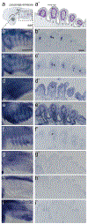
Extended Data 3.. Pharyngeal neural crest derivative expression of cranial circuit orthologues in stage 25 Leucoraja erinacea embryos.
a.) Dashed box on the illustration represents the region of the head for each embryo imaged in figures b-i, and the purple dashed line depicts the location of the frontal section for figures a’-i’. Pharyngeal neural crest derivative expression of cranial circuit orthologues is seen in panels b-f’. Dmbx1, Lhx5, and Brn3c are absent in pharyngeal arch derivatives at stage 25 (g-i’). b-i, scale bar=500μm. b’-i’, scale bar= 100μm. in situs were reproducible on n≥2 embryos.
Extended Data 4.
Extended Data 4.. Expression of cranial circuit genes in the neural crest of the zebrafish.
a.) Schematic of a 14ss Danio rerio embryo with the neural crest illustrated as blue (cranial) and red (trunk). Placement of cross-sections depicted on the illustration for figures b-h. nc=neural crest, n=notochord, scale bars= 50μm. in situs were reproducible on n≥10 embryos.
Extended Data 5.
Extended Data 5.. Expression of cranial circuit orthologues in pharyngeal neural crest derivatives of 3dpf Danio rerio embryos.
a.) Purple dashed line depicts the location of the frontal section for figures e’-h’. Expression of cranial circuit orthologues in pharyngeal arches is seen in panels e-h’. Dmbx1, Lhx5, and Brn3c are absent from pharyngeal arch derivatives at 3dpf (b-d). Scale bar= 150μm. in situs were reproducible on n≥10 whole mount and cryosectioned embryos.
Extended Data 6.
Extended Data 6.. P. marinus Dmbx is homologous to gnathostome Dmbx genes.
a.) Truncated alignment of Dmbx protein sequences. An alignment of full length Dmbx protein sequences was assembled using TCoffee and contiguous regions tagged by the program as poorly or moderately well-aligned were removed, leaving 218 well-aligned residues. b.) Bayesian consensus phylogenetic tree, with posterior probabilities are shown at corresponding nodes.
Figure 1.
Figure 1.. Lamprey cranial neural crest lacks most components of a chick “cranial crest circuit”.
a.) Biotapestry model of cranial specific gene regulatory circuit driving skeletal differentiation in amniotes. b.) Expression of lamprey orthologues of amniote cranial specific genes at T21 and T23. Blue arrows represent expression in the cranial neural crest (CNC), and red arrows represent expression in the trunk neural crest (TNC). c.) Late expression of cranial specific orthologues in the pharyngeal arch neural crest derivatives (black arrow). d.) Biotapestry model of the lamprey circuit with the addition of late module expression of markers in the pharyngeal arch neural crest derivatives. TGG, trigeminal ganglia. Scale bars, 250μm. Reproducible on n≥5 embryos per time point for n≥10 experiments.
Figure 2.
Figure 2.. Nodes of an early cranial-specific circuit were acquired in and restricted to the cranial neural crest progressively throughout gnathostome evolution.
a.) Expression of cranial-specific orthologues in the Little Skate, Leucoraja erinacea at stage (S) 17 and 18. Expression of orthologues in the cranial neural crest (CNC) are depicted with a blue arrow and rostral trunk neural crest (TNC) with red arrows. b.) Biotapestry model of the skate circuit with the addition of a novel node, Ets1. c.) Expression of cranial-specific orthologues in the zebrafish, Danio rerio at 5-9 somite stage (ss) and 14ss. d.) Biotapestry model of the zebrafish circuit with the addition of novel early nodes, lhx5 and dmbx1. FB/MB, forebrain/midbrain. St, stomodeum. Scale bars, 250μm. For skates, in situs were reproducible on n≥2 embryos for n≥2 experiments. For zebrafish, in situs were reproducible on n≥5 embryos per time point for n≥10 experiments.
Figure 3.
Figure 3.. Tissue-specific RNA-seq comparisons between lamprey and chicken reveal ancestral neural crest had a more trunk-like identity.
a.) Volcano plot showing lamprey differential enrichments of cranial (blue) and trunk (red) genes by population RNAseq (100 embryos were dissected for each of n=2 biological replicates) (adjusted p-value<0.05). b.) Volcano plot showing enrichment of genes in the cranial (blue) versus the trunk (red) neural crest in chicken, Gallus gallus (≥15 heads and 5 trunks were dissected and prepared for FAC-sorting for each of n=3 biological replicates) (adjusted p-value<0.05). c.) Hierarchical clustering analysis of all RNAseq libraries focused on the neural crest GRN reveals similarities and differences between axial levels among species.
Figure 4.
Figure 4.. Model of the evolution of neural crest axial levels during vertebrate evolution.
a.) Our data suggest that the ancestral neural crest was a more uniform population of cells along the body axis that underwent gradual regulatory modifications during gnathostome evolution. b.) Progressive restriction of the “cranial circuit genes” to only the cranial axial level led to axial specialization of the neural crest regulatory program.

References

    1. Simoes-Costa M & Bronner ME Reprogramming of avian neural crest axial identity and cell fate. Science 352, 1570–1573 (2016). - PMC - PubMed
    1. Gans C & Northcutt RG Neural crest and the origin of vertebrates: a new head. Science 220, 268–273 (1983). - PubMed
    1. Glenn Northcutt R The new head hypothesis revisited. J. Exp. Zool. B Mol. Dev. Evol 304B, 274–297 (2005). - PubMed
    1. Le Douarin NM Development Of The Peripheral Nervous System From The Neural Crest. Annu. Rev. Cell Dev. Biol 4, 375–404 (1988). - PubMed
    1. Le Douarin N The Neural Crest. (Cambridge University Press, 1982).

Methods References:

    1. Nikitina N, Bronner-Fraser M & Sauka-Spengler T Culturing lamprey embryos. Cold Spring Harb Protoc 2009, pdb.prot5122–pdb.prot5122 (2009). - PubMed
    1. Di Tommaso P et al. T-Coffee: a web server for the multiple sequence alignment of protein and RNA sequences using structural information and homology extension. Nucleic Acids Res. 39, W13–7 (2011). - PMC - PubMed
    1. Armougom F et al. Expresso: automatic incorporation of structural information in multiple sequence alignments using 3D-Coffee. Nucleic Acids Res. 34, W604–8 (2006). - PMC - PubMed
    1. O’Sullivan O, Suhre K, Abergel C, Higgins DG & Notredame C 3DCoffee: combining protein sequences and structures within multiple sequence alignments. J. Mol. Biol 340, 385–395 (2004). - PubMed
    1. Poirot O, Suhre K, Abergel C, O’Toole E & Notredame C 3DCoffee@igs: a web server for combining sequences and structures into a multiple sequence alignment. Nucleic Acids Res. 32, W37–40 (2004). - PMC - PubMed

Publication types