

Quantitative assessment of changes in cellular morphology at photodynamic treatment in vitro by means of digital holographic microscopy
- PMID: 31646023
- PMCID: PMC6788599
- DOI: 10.1364/BOE.10.004975
Quantitative assessment of changes in cellular morphology at photodynamic treatment in vitro by means of digital holographic microscopy
Abstract
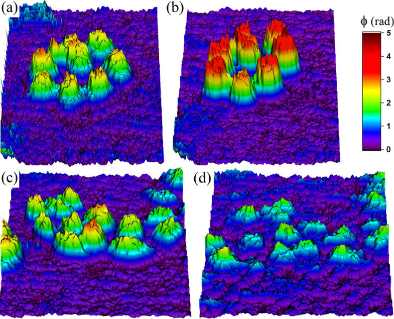
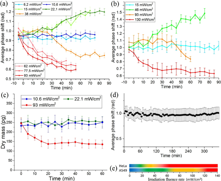
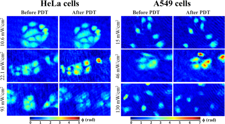
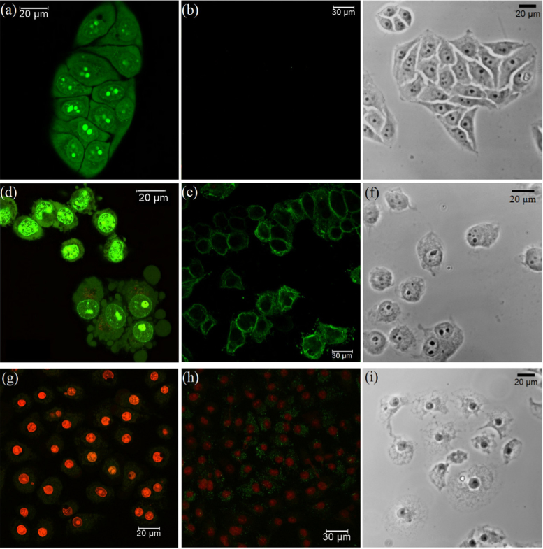
Temporal dependence of changes in the morphological characteristics of cells of two cultured lines of cancer origin, HeLa and A549, induced by photodynamic treatment with Radachlorin photosensitizer, have been monitored using digital holographic microscopy during first two hours after short-term irradiation. The observed post-treatment early dynamics of the phase shift in the transmitted wavefront indicated several distinct scenarios of cell behavior depending upon the irradiation dose. In particular the phase shift increased at low doses, which can be associated with apoptosis, while at high doses it decreased, which can be associated with necrosis. As shown, the two cell types responded differently to similar irradiation doses. Although the sequence of death scenarios with the increase of the irradiation dose was the same, each scenario was realized at substantially different doses. These findings suggest that the average phase shift of the transmitted wavefront can be used for quantitative non-invasive cell death characterization. The conclusions made were cofirmed by commonly used test assays using confocal fluorescent microscopy.
© 2019 Optical Society of America under the terms of the OSA Open Access Publishing Agreement.
Conflict of interest statement
The authors declare that there are no conflicts of interest related to this article.
Figures
References
-
- Letai A., “Apoptosis and cancer,” Annu. Rev. Cancer Biol. 1(1), 275–294 (2017). 10.1146/annurev-cancerbio-050216-121933 - DOI
LinkOut - more resources
Full Text Sources
Research Materials