. 2019 Jul 23;8(10):e1636618.
doi: 10.1080/2162402X.2019.1636618. eCollection 2019.

CD80 expression promotes immune surveillance in Barrett's metaplasia

Affiliations

CD80 expression promotes immune surveillance in Barrett's metaplasia

Melania Scarpa et al. Oncoimmunology. .

Abstract

Esophageal adenocarcinoma (EAC) is the final step of a pathway starting with esophageal reflux disease, Barrett's metaplasia and Barrett's dysplasia. Positive costimulatory ligands such as CD80 have been suggested to contribute to anti-tumor T-cell efficacy. Here we report for the first time the role of CD80 in the inflammatory esophageal carcinogenesis and characterize the immune environment of EAC. Mucosa samples from cancer were obtained during esophagectomy from patients affected by EAC. Fresh biopsies were obtained from patients who underwent endoscopy for screening or follow-up. A rodent model of reflux induced esophageal carcinogenesis was created with an esophago-gastro-jejunostomy. CD80 expression was increased in epithelial cells during metaplasia in the inflammatory esophageal carcinogenesis cascade. Cd80 null mice as well as WT mice that received antiCD80 antibodies showed a higher rate of dysplasia and KI-67+ cells. These results suggest that CD80 mediates an active immune surveillance process in early inflammation-driven esophageal carcinogenesis.

Keywords: Barrett’s esophagus; CD80; adenocarcinoma; esophageal dysplasia; immune surveillance.


Figures

Figure 1.
Figure 1.
Characterization of CD80 expression and cytotoxic T cell in human inflammatory esophageal carcinogenesis. Analysis of esophageal biopsies from healthy controls (H, n = 8), Barrett’s metaplasia (BM, n = 55), Barrett’s dysplastic esophagus (BD, n = 12) and esophageal adenocarcinoma (EAC, n = 15) for: (a) CD80 costimulatory molecule (b) HLA-ABC on esophageal epithelial cells (pan-cytokeratin+) by flow cytometry and (c) PD-L1 immune checkpoint by Real Time qRT-PCR. (d) Immunohistochemical staining and quantification of CD8. (e) Representative immunohistochemical staining of CD8 in esophageal mucosa specimen. Magnification: 20x. (f) Flow cytometric analysis for CD28 on cytotoxic T cells (CD8+) and (g) Real Time RT-PCR quantification of PD-1. Statistical differences are indicated as p value (Dunn’s multiple comparison test). IEN = intraepithelial neoplasia. Data are presented as mean ± SEM.
Figure 2.
Figure 2.
CD80 and immune microenvironment characterization in a rat model of reflux-induced esophageal carcinogenesis. (a) Archival esophageal samples (n = 7) from male Sprague Dawley rats subjected to a surgical procedure to induce gastro-esophageal reflux and sacrificed 32 weeks after surgery were used. Representative immunohistochemical staining and quantification of (b) CD8, (c) CD80 and (d) HLA type I on esophageal samples of healthy (H), Barrett’s metaplasia (BM) and esophageal adenocarcinoma (EAC) mucosa are shown. Graphs depicting number of positive cells x high power field (HPF) are shown for each staining. Statistical differences are indicated as p value (Dunn’s multiple comparison test); **p < .01 and ***p < .001 vs healthy mucosa.
Figure 3.
Figure 3.
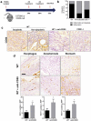
Effect of in vivo CD80 neutralization in a mouse model of reflux-induced esophageal carcinogenesis. (a) Scheme for the experimental course of the esophageal carcinogenesis model. (b) Frequency of dysplasia in CD80-/- and WT mice with esophago-gastroduodenal anastomosis subjected to administration of IgG or anti-CD80 (n = 6–8 mice per group). (c) Representative immunohistochemical staining of CD80 expression in CD80-/- and WT mice with esophago-gastroduodenal anastomosis (n = 6–8 mice per group). (d) Representative immunohistochemical staining and quantification of Ki67 expression in the mid third of crypt on WT mice with esophago-gastroduodenal anastomosis subjected to administration of IgG or anti-CD80. Statistical differences are indicated as p value (Mann–Whitney’s U test).
Figure 4.
Figure 4.
Mucosal microenvironment providing immune surveillance against esophageal carcinogenesis in Barrett’s metaplasia.

References

    1. Noffsinger A. Neoplastic risk assessment in Barrett’s esophagus: how far have come? Hum Pathol. 2003;34:965–967. - PubMed
    1. Schlemper RJ, Riddell RH, Kato Y, Borchard F, Cooper HS, Dawsey SM, Dixon MF, Fenoglio-Preiser CM, Fléjou JF, Geboes K, et al The Vienna classification of gastrointestinal epithelial neoplasia. Gut. 2000;47(2):251–255. - PMC - PubMed
    1. Sharma P, Falk GW, Weston AP, Reker D, Johnston M, Sampliner RE. Dysplasia and cancer in a large multicenter cohort of patients with Barrett’s esophagus. Clin Gastroenterol Hepatol. 2006;4:566–572. doi:10.1016/j.cgh.2006.03.001. - DOI - PubMed
    1. Schnell TG, Sontag SJ, Chejfec G, Aranha G, Metz A, O’Connell S, Seidel UJ, Sonnenberg A. Long-term nonsurgical management of Barrett’s esophagus with high-grade dysplasia. Gastroenterology. 2001;120:1607–1619. - PubMed
    1. Conio M, Blanchi S, Lapertosa G, Ferraris R, Sablich R, Marchi S, D’Onofrio V, Lacchin T, Iaquinto G, Missale G, et al Long-term endoscopic surveillance of patients with Barrett’s esophagus. Incidence of dysplasia and adenocarcinoma: a prospective study. Am J Gastroenterol. 2003;98:1931–1939. - PubMed

Publication types

LinkOut - more resources