Review
. 2019 Oct 31;47(5):1481-1488.
doi: 10.1042/BST20190331.

Towards a model of GCN2 activation

Affiliations
Review

Towards a model of GCN2 activation

Glenn R Masson. Biochem Soc Trans. .

Abstract

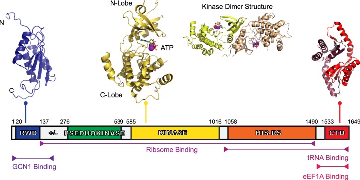
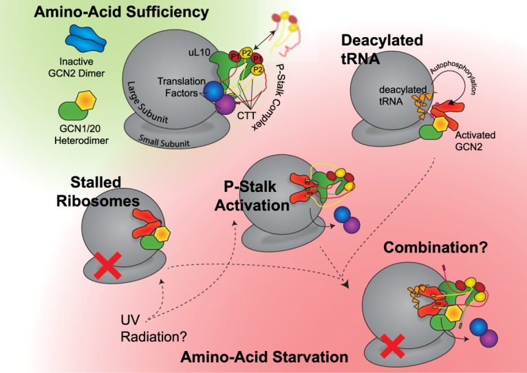
Cells must be able to sense and adapt to their surroundings to thrive in a dynamic environment. Key to adapting to a low nutrient environment is the Integrated Stress Response (ISR), a short-lived pathway that allows cells to either regain cellular homeostasis or facilitate apoptosis during periods of stress. Central to the ISR is the protein kinase General Control Non-depressible 2 (GCN2), which is responsible for sensing starvation. Upon amino acid deficiency, GCN2 is activated and initiates the ISR by phosphorylating the translation initiation factor eIF2α, stalling protein translation, and activating the transcription factor ATF4, which in turn up-regulates autophagy and biosynthesis pathways. A key outstanding question is how GCN2 is activated from an autoinhibited state. Until recently, a model of activation focussed on the increase of deacylated tRNA associated with amino acid starvation, with deacylated tRNA binding directly to GCN2 and releasing autoinhibition. However, in vivo experiments have pointed towards an alternative, deacylated-tRNA-independent mechanism of activation. Here, we review the various factors that may facilitate GCN2 activation, including recent research showing that the P-stalk complex, a ribosome-associated heteropentameric protein complex, is a potent activator of GCN2.

Keywords: GCN2; ISR; P-stalk; protein translation; ribosome.


Conflict of interest statement

The Author declares that there are no competing interests associated with this manuscript.

Figures

Figure 1.
Figure 1.. Domain organisation of human GCN2.
GCN2 is a 1649 amino acid protein with five conserved domains: the RWD, the pseudokinase domain, kinase domain, HisRS-Like domain, and CTD. Linking the RWD and pseudokinase domain is ‘charged linker’ (labelled ‘+/−’) region also. The structure of GCN2's RWD domain from mouse was determined using nuclear magnetic resonance (NMR) spectroscopy [39], showing an α + β sandwich fold (PDB ID: 1UKX). The charged linker, a stretch of arginine, lysine, glutamate, and aspartate residues, precedes the pseudokinase domain — the structure of which has yet to be determined — and was identified as a pseudokinase domain due to the sequence similarity to typical kinase domains, however, it lacks key catalytic residues. The kinase domain, shows a typical kinase domain structure (PDB ID: 1ZYD) [10], and is presented as both a monomer and dimer with its associated ATP and Mg2+. Following the kinase domain is the HisRS-like domain, a domain essential for GCN2 activity in vivo [14] which interacts directly with tRNA [15]. Finally, there is the CTD, shown here using the dimeric mouse CTD structure (PDB ID: 4OTN) [27]. The CTD is constitutively dimeric, similar to the kinase domain. There is a species-dependent interaction of the CTD with ribosomes also, which is reliant on three lysine residues found in yeast. Yeast CTDs co-migrate with ribosomes on a sucrose gradient, but this is not the case with the mouse CTD where these lysine residues are absent.
Figure 2.
Figure 2.. Potential routes to GCN2 activation.
Whilst the autophosphorylated, activated state of GCN2 is thought to be common to all routes, it is not certain what pathways take precedence, whether they are mutually exclusive, whether they are present in all eukaryotic organisms, or whether they may be facilitated in vivo by GCN20 and GCN1. In non-starvation conditions, it is assumed that the P-stalk associates both the ribosome and translation factors around the A-site, whilst GCN1, GCN2, and GCN20 remain unbound to the ribosome in the cytosol. GCN2 remains autoinhibited, whilst GCN1 and GCN20 form a stable heterodimer. From this state, there are at least three possible routes to activation of GCN2 upon depletion of amino acids. Firstly in the case of stalled ribosomes; it may be that there is an increased affinity for the GCN1/2/20 complex for the stalled ribosome, causing an association and activation of GCN2. An alternate route to a stalled ribosome may be UV radiation. Secondly, upon starvation the P-stalk C-terminal tails may be free to bind to GCN2 due to the loss of translation factors — or alternative stresses (such as UV radiation) may also cause P1/P2 proteins to associate with uL10, forming the P-stalk and activating GCN2 in this manner. Thirdly, the increase in deacylated tRNA levels may cause an associated with GCN2, causing it to become activated in a manner that involves GCN1 and GCN20. Additionally, all three of these routes may combine, or be only partially correct.

References

    1. Wek R.C., Jiang H.-Y. and Anthony T.G. (2006) Coping with stress: eIF2 kinases and translational control. Biochem. Soc. Trans. 34, 7–11 10.1042/BST0340007 - DOI - PubMed
    1. Pakos-Zebrucka K., Koryga I., Mnich K., Ljujic M., Samali A. and Gorman A.M. (2016) The integrated stress response. EMBO Rep. 17, 1374–1395 10.15252/embr.201642195 - DOI - PMC - PubMed
    1. Ye J., Kumanova M., Hart L.S., Sloane K., Zhang H., De Panis D.N. et al. (2010) The GCN2-ATF4 pathway is critical for tumour cell survival and proliferation in response to nutrient deprivation. EMBO J. 29, 2082–2096 10.1038/emboj.2010.81 - DOI - PMC - PubMed
    1. Ma T., Trinh M.A., Wexler A.J., Bourbon C., Gatti E., Pierre P. et al. (2013) Suppression of eIF2α kinases alleviates Alzheimer's disease-related plasticity and memory deficits. Nat. Neurosci. 16, 1299–1305 10.1038/nn.3486 - DOI - PMC - PubMed
    1. Eyries M., Montani D., Girerd B., Perret C., Leroy A., Lonjou C. et al. (2014) EIF2AK4 mutations cause pulmonary veno-occlusive disease, a recessive form of pulmonary hypertension. Nat. Genet. 46, 65–69 10.1038/ng.2844 - DOI - PubMed

Publication types

MeSH terms