. 2019 Dec:309:106617.
doi: 10.1016/j.jmr.2019.106617. Epub 2019 Oct 8.

First hyperpolarized [2-13C]pyruvate MR studies of human brain metabolism

Affiliations

First hyperpolarized [2-13C]pyruvate MR studies of human brain metabolism

Brian T Chung et al. J Magn Reson. 2019 Dec.

Abstract

We developed methods for the preparation of hyperpolarized (HP) sterile [2-13C]pyruvate to test its feasibility in first-ever human NMR studies following FDA-IND & IRB approval. Spectral results using this MR stable-isotope imaging approach demonstrated the feasibility of investigating human cerebral energy metabolism by measuring the dynamic conversion of HP [2-13C]pyruvate to [2-13C]lactate and [5-13C]glutamate in the brain of four healthy volunteers. Metabolite kinetics, signal-to-noise (SNR) and area-under-curve (AUC) ratios, and calculated [2-13C]pyruvate to [2-13C]lactate conversion rates (kPL) were measured and showed similar but not identical inter-subject values. The kPL measurements were equivalent with prior human HP [1-13C]pyruvate measurements.

Keywords: Brain metabolism; Hyperpolarized C13; Metabolic imaging.


Conflict of interest statement

Declaration of Competing Interest

The authors declare that they have no known competing financial interests or personal relationships that could have appeared to influence the work reported in this paper.

Figures
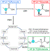
Fig. 1.
Fig. 1.
Diagram showing [2-13C]pyruvate metabolism investigated in this hyperpolarized NMR spectroscopy study of the human brain.
Fig. 2.
Fig. 2.
Representative Carbon-13 NMR summed spectrum from the brain of a healthy volunteer acquired with a 32-channel head coil following an injection of 1.43 mL/kg of 250 mM [2-13C]pyruvate. Peak identification was assigned following those by Park et al. from studies of HP [2-13C]pyruvate in the murine brain: (A) [2-13C]pyruvate, (B) [5-13C]glutamate, (C) [1-13C]citrate and/or [5-13C]glutamine, (D) [1-13C]pyruvate (natural abundance doublet), (E) [2-13C]pyruvate-hydrate, (F) [2-13C]lactate doublet [17].
Fig. 3.
Fig. 3.
Flip angle plot of the RF excitation pulse sequence with the parameters used for this study. Note the decreased excitation of the upfield [2-13C]lactate resonance versus the downfield by approximately one half.
Fig. 4.
Fig. 4.
Spectra for all four volunteers at a single timepoint 16 sec post-injection. Similar levels of [5-13C]glutamate and [2-13C]lactate reflect the underlying biochemistry of the healthy human brain of similar rates of conversion of [2-13C]pyruvate to [2-13C]lactate catalyzed by LDH as [2-13C]pyruvate to [5-13C]glutamate catalyzed by PDH.
Fig. 5.
Fig. 5.
Dynamic plots of metabolite kinetics for each of the four volunteers. Results were consistent noting minor differences in intensity scale. As shown in the corresponding Fig. 4, the rates of conversion of [2-13C]pyruvate to [2-13C]lactate and [5-13C]glutamate are similar in the normal human brain.
Fig. 6.
Fig. 6.
Plots showing kPL analysis demonstrated similar results between a previously acquired [1-13C]pyruvate dataset from a volunteer (left) and [2-13C]pyruvate volunteer dataset acquired in this study with the mean ± standard error for all 4 volunteers (right).
Fig. 7.
Fig. 7.
Images acquired using a metabolite-specific flip angle schedule and echo planar imaging (EPI) readout. From left to right 1H proton reference image, overlaid [2-13C] pyruvate, and [5-13C]glutamate images of a volunteer’s brain are shown. The single-shot HP 13C EPI images were acquired with: in-plane resolution = 2.5 × 2.5 cm2, slice thickness = 5 cm, bandwidth = 8.06 kHz, TR = 100 msec, temporal resolution = 3 sec, TE = 19 msec, and flip angles θPyr = 10°, θGlu = 60°. Average SNR of the pyruvate signal = 682 and glutamate signal = 31.1.

References

    1. Ardenkjaer-Larsen JH, On the present and future of dissolution-DNP, J. Magn. Reson (2016), 10.1016/j.jmr.2016.01.015. - DOI - PubMed
    1. Ardenkjaer-Larsen JH, Fridlund B, Gram A, et al., Increase in signal-to-noise ratio of > 10,000 times in liquid-state NMR, Proc. Natl. Acad. Sci 100 (18) (2003) 10158–10163. - PMC - PubMed
    1. Nelson SJ, Kurhanewicz J, Vigneron DB, et al., Metabolic imaging of patients with prostate cancer using hyperpolarized [1–13C] pyruvate, Sci Transl Med. (2013;5(198):) 198ra108, 10.1126/scitranslmed.3006070. - DOI - PMC - PubMed
    1. Park Ilwoo, Larson Peder E.Z., Gordon Jeremy W., Carvajal Lucas, Chen Hsin-Yu, Bok Robert, Van Criekinge Mark, Ferrone Marcus, Slater James B., Xu Duan, Kurhanewicz John, Vigneron Daniel B., Chang Susan, Nelson Sarah J., Development of methods and feasibility of using hyperpolarized carbon-13 imaging data for evaluating brain metabolism in patient studies: hyperpolarized Carbon-13 metabolic imaging of patients with brain tumors, Magn. Reson. Med 80 (3) (2018) 864–873, 10.1002/mrm.v80.310.1002/mrm.27077. - DOI - PMC - PubMed
    1. Golman K, Zandt R.i., Lerche M, Pehrson R, Ardenkjaer-Larsen JH, Metabolic imaging by hyperpolarized 13C magnetic resonance imaging for in vivo tumor diagnosis, Cancer Res. 66 (22) (2006) 10855–10860, 10.1158/0008-5472.CAN-06-2564. - DOI - PubMed

Publication types

MeSH terms