Review
. 2019 Dec;29(12):964-973.
doi: 10.1016/j.tcb.2019.09.002. Epub 2019 Oct 21.

Cell Fusion: Merging Membranes and Making Muscle

Affiliations
Review

Cell Fusion: Merging Membranes and Making Muscle

Michael J Petrany et al. Trends Cell Biol. 2019 Dec.

Abstract

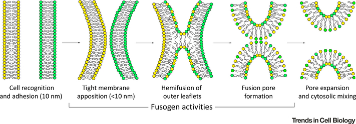
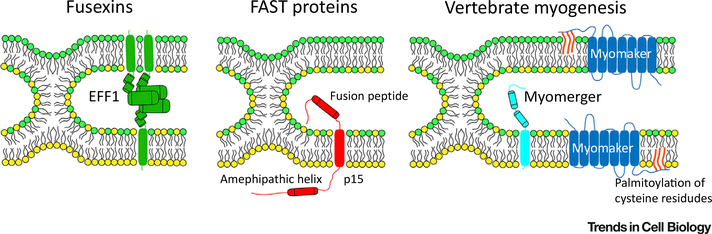
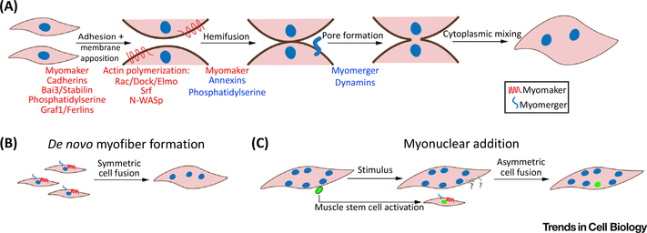
Cell fusion is essential for the development of multicellular organisms, and plays a key role in the formation of various cell types and tissues. Recent findings have highlighted the varied protein machinery that drives plasma-membrane merger in different systems, which is characterized by diverse structural and functional elements. We highlight the discovery and activities of several key sets of fusion proteins that together offer an evolving perspective on cell membrane fusion. We also emphasize recent discoveries in vertebrate myoblast fusion in skeletal muscle, which is composed of numerous multinucleated myofibers formed by the fusion of progenitor cells during development.

Keywords: cell fusion; fusogens; myoblast fusion; myomaker; myomerger; skeletal muscle.


Figures

Figure 1.
Figure 1.. Essential Steps of the Hemifusion Stalk/Fusion-Pore Model of Cell Membrane Fusion.
A series of distinct membrane events must occur for two cells to fuse and merge their cytoplasmic contents. First, two cells must recognize and adhere to one another, followed by close membrane adhesion (to within 10 nm). The outer-membrane leaflets must then fuse, which results in mixing of lipids and the formation of an unstable membrane stalk intermediate. The formation and expansion of a fusion pore within the hemifused membrane then completes the reaction.
Figure 2.
Figure 2.. Diversity of Protein Machineries Driving Cell–Cell Fusion.
Representative fusogenic proteins from three classes are shown at the stage of the unstable hemifusion-stalk intermediate. EFF1, representing the fusexin family, acts in a trans-trimerization form to drive membrane fusion. The transmembrane domains perform a zippering-like action to bring opposing membranes together and drive pore formation. Fusion-associated small transmembrane (FAST) proteins act unilaterally to drive fusion of reovirus-infected cells. FAST proteins harness endogenous cellular machinery to promote cell adhesion. An extracellular fusion peptide inserts into the trans membrane, and an intracellular amphipathic helix is postulated to drive fusion pore formation. Myomaker and myomerger act independently at the membrane to drive a bipartite myoblast fusion mechanism. Myomaker is required for hemifusion to occur, and must be palmitoylated in its C-terminal region to function. Myomerger mediates pore formation through the activity of one or both extracellular helices.
Figure 3.
Figure 3.. Mechanisms of Vertebrate Myoblast Fusion.
(A) Myoblast fusion involves a series of membrane modification steps. Key protein and lipid regulators are listed beneath each step. Knowledge of the precise stage at which a given factor regulates fusion is frequently absent, therefore factors are color-coded according to the strength of evidence that they act at a particular step (blue, high confidence; red, further clarification is needed). Highlighted are the bilateral requirement for myomaker and the unilateral requirement for myomerger. (B) Symmetric fusion between cells in similar states (myocytes to myocytes) occurs to form myofibers de novo during early development and adult regeneration. Myomaker and myomerger are shown bilaterally to highlight their expected coexpression in fusogenic myocytes. (C) Myonuclear addition to existing myofibers can also occur during postnatal development after myofiber number has been established, and after a stimulus (such as exercise or injury) in the adult. In this case, muscle stem cells become activated, and undergo an asymmetric fusion event (fusion between cells in different states – myocytes and myofibers). Whether fusion between myocytes and myofibers progresses through a unilateral or a bilateral mechanism, with respect to the requirement of myomaker or myomerger on each of the fusing cells, is not fully understood. Although myomaker/myomerger are required on the myocyte, it is not known whether they are active on the myofiber. Note that, in adult regenerative contexts, myocyte-to-myocyte fusion can also occur between this activated muscle stem-cell pool to generate de novo myofibers (not shown).

References

    1. Yin H et al. (2013) Satellite cells and the muscle stem cell niche. Physiol. Rev. 93, 23–67 - PMC - PubMed
    1. Chal J and Pourquie O (2017) Making muscle: skeletal myogenesis in vivo and in vitro. Development 144, 2104–2122 - PubMed
    1. Chernomordik LV et al. (2006) Membranes of the world unite! J. Cell Biol. 175, 201–207 - PMC - PubMed
    1. Katsov K et al. (2006) Field theoretic study of bilayer membrane fusion. II. Mechanism of a stalk-hole complex. Biophys. J. 90, 915–926 - PMC - PubMed
    1. Haldar S et al. (2018) Lipid-dependence of target membrane stability during influenza viral fusion. J. Cell Sci. 132, jcs218321 - PMC - PubMed

Publication types