. 2019 Dec 6;294(49):18909-18922.
doi: 10.1074/jbc.RA119.010384. Epub 2019 Oct 24.

Protochromic absorption changes in the two-cysteine photocycle of a blue/orange cyanobacteriochrome

Affiliations

Protochromic absorption changes in the two-cysteine photocycle of a blue/orange cyanobacteriochrome

Teppei Sato et al. J Biol Chem. .

Erratum in

Abstract

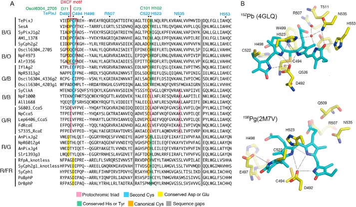
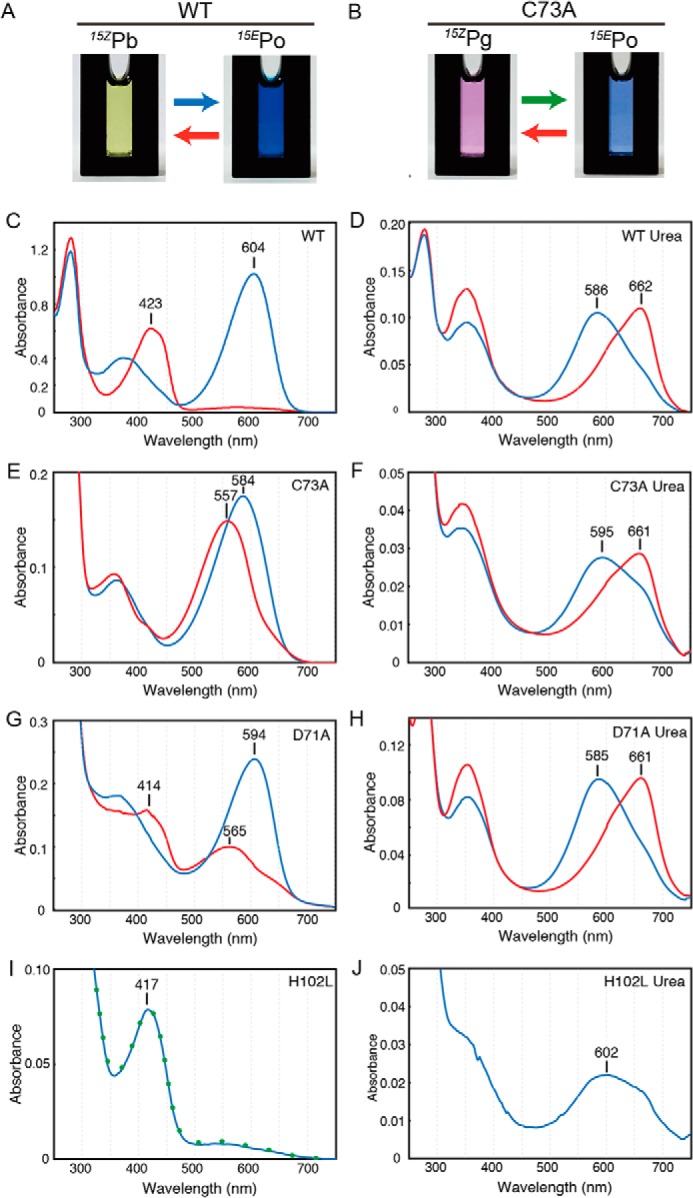
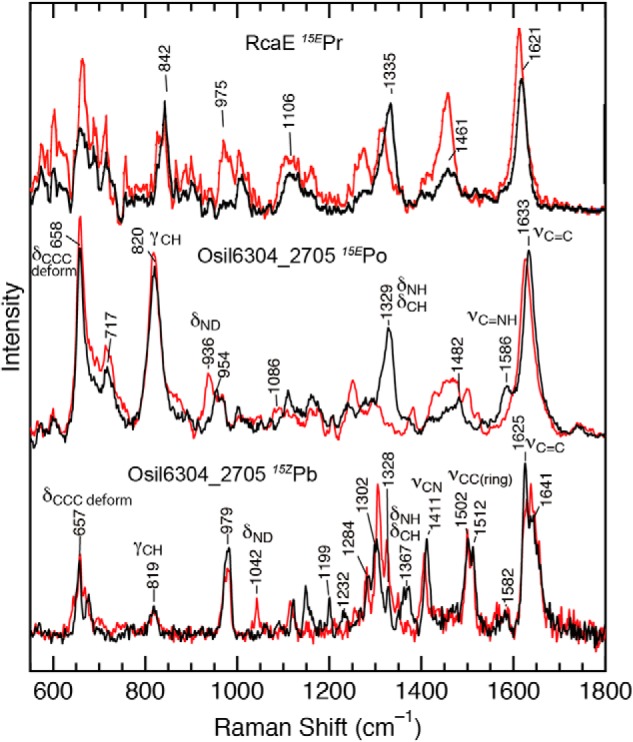
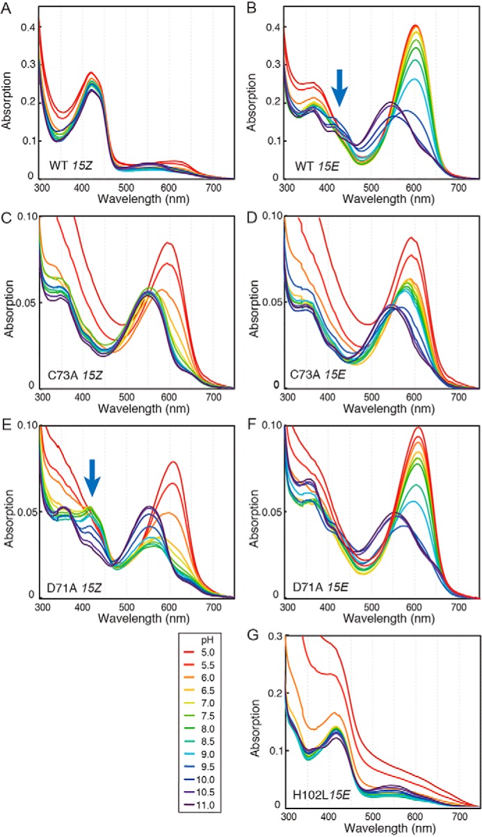
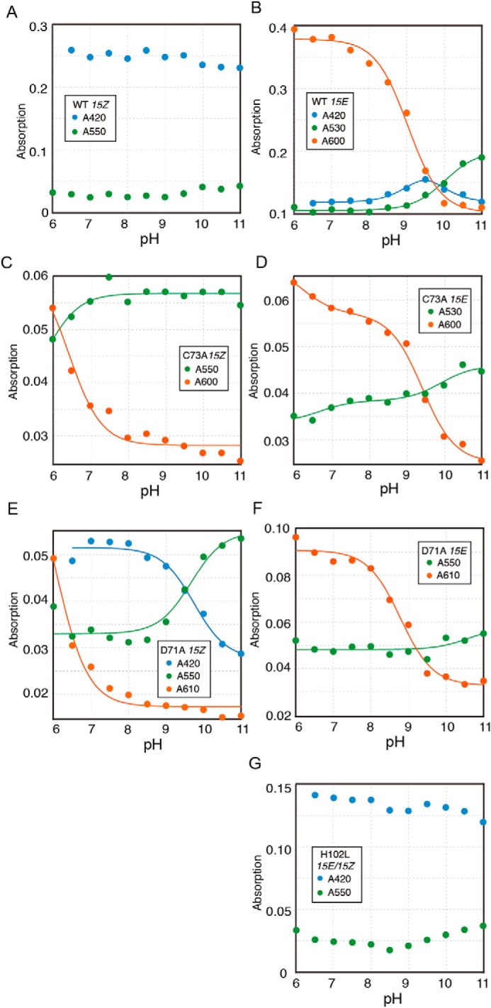
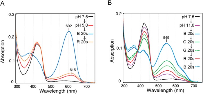
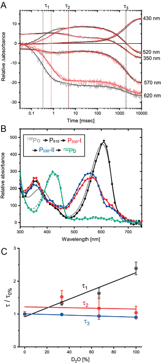
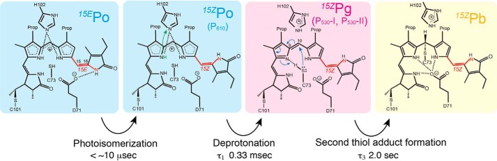
Cyanobacteriochromes (CBCRs) are phytochrome-related photosensors with diverse spectral sensitivities spanning the entire visible spectrum. They covalently bind bilin chromophores via conserved cysteine residues and undergo 15Z/15E bilin photoisomerization upon light illumination. CBCR subfamilies absorbing violet-blue light use an additional cysteine residue to form a second bilin-thiol adduct in a two-Cys photocycle. However, the process of second thiol adduct formation is incompletely understood, especially the involvement of the bilin protonation state. Here, we focused on the Oscil6304_2705 protein from the cyanobacterium Oscillatoria acuminata PCC 6304, which photoconverts between a blue-absorbing 15Z state ( 15Z Pb) and orange-absorbing 15E state ( 15E Po). pH titration analysis revealed that 15Z Pb was stable over a wide pH range, suggesting that bilin protonation is stabilized by a second thiol adduct. As revealed by resonance Raman spectroscopy, 15E Po harbored protonated bilin at both acidic and neutral pH, but readily converted to a deprotonated green-absorbing 15Z state ( 15Z Pg) at alkaline pH. Site-directed mutagenesis revealed that the conserved Asp-71 and His-102 residues are required for second thiol adduct formation in 15Z Pb and bilin protonation in 15E Po, respectively. An Oscil6304_2705 variant lacking the second cysteine residue, Cys-73, photoconverted between deprotonated 15Z Pg and protonated 15E Pr, similarly to the protochromic photocycle of the green/red CBCR subfamily. Time-resolved spectroscopy revealed 15Z Pg formation as an intermediate in the 15E Pr-to- 15Z Pg conversion with a significant solvent-isotope effect, suggesting the sequential occurrence of 15EP-to-15Z photoisomerization, deprotonation, and second thiol adduct formation. Our findings uncover the details of protochromic absorption changes underlying the two-Cys photocycle of violet-blue-absorbing CBCR subfamilies.

Keywords: Raman spectroscopy; bilin; cyanobacteria; cyanobacteriochrome; photobiology; photoreceptor; phytochrome; spectroscopy; time-resolved spectroscopy.


Conflict of interest statement

The authors declare that they have no conflicts of interest with the contents of this article

Figures

Figure 1.
Figure 1.
Sequence and structural information of CBCR GAF domains. A, multiple alignment of the bilin-binding pocket of the GAF domain of CBCR subfamilies undergoing blue/orange (B/O), blue/green (B/G), green/blue (G/B), blue/blue (B/B), green/red (G/R), and red/green (R/G) photocycles. Sequences of the GAF domain of phytochromes (R/FR) were also included. Key residues interacting with the bilin were colored accordingly. Residue numbers of TePixJ and (cyan) corresponding Oscil6304_2705 (green) are indicated. B, PVB chromophore and its associated hydrogen bond network in the structures of TePixJ of 15ZPb (upper) and 15EPg (lower).
Figure 2.
Figure 2.
Spectral characterization of WT and single amino acid variants. Photographs of protein solution of WT undergoing blue/orange photocycle (A) and C73A variant undergoing green/orange photocycle (B) are shown. C–J, native absorption spectra of WT (C), C73A (E), D71A (G), and H102L (I) and acid-denaturation absorption spectra of WT (D), C73A (F), D71A (H), and H102L (J). For H102L, absorption after blue light illumination was also included as a green dot (I). Spectra corresponding to the 15Z state (red) or 15E state (blue) of the bilin were indicated.
Figure 3.
Figure 3.
RR spectroscopy of RcaE and Osil6304_2705. The observed Raman spectra of 15EPr of RcaE (upper) in the previous study (63), and those of 15EPo (middle) and 15ZPb (lower) of Oscil6304_2705 in this study. Spectra were measured at room temperature in the buffer containing H2O (black) or D2O (red). Important Raman bands are assigned as follows: νC=C, the C=C stretching mode; νC=NH, the C=N stretching coupled with an in-plane NH bending vibration; νCC (ring), the CC stretching mode of a pyrrole ring; νCN, the CN stretching mode, δNH/δCH, the in-plane NH and CN bending mode; δND, the in-plane ND bending mode; γCH, the out-of-plane CH bending mode of a methine bridge; δCCC, the C–C–C bending deformation mode.
Figure 4.
Figure 4.
pH titration of WT and single amino acid variants. A, pH-dependent absorbance spectra of the 15Z and 15E states of WT (A and B), C73A (C and D), D71A (E and F), and H102L (G) are shown. pH values are shown between 5.0 (red) and 11.0 (purple) in 0.5 pH unit increments.
Figure 5.
Figure 5.
pKa estimation of WT and single amino acid variants. A, pH-dependent absorption changes of 15Z and 15E states of WT (A and B), C73A (C and D), D71A (E and F), and H102L (G) are shown. The peak wavelengths and bilin configuration are shown as insets. The absorption changes were fitted with one or two titrating groups of the Henderson-Hasselbalch equation to estimate pKa (solid line) (41) (B–F).
Figure 6.
Figure 6.
Photocycle of WT protein at extreme pH conditions. WT protein was photoconverted to the 15ZPb state with red light illumination at pH 7.5. Then, an excess amount of buffer of pH 5.0 (A) or pH 11.0 (B) was added. Absorption spectra were measured after pH change or each illumination of LED light shown as insets.
Figure 7.
Figure 7.
Time-resolved spectroscopy of the reverse photocycle. A, time trace of the absorption changes at selected wavelengths from 15EPo to 15ZPb conversion. Absorption changes were monitored for the samples in the buffer containing 100% H2O (black) or 100% D2O (blue). These traces were fitted with three exponentials (solid line). Time constants τ1, τ2, and τ3 are shown as vertical dotted lines. B, absorption spectra of three intermediates P610 (black), P530-I (red), and P530-II (blue) and the final product (green), which was calculated by the subtraction of absorption changes from the initial 15EPo spectrum (gray, solid line). The good agreement between the final product (green) and 15ZPb spectra (gray, dashed line) demonstrated that the final product corresponds to 15ZPb. C, effect of the H2O/D2O ratio on the time constants τ1, τ2, and τ3.
Figure 8.
Figure 8.
Hypothetical model of the reverse reaction of Oscil6304_2705. Stationary 15EPo converted to 15ZPo with the bilin 15Z/15E photoisomerization. Then, 15ZPo converted to 15ZPg with the bilin deprotonation. Finally, 15ZPg converted to 15ZPb with the formation of a second thiol adduct. Intermediates and their time constants identified by time-resolved spectroscopy are shown. Hydrogen bond networks with the bilin and chemical reactions suggested by the site-directed mutagenesis are shown.

References

    1. Rockwell N. C., Su Y. S., and Lagarias J. C. (2006) Phytochrome structure and signaling mechanisms. Annu. Rev. Plant Biol. 57, 837–858 10.1146/annurev.arplant.56.032604.144208 - DOI - PMC - PubMed
    1. Auldridge M. E., and Forest K. T. (2011) Bacterial phytochromes: more than meets the light. Crit. Rev. Biochem. Mol. Biol. 46, 67–88 10.3109/10409238.2010.546389 - DOI - PubMed
    1. Hughes J. (2013) Phytochrome cytoplasmic signaling. Annu. Rev. Plant Biol. 64, 377–402 10.1146/annurev-arplant-050312-120045 - DOI - PubMed
    1. Song C., Psakis G., Kopycki J., Lang C., Matysik J., and Hughes J. (2014) The D-ring, not the A-ring, rotates in Synechococcus OS-B′ phytochrome. J. Biol. Chem. 289, 2552–2562 10.1074/jbc.M113.520031 - DOI - PMC - PubMed
    1. Lamparter T., Carrascal M., Michael N., Martinez E., Rottwinkel G., and Abian J. (2004) The biliverdin chromophore binds covalently to a conserved cysteine residue in the N terminus of Agrobacterium phytochrome Agp1. Biochemistry 43, 3659–3669 10.1021/bi035693l - DOI - PubMed

Publication types

MeSH terms

Supplementary concepts

LinkOut - more resources