

Review
doi: 10.1136/heartjnl-2019-315560.
Epub 2019 Oct 24.
Myocardial fibrosis: why image, how to image and clinical implications
Affiliations
- PMID: 31649047
- PMCID: PMC6900237
- DOI: 10.1136/heartjnl-2019-315560
Item in Clipboard
Review
Myocardial fibrosis: why image, how to image and clinical implications
Heart.
2019 Dec.
No abstract available
Keywords: cardiac magnetic resonance (CMR) imaging; myocardial disease.
Conflict of interest statement
Competing interests: None declared.
Figures
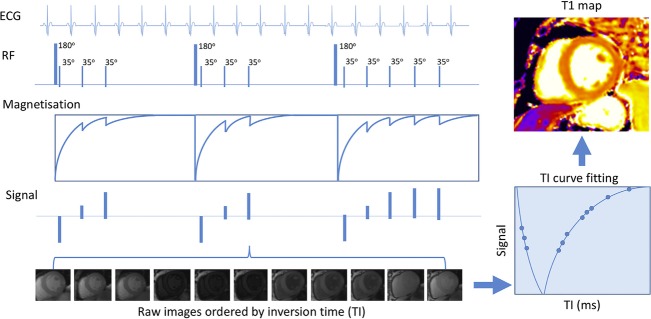
T1 mapping in cardiovascular magnetic resonance modified Look-Locker inversion recovery sequence. A sequence of three inversion recovery experiments are performed with images acquired and ordered according to inversion times. Signals are then used to plot a T1 recovery curve. The T1 value is the time when T1 recovery is 63% complete. T1 values are then used to create a voxel map. Adapted from Everett et al.
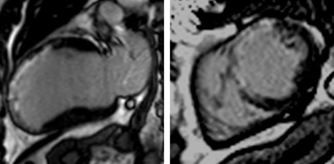
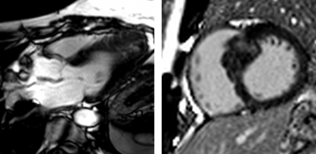
Ischaemic cardiomyopathy extensive anteroseptal myocardial infarction. Two-chamber (left) and short-axis (right) views demonstrate transmural late gadolinium enhancement in the left anterior descending artery territory with associated wall thinning, suggesting no viability.
Non-ischaemic dilated cardiomyopathy. Four-chamber (left) and short-axis (right) views demonstrate anteroseptal and inferoseptal late gadolinium enhancement in a typical non-ischaemic (mid-wall) distribution. Note sparing of the subendocardium.
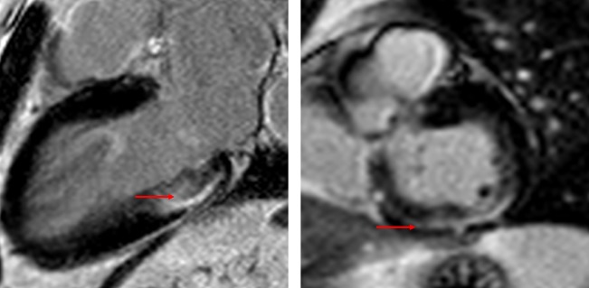
Aortic stenosis myocardial fibrosis in aortic stenosis. There is non-ischaemic late gadolinium enhancement in the basal inferolateral and inferior wall, where the subendocardium is spared (red arrow).
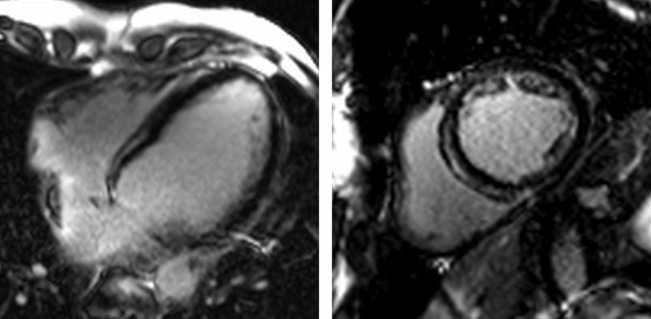
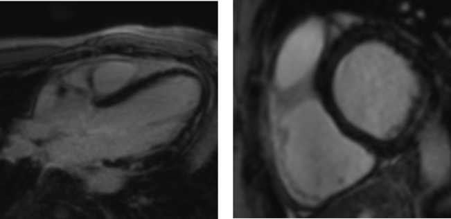
Hypertrophic cardiomyopathy examples of hypertrophic cardiomyopathy with typical septal hypertrophy (right) and an apical variant (left). Patchy non-infarct late gadolinium enhancement is seen within the regions of wall thickening.
Myocarditis. Three-chamber (left) and short-axis (right) examples of patchy, non-infarct, mid-wall late gadolinium enhancement in the anterolateral and inferolateral walls of a patient with chronic myocarditis (3 months after the onset of symptoms). Note that these findings are non-specific.
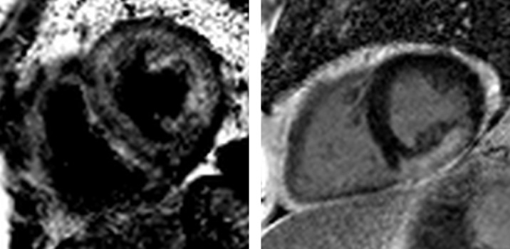
Cardiac amyloidosis and sarcoidosis. Left: cardiac amyloidosis. Note the black blood pool and diffuse late gadolinium enhancement within the abnormal myocardium. Right: cardiac sarcoidosis. The distribution of late gadolinium enhancement in cardiac sarcoidosis is variable. Here, there is a large burden of confluent enhancement in the inferior and inferolateral wall.
References
-
- Messroghli DR, Moon JC, Ferreira VM, et al. . Clinical recommendations for cardiovascular magnetic resonance mapping of T1, T2, T2* and extracellular volume: a consensus statement by the Society for (SCMR) endorsed by the European (EACVI). J Cardiovasc Magn Reson 2017;19 10.1186/s12968-017-0389-8 - DOI - PMC - PubMed
Publication types
MeSH terms
Grants and funding
LinkOut - more resources
Full Text Sources
Medical