. 2019 Oct 24;9(1):15263.
doi: 10.1038/s41598-019-51336-0.

An uncharacterized region within the N-terminus of mouse TMC1 precludes trafficking to plasma membrane in a heterologous cell line

Affiliations

An uncharacterized region within the N-terminus of mouse TMC1 precludes trafficking to plasma membrane in a heterologous cell line

D C Soler et al. Sci Rep. .

Abstract

Mechanotransduction by hair cell stereocilia lies at the heart of sound detection in vertebrates. Considerable effort has been put forth to identify proteins that comprise the hair cell mechanotransduction apparatus. TMC1, a member of the transmembrane channel-like (TMC) family, was identified as a core protein of the mechanotransduction complex in hair cells. However, the inability of TMC1 to traffic through the endoplasmic reticulum in heterologous cellular systems has hindered efforts to characterize its function and fully identify its role in mechanotransduction. We developed a novel approach that allowed for the detection of uncharacterized protein regions, which preclude trafficking to the plasma membrane (PM) in heterologous cells. Tagging N-terminal fragments of TMC1 with Aquaporin 3 (AQP3) and GFP fusion reporter, which intrinsically label PM in HEK293 cells, indicated that residues at the edges of amino acid sequence 138-168 invoke intracellular localization and/or degradation. This signal is able to preclude surface localization of PM protein AQP3 in HEK293 cells. Substitutions of the residues by alanine or serine corroborated that the information determining the intracellular retention is present within amino acid sequence 138-168 of TMC1 N-terminus. This novel signal may preclude the proper trafficking of TMC1 to the PM in heterologous cells.


Conflict of interest statement

The authors declare no competing interests.

Figures

Figure 1
Figure 1
Heterologous expression of TMC1 and AQP3. (A) When expressed in HEK293 cells, TMC1 is retained in ER and no PM localization is observed (left column, top and bottom panels), when compared to membrane immunolabeling of Na/K ATPase (left column, middle panel). Co-labeling with ER-Tracker (right column) confirms predominant ER localization of TMC1-YFP in live HEK293 cells. (B) Palmitoylated-GFP displays both intense cytoplasmic and membrane localization. (C) Expression of AQP3-GFP in HEK293 cells produces unequivocal strong membrane labeling. (D) Co-labeling with wheat germ agglutinin (WGA) conjugate demonstrates prevalent membrane localization of AQP3-GFP in live HEK293 cells. Scale bars: 8 µm.
Figure 2
Figure 2
AQP3-GFP reports intracellular retention quality of TMC1. (A) Schematic representation of AQP3-GFP reporter construct. A protein sequence of interest could be fused to the C-terminus of AQP3-GFP and tested for its ability to prevent AQP3-GFP from trafficking to the PM. (B) When fused to the C-terminus of AQP3-GFP, expression of full-length TMC1 results in an ER-like labeling pattern in HEK293 cells. (C) When only the N-terminus of TMC1 was tagged with AQP3-GFP, an ER-like labeling pattern was observed as well, without any noticeable PM fluorescence. (D) C-terminus of TMC1, when fused to AQP-GFP, does not preclude PM localization of the reporter (arrows). Scale bars: 8 µm.
Figure 3
Figure 3
Discerning the region within TMC1 N-terminus that precludes reporter trafficking to the PM in HEK293 cells. (A) N-terminal region of TMC1 composed of amino acids (aa) 1-46 (TMC11-46) does not preclude trafficking of AQP3-GFP to the PM (top row, white arrows). (B,C) TMC1 N-terminal aa sequences 47-92 (B, white arrows) and 93-137 (C, white arrows) do not preclude trafficking of the reporter to the PM. (D) TMC1 N-terminal aa sequence 138-183 (TMC1138-183) prevents AQP3-GFP from reaching the PM with a concomitant decrease in reporter fluorescence intensity (intensity of green channel is enhanced). (E) Quantification of AQP3-GFP-TMC1138-183 signal intensity as compared to AQP3-GFP-TMC11-46. When tagged with AQP3-GFP, TMC1138-183 induces a substantial decrease in reporter intensity. Fluorescent signal of AQP3-GFP-TMC1138-183 only becomes apparent if excitation light intensity is increased from 2% of maximal value (top micrograph) to 15% (bottom micrograph, the same field of view). ****p < 0.0001, ANOVA with Tukey post hoc test. Control – identically treated untransfected cells. (F) When tagged along with GFP alone, the TMC1138-183 fragment reduces the signal intensity and reproduces a reticular intracellular pattern (white arrow). (G,H) When tagged with AQP3-GFP, first half of TMC1 N-terminus, TMC11-92 (G), and fragment containing two thirds of N-terminus, TMC11-137 (H), were able to reach the PM (white arrows), although a strong ER pattern was evident. Scale bars: 8 µm (AD,G,H); 5 µm (E); 3 µm (F).
Figure 4
Figure 4
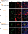
TMC1 N-terminus region between amino acids 138-168 precludes trafficking to the PM. (AC). Fragments TMC1138-152, TMC1153-168, and TMC1169-183 are able to reach the PM, when tagged with AQP3-GFP. (D) Fragment TMC1138-168, when tagged with AQP3-GFP, is sufficient to cause ER localization pattern and a decrease in reporter intensity. (E) When the overlapping 31 amino acids TMC1153-183 are tagged to AQP3-GFP, membrane localization and absence of ER staining are evident (white arrows). Scale bars: 8 µm (AC); 3 µm (D,E).
Figure 5
Figure 5
Amino acid residues at either ends of TMC1138-168 are critical for its activity. (A) Multiple alanine substitutions within the middle segment of TMC1138-168 failed to affect the ability of this region to prevent reporter trafficking to the PM. (B) A scrambled peptide of TMC1138-168 fragment is not able to prevent AQP3-GFP trafficking to the PM. (C,D) When residues WENKI (C) or AFKMM (D) at the ends of TMC1138-168 are removed, the resulting TMC1138-163 (C) or TMC1142-168 (D) fragments were able to reach the PM when tagged with AQP3-GFP. Scale bars: 3 µm.
Figure 6
Figure 6
Alanine and serine substitutions at the ends of TMC1136-168 sequence invoke N-terminus of TMC1 trafficking to the PM. (A) When key residues AFKMM and WENKI at either ends of TMC1138-168 are substituted by alanine and serine, the resulting mutant TMC1138-168 (mTMC1138-168) is able to reach the PM when tagged with AQP3-GFP. (B) Likewise, reporter tagged mutant full N-terminus of TMC1 (mTMC1138-168), harboring AFKMM and WENKI substitutions, is able to traffic to the PM. Scale bars: 8 µm

References

    1. Corey DP, Holt JR. Are TMCs the Mechanotransduction Channels of Vertebrate Hair Cells? The Journal of neuroscience: the official journal of the Society for Neuroscience. 2016;36:10921–10926. doi: 10.1523/JNEUROSCI.1148-16.2016. - DOI - PMC - PubMed
    1. Kurima K, et al. Dominant and recessive deafness caused by mutations of a novel gene, TMC1, required for cochlear hair-cell function. Nature genetics. 2002;30:277–284. doi: 10.1038/ng842. - DOI - PubMed
    1. Kitajiri SI, et al. Identities, frequencies and origins of TMC1 mutations causing DFNB7/B11 deafness in Pakistan. Clin Genet. 2007;72:546–550. doi: 10.1111/j.1399-0004.2007.00895.x. - DOI - PubMed
    1. Manji SS, Miller KA, Williams LH, Dahl HH. Identification of three novel hearing loss mouse strains with mutations in the Tmc1 gene. Am J Pathol. 2012;180:1560–1569. doi: 10.1016/j.ajpath.2011.12.034. - DOI - PubMed
    1. Vreugde S, et al. Beethoven, a mouse model for dominant, progressive hearing loss DFNA36. Nature genetics. 2002;30:257–258. doi: 10.1038/ng848. - DOI - PubMed

Publication types

LinkOut - more resources