. 2019 Sep;9(5):1021-1034.
doi: 10.1016/j.apsb.2019.02.001. Epub 2019 Feb 10.

MCC1019, a selective inhibitor of the Polo-box domain of Polo-like kinase 1 as novel, potent anticancer candidate

Affiliations

MCC1019, a selective inhibitor of the Polo-box domain of Polo-like kinase 1 as novel, potent anticancer candidate

Sara Abdelfatah et al. Acta Pharm Sin B. 2019 Sep.

Abstract

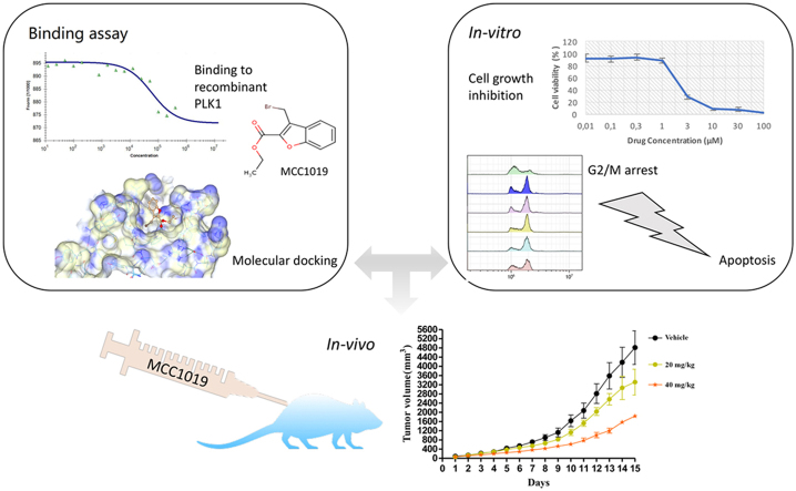
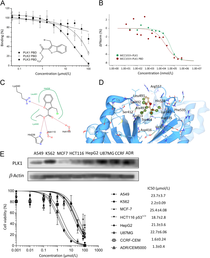
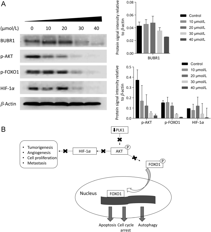
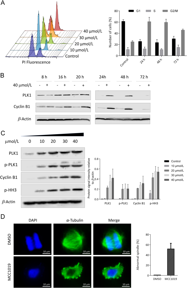
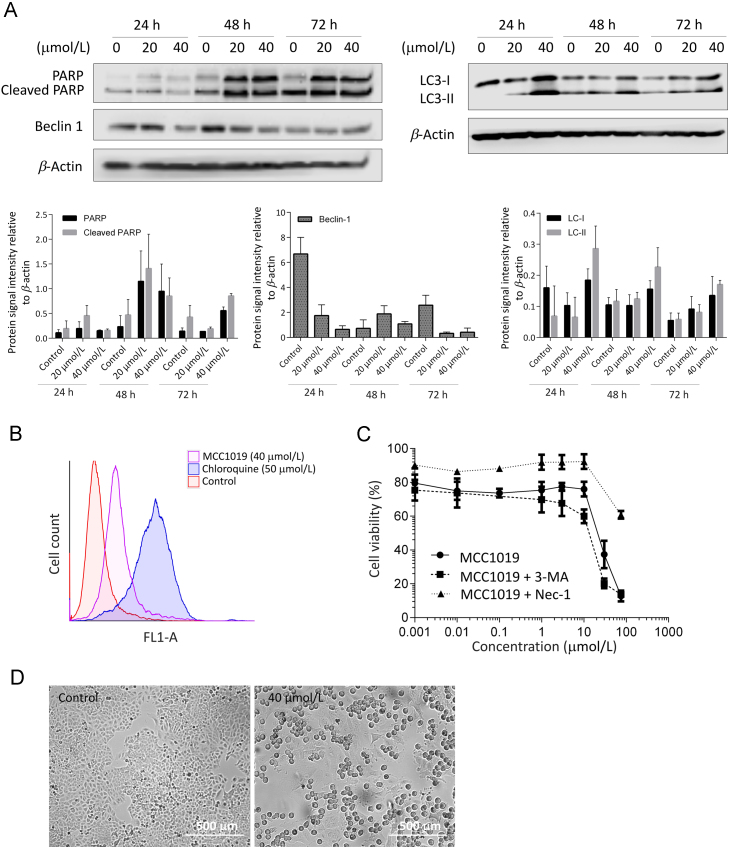
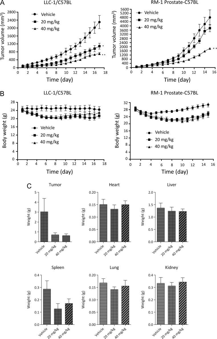
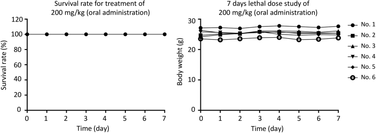
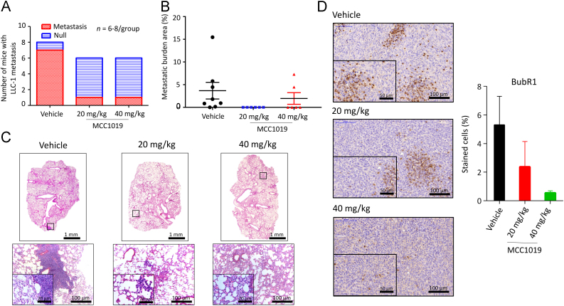
Polo-like kinase (PLK1) has been identified as a potential target for cancer treatment. Although a number of small molecules have been investigated as PLK1 inhibitors, many of which showed limited selectivity. PLK1 harbors a regulatory domain, the Polo box domain (PBD), which has a key regulatory function for kinase activity and substrate recognition. We report on 3-bromomethyl-benzofuran-2-carboxylic acid ethyl ester (designated: MCC1019) as selective PLK1 inhibitor targeting PLK1 PBD. Cytotoxicity and fluorescence polarization-based screening were applied to a library of 1162 drug-like compounds to identify potential inhibitors of PLK1 PBD. The activity of compound MC1019 against the PLK1 PBD was confirmed using fluorescence polarization and microscale thermophoresis. This compound exerted specificity towards PLK1 over PLK2 and PLK3. MCC1019 showed cytotoxic activity in a panel of different cancer cell lines. Mechanistic investigations in A549 lung adenocarcinoma cells revealed that MCC1019 induced cell growth inhibition through inactivation of AKT signaling pathway, it also induced prolonged mitotic arrest-a phenomenon known as mitotic catastrophe, which is followed by immediate cell death via apoptosis and necroptosis. MCC1019 significantly inhibited tumor growth in vivo in a murine lung cancer model without affecting body weight or vital organ size, and reduced the growth of metastatic lesions in the lung. We propose MCC1019 as promising anti-cancer drug candidate.

Keywords: 3-MA, 3-methyladenine; ABC, avidin-biotin complex; APC/C, anaphase-promoting complex/cyclosome; BUBR1, budding uninhibited by benzimidazole-related 1; CDC2, cell division cycle protein 2 homolog; CDC25, cell division cycle 25; CDK, cyclin-dependent kinase; Cell cycle; DAPI, 4′,6-diamidino-2-phenylindole; DAPKs, death-associated protein kinase; FBS, fetal bovine serum; FOXO, forkhead box O; HIF-1α, hypoxia-inducible factor 1 α; IC50, 50% inhibition concentration; IHC, immunohistochemistry; Kd, the dissociation constant; LC3, light chain 3; MFP, M phase promoting factor; MST, microscale thermophoresis; MTD, maximal tolerance dose; Mono-targeted therapy; Nec-1, necrostatin 1; Necroptosis; PARP-1, poly(ADP-ribose) polymerase-1; PBD, Polo box domain; PDB, Protein Data Bank; PI, propidium iodide; PLK1; PLK1, Polo-like kinase; Polo box domain; Polo-like kinase; SAC, spindle assembly checkpoint; Spindle damage.


Figures

fx1
Graphical abstract
Fig. 1
Figure 1
Inhibition of PLK1 PBD by MCC1019. (A) Inhibition of the binding of fluorescein-labeled phosphopeptides to PLK1, PLK2 or PLK3 PBDs by MCC1019 using a fluorescence polarization assay. The data are represented as mean ± SD of three independent experiments. (B) Thermophoresis binding curve of MCC1019 to the PLK1 and PLK1 PBD obtained by microscale thermopheresis. (C) Surface visualization of molecular docking of MCC1019 to the PLK1 PBD (PDB: 4X9R). (D) SeeSAR visualization of MCC1019 binding to human PLK1 (PDB: 4X9R): HYDE corona coloring is based on atomic affinity: green for favourable and red for unfavourable predicted affinities between atoms and amino acids. (E) Dose response curves of MCC1019 for different cancer cell lines obtained by cytotoxicity resazurin reduction assay, with westernblot analysis of PLK1 expression on each cell. The data are represented as mean ± SD of three independent experiments.
Fig. 2
Figure 2
MCC1019 inhibit downstream effector proteins of PLK1. (A) Western blot analysis of BUBR1, p-AKT, p-FOXO1 and HIF-1α after treatment with different concentrations of MCC1019 for 24 h. Data represent relative expression intensity to β-actin, Error bars are ± SD of three independent experiments. (B) Diagram showing molecular effects of PLK1 inhibition on AKT signaling pathway that appeared in the study.
Fig. 3
Figure 3
Induction of mitotic arrest by MCC1019. (A) Flow cytometric cell cycle analysis of exponentially growing A549 cells treated with MCC1019 for 24 h in a concentration range from 10 to 40 μmol/L. The data are represented as mean ± SD of three independent experiments. (B) Western blot analysis of A549 cell lysates treated with MCC1019 for 8, 16, 20, 24, 48 or 72 h. Increased expression levels of PLK1 and cyclin B1 were seen after 24 h treatment. (C) Western blot analysis of the mitotic markers PLK1, cyclin B1 and p-HH3 after treatment with different concentrations of MCC1019 for 24 h. Data represent relative expression intensity to β-actin, Error bars are ± SD of three independent experiments. (D) Immunofluorescence analysis of A549 treated with MCC1019 and DMSO (control) and stained for α-tubulin (green) and DNA (blue). The data are represented as mean ± SD of cells undergoing mitosis.
Fig. 4
Figure 4
Induction of apoptosis and necroptosis by MCC1019. (A) Western blot analysis of the apoptosis marker PARP and the autophagy markers LC3B and Beclin-1 in A549 cells treated with MCC1019 (20 or 40 μmol/L) for 24, 48 or 72 h. Data represent relative expression intensity to β-actin, Error bars are ± SD of three independent experiments. (B) Flowcytometric analysis of green dye signal for the detection of autophagy in A549 cells treated with MCC1019 (40 μmol/L) or chloroquine (50 μmol/L) as positive control. (C) Dose response curves of A549 treated with MCC1019 alone or in combination with 3 methyladenine (3-MA) or necrostatin-1 (Nec-1) obtained by cytotoxicity resazurin reduction assay. The data are represented as mean ± SD of three independent experiments. (D) Images of A549 cells after 24 h treatment with MCC1019 (40 μmol/L) taken with Juli Br live cell analyzer.
Fig. 5
Figure 5
Inhibition of tumour growth in vivo by MCC1019. (A) LLC-1 lung tumor bearing mice and RM-1 prostate tumor bearing mice models were used to determine the in vivo anti-tumor efficacy of MCC1019. (B) Total animal body weight changes were monitored after treatment with MCC1019 in both models. (C) Tumors and organs weight changes after MCC1019 treatment in LLC-1. The data are represented as mean ± SD (n = 6–8/group).
Fig. 6
Figure 6
Determination of the sub-chronic toxic dose of MCC1019. C57/BL/6 mice were orally administrated with 200 mg/kg MCC1019 for 7 consecutive days (n = 6). The survival and body weight of mice were monitored and recorded.
Fig. 7
Figure 7
MCC1019 inhibits tumor metastasis in vivo. MCC1019 (20 and 40 mg/kg/day) suppressed the lung metastasis frequency. Lung tissue sections from MCC1019 or vehicle control-treated mice (LLC-1 bearing mice model) were stained by hematoxylin. Metastasized LLC-1 cancer cells with larger nuclei were visualized in lung tissues and captured from 6–8 animals of each group. Normal images, 10× magnifications; enlarged images, 40 × magnification. (A) The bar chart represents the number of mice that survived in each group. Number of mice with lung metastatic lesions (red area) and mice without metastatic lesion (blue area) are shown. (B) MCC1019 (20 and 40 mg/kg/day) reduced the metastatic burden area. The percentage of metastatic burden area is displayed. Each dot represents one mouse. Data represent mean ± SEM. **P≤0.01, Mann–Whitney t-test for comparison between vehicle and MCC1019 (20 mg/kg/day). (C) Representative H&E stained lung sections images with metastatic lesions. Infiltrating metastatic cells are detailed at higher magnification. (D) Immunohistochemical determination of BUBR1 expression in formalin-fixed, paraffin-embedded LLC-1 cancer cells. The data are represented as mean ± SD of six randomly selected sections.

References

    1. Golsteyn R.M., Schultz S.J., Bartek J., Ziemiecki A., Ried T., Nigg E.A. Cell cycle analysis and chromosomal localization of human Plk1, a putative homologue of the mitotic kinases Drosophila polo and Saccharomyces cerevisiae Cdc5. J Cell Sci. 1994;107:1509–1517. - PubMed
    1. Nigg E.A. Polo-like kinases: positive regulators of cell division from start to finish. Curr Opin Cell Biol. 1998;10:776–783. - PubMed
    1. Jackman M., Lindon C., Niggt E.A., Pines J. Active cyclin B1–Cdk1 first appears on centrosomes in prophase. Nat Cell Biol. 2003;5:143–148. - PubMed
    1. Descombes P., Nigg E.A. The polo-like kinase Plx1 is required for M phase exit and destruction of mitotic regulators in Xenopus egg extracts. EMBO J. 1998;17:1328–1335. - PMC - PubMed
    1. Bosco A., Golsteyn R.M. Emerging anti-mitotic activities and other bioactivities of sesquiterpene compounds upon human cells. Molecules. 2017;22 :459-48. - PMC - PubMed