

Necrotic uveal melanoma presenting as orbital cellulitis with intraocular hemorrhage: A case report
- PMID: 31650085
- PMCID: PMC6804517
- DOI: 10.1016/j.ajoc.2019.100557
Necrotic uveal melanoma presenting as orbital cellulitis with intraocular hemorrhage: A case report
Abstract
Purpose: To report a case of necrotic uveal melanoma presenting as orbital cellulitis with an intraocular hemorrhage.
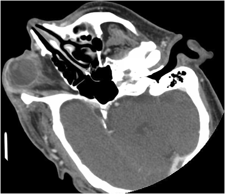
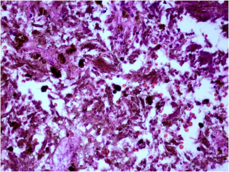
Observations: A 61 year-old non-verbal male presented with a two-week history of right eyelid swelling and erythema unresponsive to antibiotics. In addition to these signs of orbital cellulitis, he presented with an opaque media precluding fundus visualization. He was later found to have a collar-button shaped mass consistent with uveal melanoma on B scan ultrasonography during an exam under anesthesia. The patient underwent enucleation with histopathology confirming a necrotic uveal melanoma.
Conclusion and importance: This case demonstrates how necrotic uveal melanoma can present as orbital cellulitis and the importance of keeping the diagnosis on the differential.
Keywords: Necrotic uveal melanoma; Orbital cellulitis.
© 2019 The Authors.
Figures


References
-
- Fraser D.J., Font R.L. Ocular inflammation and hemorrhage as initial manifestations of uveal malignant melanoma incidence and prognosis. Arch Ophthalmol. 1979;97(7):1311–1314. - PubMed
-
- Nalcaci S., Melis P., Banu Y. Choroidal malignant melanoma with no extraocular extension presenting as orbital cellulitis. Orbit. 2016;35(5):285–287. - PubMed
-
- Nair A.G., Kaliki S., Ali M.J. Intraocular malignant melanoma of the choroid presenting as orbital cellulitis. Int Ophthalmol. 2014;34:647–650. - PubMed
-
- Biswas J., Ahuja V.K., Shanmugam M.P. Malignant melanoma of the choroid presenting as orbital cellulitis: report of two cases with a review of the literature. Orbit. 1999;18(2):123–130. - PubMed
Publication types
LinkOut - more resources
Full Text Sources
